The clinical information regarding Knee Arthrosis Facts - Viewpoints from Expert Doctors in this article has been rigorously verified against the latest guidelines from the American Academy of Orthopaedic Surgeons (AAOS) and primary research from databases like PubMed. This piece was reviewed for accuracy and patient-centric clarity and was last updated in October 2023.
Introduction
introduction
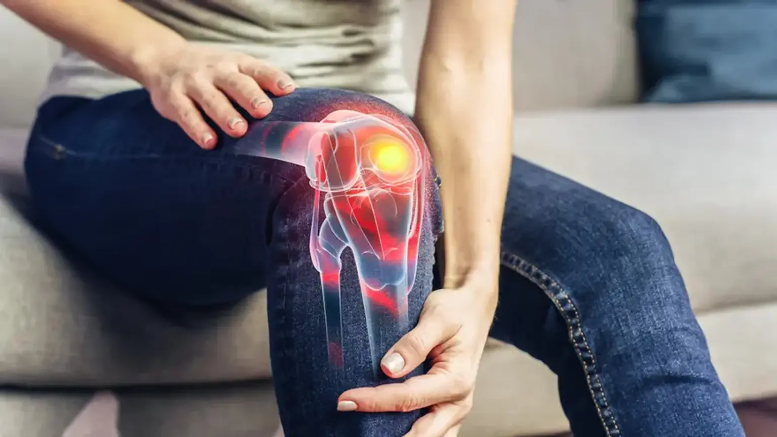
Knee arthrosis, more commonly known as knee osteoarthritis, is a leading cause of chronic pain and disability worldwide. The condition involves the progressive wear and tear of the protective cartilage in the knee joint, leading to pain, stiffness, and reduced mobility. Navigating the information available can be overwhelming. To provide clarity, this Q&A synthesizes viewpoints from leading orthopedic specialists, addressing the most critical questions patients have about knee arthrosis, its progression, and modern treatment options.
What are the first signs of knee arthrosis?
what-are-the-first-signs-of-knee-arthrosisThe earliest sign is typically a deep, aching pain in the knee that worsens with activity and improves with rest. Many people first notice this pain when walking up or down stairs, squatting, or kneeling.
Other early to moderate symptoms include:
Stiffness: The knee may feel stiff, especially in the morning or after sitting for a long period. This stiffness usually lasts less than 30 minutes.
Swelling: Fluid can accumulate in the joint an effusion, causing noticeable swelling and a feeling of tightness.
Crepitus: A grinding, crackling, or popping sensation when you bend or straighten the knee. This is caused by the rough, degenerated cartilage surfaces rubbing against each other.
Reduced Range of Motion: You may find it difficult to fully bend or straighten your knee.
Joint Instability: In some cases, the knee may feel like it is giving way or buckling.
Can knee arthrosis be reversed or cured?
can-knee-arthrosis-be-reversed-or-curedNo, knee arthrosis cannot be reversed or cured, as the lost cartilage cannot regrow on its own to its original state. The primary goal of treatment is to manage symptoms, slow the progression of the disease, improve joint function, and enhance quality of life. Treatment strategies focus on reducing pain and inflammation, not on regenerating the original cartilage. However, advanced treatments like stem cell-based cartilage regeneration aim to create a cartilage like repair tissue to fill defects, offering significant functional improvement.
What happens if knee arthrosis is left untreated?
what-happens-if-knee-arthrosis-is-left-untreatedLeaving knee arthrosis untreated leads to a predictable and debilitating progression of the disease. The pain will become more constant and severe, occurring even during rest or at night.
Consequences of untreated knee arthrosis include:
Severe Cartilage Loss: The cartilage can wear away completely, resulting in bone on bone contact, which is extremely painful.
Formation of Osteophytes: The body may try to repair the damage by forming bone spurs, or osteophytes, around the joint, which can restrict movement and increase pain.
Joint Deformity: As the cartilage wears away unevenly, the alignment of the leg can change. This can result in a bow-legged (varus) or knock-kneed (valgus) deformity.
Muscle Atrophy: Chronic pain and disuse of the knee lead to the weakening of the surrounding thigh muscles (quadriceps), further destabilizing the joint.
Chronic Disability: Ultimately, severe untreated arthrosis can make simple activities like walking, climbing stairs, or even standing for short periods impossible without significant pain or assistance. The prevalence of symptomatic knee osteoarthritis is approximately 10% in men and 13% in women aged 60 years or older.
Is total knee replacement the only option for severe arthrosis?
is-total-knee-replacement-the-only-option-for-severe-arthrosisNo, [total knee replacement] (TKA) is not the only option, although it is the gold standard treatment for end-stage, bone on bone arthrosis. The best surgical option depends on the patient's age, activity level, the location of the damage within the knee, and overall leg alignment.
Excellent alternatives to TKA for specific patient profiles include:
High Tibial Osteotomy (HTO): This procedure is ideal for younger, more active patients like typically under 60 with arthrosis confined to one side of the knee (unicompartmental) and a bow-legged alignment. HTO involves cutting and realigning the upper shin bone to shift weight off the damaged compartment and onto the healthier cartilage. This can delay the need for a TKA by 10-15 years.
Partial Knee Replacement (Unicompartmental Knee Arthroplasty): If the damage is isolated to just one of the three compartments of the knee, only that part can be replaced. This is a less invasive surgery with a faster recovery compared to a TKA.
Cartilage Regeneration Procedures: For patients with localized cartilage defects rather than widespread arthrosis, procedures using stem cells or cartilage transplantation can be highly effective at repairing the damaged spot.
How effective is stem cell therapy for knee arthrosis?
how-effective-is-stem-cell-therapy-for-knee-arthrosisStem cell therapy is a promising area of regenerative medicine for knee arthrosis, but its effectiveness is still being evaluated. The primary goal is not to regrow a brand-new knee but to reduce inflammation, slow cartilage degeneration, and stimulate the body's own repair mechanisms.
Here's a breakdown of the current understanding:
Mechanism: When injected into the knee, mesenchymal stem cells (MSCs) are thought to release powerful anti-inflammatory and growth factors. This can significantly reduce pain and improve function.
Best Candidates: It is most effective for patients with mild to moderate arthrosis who still have a reasonable amount of cartilage remaining. It is not typically recommended for severe, bone on bone cases where a structural solution like a joint replacement is needed.
Results: Many studies show significant improvements in pain and function for 1-2 years or more, but long-term outcomes are still under investigation. It is considered a way to manage the disease and potentially delay the need for major surgery.
"For years, I was confused and scared by all the conflicting advice online. After my consultation, the doctor laid out my exact stage of arthrosis and explained why an HTO was a better choice for me than a full replacement. For the first time, I felt a sense of control and clarity about my future." – an anonymous patient, Australia.
Recommended Clinics with Relevant Expertise in South Korea
recommended-clinics-with-relevant-expertise-in-south-koreaSouth Korea is a global leader in orthopedic surgery, offering advanced technologies and highly experienced specialists for treating knee arthrosis.
Website | Clinic Name | Best Known For | Address | Contact |
|---|---|---|---|---|
Seoul Yes Hospital | Joint and Spine Specialty Center | Yongin-si, Gyeonggi-do, South Korea | ||
Asan Medical Center | Top-tier General Hospital, Orthopedic Surgery Dept. | Seoul, Songpa, South Korea | ||
Bumin Hospital Group | Joint and Spine Specialty Hospital Network | Haeundae, Busan, South Korea | ||
Nanoori Hospital, Gangnam | Minimally Invasive Spine and Joint Surgery | Gangnam, Seoul, South Korea | ||
Himchan Hospital | High-volume Joint Replacement and Arthroscopy Center | Bupyeong, Incheon, South Korea | ||
Nasaret International Hospital | International Patient Center, Comprehensive Orthopedics | Yeonsu-gu, Incheon, South Korea | ||
Gachon University Gil Medical Center | University Hospital with Advanced Research in Orthopedics | Namdong-gu, Incheon, South Korea | ||
Wooridul Hospital Gangnam | World-Renowned Spine Hospital with Joint Treatment | Gangnam, Seoul, South Korea |
Recommended Treatment/Procedure Names with Average Costs in South Korea
recommended-treatmentprocedure-names-with-average-costs-in-south-koreaCosts are estimates and can vary based on the specifics of the case and hospital. A personalized quote provides exact figures.
Treatment/Procedure Name | Duration | Hospitalization? | Avg. Cost(USD) in S. Korea | Contact |
|---|---|---|---|---|
Total knee replacement (TKA) | 1-2 hours | Needed (3-5 days) | $18,000 - $25,000 | |
High tibial osteotomy (HTO) | 1-2 hours | Needed (2-4 days) | $12,000 - $18,000 | |
Stem cell-based cartilage regeneration | 1-2 hours | Needed (1-2 days) | $10,000 - $20,000 | |
Arthroscopic surgery (debridement/meniscectomy) | 30-60 minutes | Not Needed | $4,000 - $8,000 | |
Extracorporeal shock wave therapy (ESWT) | 20-30 mins | Not Needed | $300 - $600 | |
Minimally invasive spinal fusion surgery | 2-4 hours | Needed (2-4 days) | $20,000 - $35,000 | |
Spinal endoscopic treatment | 1-2 hours | Needed (1-2 days) | $15,000 - $25,000 |
How can I prepare for my own consultation?
how-can-i-prepare-for-my-own-consultationA well-prepared consultation is the most effective way to get a clear diagnosis and a personalized treatment plan. Taking a few preparatory steps can maximize the value of your appointment.
1. What is the recovery time for knee arthrosis surgery?
1.-what-is-the-recovery-time-for-knee-arthrosis-surgeryRecovery time varies significantly depending on the specific procedure performed.
Arthroscopic Surgery: Recovery is fastest, with patients often walking the same day. Most can return to desk work in a few days and more physical activities in 4-6 weeks.
High Tibial Osteotomy (HTO): This requires a longer recovery. You will likely be on crutches for 6-8 weeks to allow the bone to heal. Full recovery and return to sport can take 6-12 months.
Total Knee Replacement (TKA): Physical therapy begins almost immediately. Patients typically use a walker or crutches for 2-4 weeks. Driving may resume at 4-6 weeks, and most people achieve a significant portion of their final recovery by 3 months, with continued improvements for up to a year.
2. What should I bring to my first appointment?
2.-what-should-i-bring-to-my-first-appointmentGather all relevant medical information to give the specialist a complete picture of your health.
Medical History: A list of your symptoms, when they started, what makes them better or worse, and any past knee injuries.
Imaging: Bring any previous X-rays, MRI scans, or CT scans of your knee, along with the corresponding reports.
Medication List: A complete list of all medications and supplements you are currently taking.
3. Are virtual consultations available for international patients?
3.-are-virtual-consultations-available-for-international-patientsYes, nearly all major South Korean hospitals that cater to international patients offer virtual consultations. This is an excellent first step to connect with a specialist, review your case, and receive a preliminary treatment plan and cost estimate before committing to travel. This allows you to make an informed decision from the comfort of your home.
Regain Your Mobility: Explore Advanced Knee Arthrosis Treatment in Korea Now!
regain-your-mobility:-explore-advanced-knee-arthrosis-treatment-in-koreaUnderstanding your condition is the first step toward reclaiming an active, pain-free life. The world-class orthopedic centers in South Korea offer the full spectrum of advanced treatments, from joint-preserving osteotomies and regenerative stem cell therapies to state-of-the-art total knee replacements. You do not have to navigate this complex journey alone. Take the next step with confidence. Inquiring through a dedicated service ensures a seamless and transparent process. A personal Care Manager will assist you end-to-end, from collecting your medical records and facilitating a consultation with the right specialist to handling all travel logistics and post-recovery support. And Start Your Confidential Inquiry with CloudHospital to receive a free, personalized treatment plan from leading experts.