The clinical information regarding How to Prevent and Treat Retinal Detachment in Korea? in this article has been rigorously verified against the latest Preferred Practice Pattern guidelines from the American Academy of Ophthalmology (AAO) and primary research from databases like PubMed. This piece was reviewed for accuracy and patient-centric clarity and was last updated in October 2023.
Introduction
introduction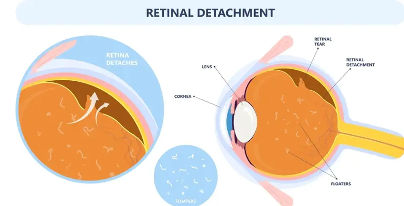
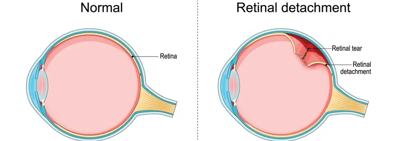
A retinal detachment is a serious medical emergency where the retina the light-sensitive layer of tissue at the back of the eye pulls away from its normal position. If not treated promptly, it can lead to permanent vision loss. Recognizing the symptoms and knowing your treatment options are critical. This guide provides a clear, step by step plan for preventing, identifying, and treating retinal detachment, with a specific focus on the advanced medical landscape of South Korea.
How Can I Reduce My Risk of Retinal Detachment?
how-can-i-reduce-my-risk-of-retinal-detachmentYou can reduce your risk by managing underlying health conditions, protecting your eyes from injury, and attending regular comprehensive eye exams. While not all cases are preventable, proactive measures are crucial, especially for high-risk individuals.
Regular Eye Exams: Annual dilated eye exams are the single most effective preventive step. An ophthalmologist can detect retinal tears or weaknesses before they progress to a full detachment.
Protective Eyewear: Use safety glasses or sports goggles during activities that pose a risk of eye injury, such as contact sports, construction work, or racquet sports.
Control Medical Conditions: Effectively manage conditions like diabetes and high blood pressure, as they can affect the blood vessels in your eyes.
Know Your Risk Factors: Be aware if you have a high degree of nearsightedness myopia, a family history of retinal detachment, have had previous eye surgery like Cataract Surgery, or have experienced a prior serious eye injury.
What Are the Early Warning Signs of a Retinal Detachment?
what-are-the-early-warning-signs-of-a-retinal-detachmentThe primary warning signs of a retinal detachment are the sudden appearance of many floaters, sudden flashes of light photopsia, and a shadow or curtain appearing in your field of vision. These symptoms signal a medical emergency requiring immediate attention from an ophthalmologist.
Sudden Increase in Floaters: A sudden shower of tiny specks or cobweb like shapes drifting through your field of vision.
Flashes of Light (Photopsia): Brief, lightning like flashes in your peripheral vision, often more noticeable in dim lighting. This is caused by the vitreous humor gel tugging on the retina.
A Curtain or Shadow: A dark, curtain like shadow that starts in your peripheral vision and slowly moves towards the center.
Blurred Vision: A sudden, significant decrease in the clarity of your vision.
"I noticed a few extra floaters one afternoon but ignored them. The next morning, it was like a dark curtain was drawn over the side of my eye. I went to the emergency room immediately, and the doctors said that quick action saved my sight." – An anonymous patient, United States.
How is a Retinal Detachment Diagnosed?
how-is-a-retinal-detachment-diagnosedA retinal detachment is diagnosed through a comprehensive dilated eye examination performed by an ophthalmologist, often supplemented with imaging tests like an ocular ultrasound. The ophthalmologist uses a special instrument with a bright light and lens to view the back of your eye, including the retina, to identify any tears, holes, or detachment.
Dilated Eye Exam: This is the standard and most crucial diagnostic step. Eye drops are used to widen your pupils, giving the doctor a clear view of the entire retina.
Ocular Ultrasound (B-scan): If the view of the retina is obscured by blood or a dense cataract, an ultrasound can create a detailed image of the back of the eye to confirm the detachment.
Optical Coherence Tomography (OCT): This non-invasive imaging test provides a high resolution cross sectional image of the retina, which can identify if fluid has accumulated underneath it.
What Are the Main Surgical Treatments for Retinal Detachment in Korea?
what-are-the-main-surgical-treatments-for-retinal-detachment-in-koreaThe three primary surgical treatments for a detached retina are vitrectomy, scleral buckle, and pneumatic retinopexy, often combined with laser or freezing therapy to seal the retinal tear. The choice of procedure depends on the type, location, and severity of the detachment, and South Korean vitreoretinal surgeons are highly skilled in all three techniques.
Vitrectomy: This is the most common procedure performed today. The surgeon removes the vitreous gel that is pulling on the retina and replaces it with a gas bubble or silicone oil. The bubble pushes the retina back into place, allowing it to heal. The success rate for a single vitrectomy procedure is generally high, often reported between 80-90% for reattaching the retina.
Scleral Buckle: In this procedure, the surgeon places a flexible band usually made of silicone around the eye. This band gently pushes the wall of the eye inward to meet the detached retina, relieving the traction that caused the tear and allowing it to reattach.
Pneumatic Retinopexy: A less invasive option suitable for smaller, uncomplicated detachments. The surgeon injects a small gas bubble into the eye, which floats up and presses the retina back into place. The patient must maintain a specific head position for several days to keep the bubble correctly positioned. This is often combined with cryopexy freezing or laser photocoagulation to seal the retinal tear.
What is the Recovery Process Like After Retinal Detachment Surgery?
what-is-the-recovery-process-like-after-retinal-detachment-surgeryRecovery after retinal detachment surgery involves a period of restricted activity, the use of medicated eye drops, and potentially maintaining a specific head position for days or weeks. The eye will be sore and blurry initially, with vision gradually improving over several months.
Head Positioning: If a gas bubble was used vitrectomy or pneumatic retinopexy, you will be required to maintain a face down or side lying position for a specific period to ensure the bubble properly supports the retina. This is a critical part of the healing process.
Activity Restrictions: Strenuous activity, heavy lifting, and bending are prohibited for several weeks to prevent pressure changes in the eye.
Eye Drops: You will be prescribed antibiotic and anti-inflammatory eye drops to prevent infection and reduce swelling.
Vision Return: Vision is typically very poor immediately after surgery but improves slowly over weeks to months. The final visual outcome depends on whether the macula the center of the retina was detached before the surgery.
Travel Restrictions: You cannot fly in an airplane or travel to high altitudes until the gas bubble is completely gone, as changes in atmospheric pressure can cause a dangerous spike in eye pressure.
Recommended Clinics with Retinal Disease Expertise in South Korea
recommended-clinics-with-retinal-disease-expertise-in-south-koreaSouth Korea is home to world class medical facilities and highly experienced ophthalmologists specializing in complex retinal diseases. The clinics listed below are recognized for their advanced diagnostic and surgical capabilities.
Website | Clinic Name | Best Known For | Address | Contact |
|---|---|---|---|---|
SNU Eye Clinic | University based comprehensive eye care, corneal disease research | Gangnam-gu, Seoul, South Korea | ||
Gangnam Joeunnun Vision Clinic | Advanced vision correction surgery, ICL, LASIK | Gangnam-gu, Seoul, South korea | ||
Jryn Eye Clinic | LASIK, LASEK, Smile, and Presbyopia Correction | Busanjin-gu, Busan, South Korea | ||
Global Ubal Eye Center | Refractive surgery and specialized eye treatments | Jung-gu, Incheon, South Korea | ||
Cha University Bundang Medical Center | Comprehensive hospital with specialized ophthalmology department | Bundang-gu, Seongnam-si, South Korea | ||
Chosun University Hospital | University hospital with extensive eye care services | Gwangju, Dong-gu, South Korea | ||
Kangdong Sacred Heart Hospital | Full service hospital with advanced ophthalmology diagnostics | Gangdong-gu ,Seoul, South Korea | ||
Samyook Busan Adventist Hospital | General hospital with ophthalmology and patient care services | Busan, Seo-gu, South Korea |
Retinal Detachment Treatment Costs in South Korea
retinal-detachment-treatment-costs-in-south-koreaThe cost of retinal detachment surgery varies based on the specific procedure, the complexity of the case, and the chosen hospital. The following table provides estimated costs for common treatments in South Korea.
Treatment/Procedure Name | Duration | Hospitalization? | Avg. Cost (USD) in S. Korea | Contact |
|---|---|---|---|---|
Vitrectomy | 1-3 hours | Needed (1-2 days) | $6,000 - $12,000 | |
Scleral Buckling | 1-2 hours | Needed (1-2 days) | $5,000 - $10,000 | |
Pneumatic Retinopexy | 30-60 minutes | Not Needed | $3,000 - $6,000 | |
Laser Photocoagulation / Cryopexy (for tears) | 15-30 minutes | Not Needed | $800 - $2,500 |
Frequently Asked Questions About Retinal Detachment
frequently-asked-questions-about-retinal-detachmentThis section answers common follow-up questions about retinal detachment and its treatment.
1. What happens if retinal detachment is not treated?
1.-what-happens-if-retinal-detachment-is-not-treatedIf left untreated, a retinal detachment will almost always lead to complete and permanent blindness in the affected eye. The retinal cells lose their blood supply and begin to die, a process that is irreversible. This is why it is considered a true medical emergency.
2. Can I fly on an airplane after my surgery?
2.-can-i-fly-on-an-airplane-after-my-surgeryYou absolutely cannot fly if a gas bubble was placed in your eye during a vitrectomy or pneumatic retinopexy. The change in cabin pressure can cause the bubble to expand, leading to a catastrophic increase in eye pressure that can cause blindness. You must wait until the doctor confirms the bubble has fully dissolved.
3. Will my vision be perfect after a successful surgery?
3.-will-my-vision-be-perfect-after-a-successful-surgeryThe goal of surgery is to reattach the retina and prevent total blindness. While vision is saved, it may not return to the level it was before the detachment, especially if the macula was involved. Some degree of permanent blurriness or distortion is common, but most patients regain useful vision.
4. Are both eyes usually affected?
4.-are-both-eyes-usually-affectedUsually, only one eye is affected at a time. However, if you have a detachment in one eye, you are at a significantly higher risk of developing one in the other eye. The lifetime risk for the fellow eye can be as high as 10%. Your ophthalmologist will carefully examine your other eye for any signs of weakness.
5. What are the risks associated with retinal detachment surgery?
5.-what-are-the-risks-associated-with-retinal-detachment-surgeryWhile surgery is highly effective, it carries risks. These include infection, bleeding, increased eye pressure glaucoma, cataract formation very common after vitrectomy, and failure of the retina to reattach, which may require additional surgery.
6. Why is face-down positioning required after some surgeries?
6.-why-is-face-down-positioning-required-after-some-surgeriesFace down positioning is required to ensure the gas bubble injected into the eye floats up to press against the retinal tear, holding it in place like an internal bandage. Proper positioning is absolutely critical for the surgery to be successful.
Take the First Step to Protecting Your Vision Now
take-the-first-step-to-protecting-your-visionFacing a diagnosis of retinal detachment is daunting, but immediate action and expert care provide the best chance for a successful outcome. The highly advanced ophthalmology centers in South Korea offer a path to preserving your sight. By inquiring, you can access a seamless and transparent process, guided by a dedicated Care Manager who will support you from the initial consultation to your post-recovery care. Don't wait Get a Free Treatment Plan & Quote from CloudHospital today!