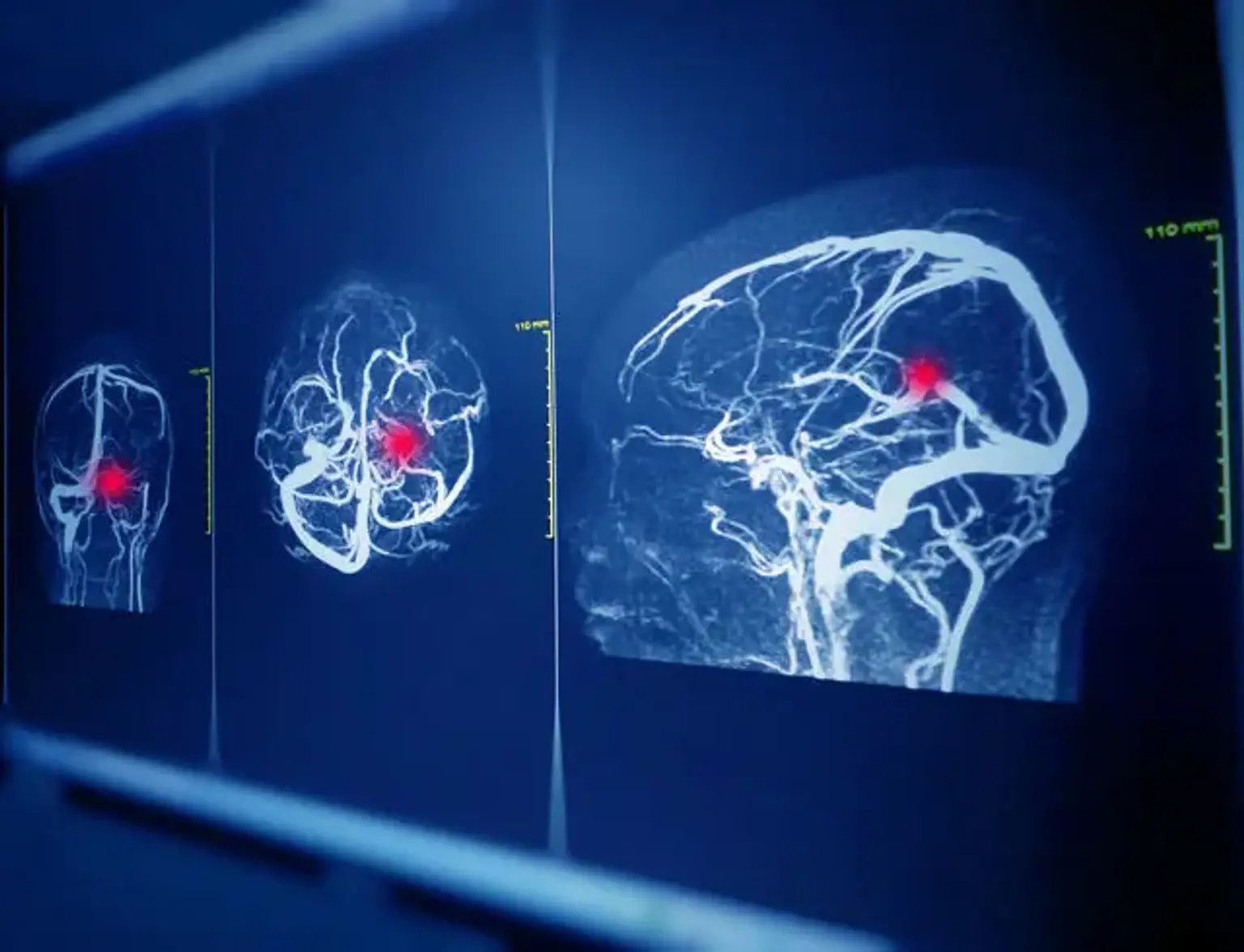
Blood vessel blockages or abnormalities can lead to a stroke or bleeding in the brain. Cerebral angiography is a procedure originally pioneered in 1927 by the Portuguese neurologist Egas Moniz at the University of Lisbon that today uses a catheter, X-ray imaging, and an injection of contrasting material to examine blood vessels in the brain for abnormalities such as aneurysms and disease such as atherosclerosis. The use of a catheter makes it possible to combine diagnosis and treatment in a single procedure. Cerebral angiography produces very detailed, clear, and accurate pictures of blood vessels in the brain and may eliminate the need for surgery.
Doctors use cerebral angiography to detect problems within the blood vessels in the brain, including:
- aneurysm, a bulge, or sac that develops in an artery due to weakness of the arterial wall.
- atherosclerosis, a narrowing of the arteries
- arteriovenous malformation, a tangle of dilated blood vessels that prevents normal blood flow in the brain.
- vasculitis (inflammation of the blood vessels)
- brain tumor
A cerebral angiogram may be performed to:
- evaluate arteries of the head and neck before surgery
- provide additional information on abnormalities seen on MRI or CT of the head
- prepare for medical treatments, such as in the surgical removal of a tumor
- prepare for minimally invasive treatment of a vessel abnormality
