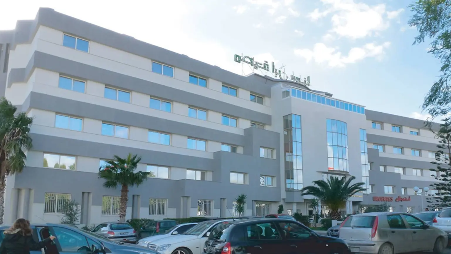
This page is been managed directly by the hospital. Any communications initiated will be directly responded by the hospital only.

    Avicenna Clinic

    Tunis, Tunisia

    1974

    Foundation year

    80

    Doctors

    All / Top Specialties

    • Varicose Veins

    • Bladder cancer

    • Tympanoplasty

    All / Top Services

    • Thrombosis

    • Venous thrombosis

    • Bladder Cancer

    • Tympanoplasty

    • Genioplasty

    • Osteotomy

    • Hydrocelectomy

    • Anal Fistula

    • Gynecomastia

    • Enlargement of Breast in Men (Gynecomastia)

    • Abdominoplasty (Tummy Tuck)

    • Sleeve bariatric surgery

    • Buttock Lipofilling

    • Total Hip Replacement

    • Ischemic stroke

    • Embolic Stroke

    • Cervico-facial facelift

    • Facial lipofilling

    • Face Lift

    • Cervico-facial Lift

    About

    A leader in its field since 1974, the Avicenne TUNISIA clinic is a multidisciplinary high standing clinic which has a state-of-the-art technical platform in accordance with international standards. Avicenne clinic owes its reputation to the experience of its paramedical staff and the competence of its highly qualified doctors in their specialties. Gynecology and Obstetrics, Neonatology, Pediatrics, Medical and Surgical Oncology, General Surgery, Orthopedic Surgery, ENT, Neurosurgery, Urology, Thyroid Surgery and Cosmetic Surgery. The Avicenne clinic is distinguished by Its Neonatology department where it ensures the best care and well-being of fragile or premature newborns as well as the management of congenital malformations (hare lip, spina bifida, etc.). Its orthopedic-traumatology unit which constitutes one of the prides of the Clinic, where the activity is based both on primary surgery but also on complex recovery surgeries, requiring highly specialized care (rupture of the cruciate ligaments, hip prosthesis, knee prosthesis, shoulder arthroscopy, herniated disc, scoliosis). Its urological surgery department which is among the best for laser treatment of the prostate. Cancer center The Avicenne clinic, through its cancerology center, is recognized as a "center specializing in the treatment of cancer" authorized to treat most cancers by surgery or chemotherapy. The Avicenne clinic offer the patient comprehensive care that begins with cancer screening in the medical imaging department or the endoscopy center and continues with surgery and/or chemotherapy treatments. As far as radiotherapy and hormone therapy are concerned, the Avicenne clinic collaborates with the best centers in the capital. Clinique Avicenne has a multidisciplinary team made up of oncologists, surgeons, radiologists, doctors from hospital departments, pathologists and specialists. A wide range of supportive care is offered to patients. Obstetrics Gynecology Center The medical team of the Gynecology-Obstetrics Center of the Avicenne clinic takes care of the various gynecological problems that may arise: annual follow-up, control, contraception (in particular implant and IUD), menopause, fertility, pregnancy and childbirth, gynecological and breast surgery and gynecological oncology. Vascular surgery center The Avicenne clinic has a team of medical specialists who provide interventions affecting both arteries and veins, such as varicose veins or deep vein thrombosis. Adult and Pediatric Orthopedic Center Total Hip prosthesis The placement of a total hip prosthesis is suggested by the doctor when the disability has become severe and when the joint damage is very advanced. The patient must know what the intervention consists of and what he is exposed to in the aftermath. After having been well informed, it is he who decides. "Total" means that the prosthesis concerns the 2 parts of the hip joint: the part of the pelvis (the acetabulum) and the part of the femur (the head). The models of total prosthesis are not lacking. They vary according to the shape, the material and the method of fixation of the prosthesis. Pain that is insufficiently relieved by analgesic drugs and the resulting disability (inconvenience in walking, reduced activity) are good reasons for considering the installation of a total hip prosthesis. If, in addition, the osteoarthritis lesions are very advanced on the most recent X-rays, this is an additional reason to think about it. The lifespan of a prosthesis varies from 15 to 25 years depending on the age, weight and activity of the patient. Crossed ligaments The ligament breaks most often in sport (football, skiing, basketball, etc). Unlike other ligaments, it does not repair itself, even with a cast. This is the most important ligament of the knee since it connects the femur to the tibia by allowing these two bones to articulate with each other harmoniously. Surgery It will make it possible to remake a new anterior cruciate ligament. Apart from the opening of the skin necessary to take this or these tendons, all the rest of the operation takes place under arthroscopy and video control by a miniaturized camera. This arthroscopy will allow at the same time to assess the interior of the knee (cartilage, meniscus, other ligaments) and treat any torn menisci. The new ligament will be fixed by special screws in absorbable product. Cervical disc herniation The cervical spine (or cervical spine) is made up of 7 stacked vertebrae separated by discs, except between the 1st and 2nd cervical vertebra (C1 and C2). In the middle of the cervical spine is the spinal canal containing the spinal cord. The normal intervertebral disc is a flattened, cylindrical structure uniting the two vertebrae and acting as a shock absorber. Disc deterioration is characterized by tearing or cracking of the annulus fibrosis. The causes are dehydration due to aging, microtraumas due to mobility constraints and sometimes also traumas such as cervical sprains. What Are The Objectives Of The Surgery? The goal of the operation is to remove the herniated disc to release the nerve root (or even the spinal cord) and eliminate the pain in the upper limb. This goal is achieved in approximately 90% of cases. On the other hand, neck pain may persist Carpal Tunnel Carpal tunnel syndrome is the clinical manifestation of compression of the median nerve at the wrist. This nerve innervates the hand on the sensory level (thumb, index, middle finger and half of the ring finger) and motor (muscles at the base of the thumb). The flexor nerve and tendons pass through a bony canal (the carpal tunnel) which is covered by a thick ligament (the anterior annular ligament). Any increase in pressure in the carpal tunnel causes pain in the nerve. Treatment Symptoms can be reduced with medical treatment. In the event of an associated pathology (diabetes, hypothyroidism, etc.), the treatment of the disease generally relieves the carpal tunnel syndrome. Resting the affected hand, a correct working position, or even wearing a resting orthosis can help reduce compression of the median nerve. Intraductal infiltration can also relieve symptoms. In case of persistence or aggravation of the signs, it is necessary to resort to a surgical treatment. The goal of the surgery is to cut the ligament covering the nerve, to reduce the pressure. The consequences are usually simple, the patient can resume a light activity in the week following the intervention. Strength recovers in 6-12 weeks. The healing of the incision is complete in 15 to 21 days. Narrow Lumbar Canal The vertebral column (or rachis) is made up of vertebrae in the middle of which there is a canal: the spinal canal. This contains the spinal cord and nerves. At the lumbar level, the spinal cord stops and the canal only contains the nerves of the “cauda equina” which innervate the lower limbs and the perineum. This channel is usually wide enough to contain all the nerve roots without problems. Depending on the constitution of the patient or due to natural wear phenomena (osteoarthritis), a narrowing of the spinal canal (lumbar canal stenosis or narrowed lumbar canal) may occur. This narrowing or “stenosis” of the spinal canal will cause compression of the nervous elements contained in the spinal canal. Only surgery can expand the diameter of the spinal canal. An intervention is considered when the patient has had a complete medical treatment, when he is “fed up with pain” or in an emergency: Motor deficit (paralyzing sciatica); Intolerable pain not relieved by opioids (hyperalgesic sciatica); Cauda equina syndrome (perineal disorders, sphincter dysfunctions in particular urinary). Shoulder surgery What Is Shoulder Arthroscopy? At the Clinique Avicenne, most shoulder conditions are treated arthroscopically. Via arthroscopy or "endoscopy", a camera is introduced into the cartilage. The surgeon then treats the source of your shoulder problem by penetrating the joint using a microsurgical technique. This technique makes it possible to achieve a permanent solution with a minimum of side effects. To do this, a few small incisions of 5 to 10 mm are made around the shoulder joint. These rarely, if ever, result in the formation of a scar. What Conditions Can Be Treated With Shoulder Arthroscopy? Most shoulder conditions can be treated with arthroscopic surgery. It is only in case of necessity of placement of a prosthesis due to an advanced osteoarthritis that this intervention must be carried out in open technique. Most bone fractures also need to be treated with open surgery. With rare exceptions, ALL shoulder operations are performed arthroscopically. Shoulder instability operations (following shoulder dislocations) have only been performed arthroscopically since 1998. The success rate of these operations is at least as high as surgical operations. open made in the past. Arthroscopy What Is Arthroscopy? Arthroscopy literally means "looking into the joint". A surgical intervention where observe and or they work inside a joint using a mini-camera. It is for this reason that arthroscopic procedures are also sometimes called endoscopies. They don't just observe during an arthroscopy or an endoscopy. Surgical operations are performed within the joint directly via the camera. It is therefore a very broad concept. The common point of all these operations is that the joint is no longer opened. Arthroscopy is a specific joint operation. It belongs to the group of "endoscopic operations." There are also orthopedic endoscopic operations (therefore outside the joint) Which Joints Can Be Treated With Arthroscopy? In principle, all joints can be treated arthroscopically. In fact, most knee and shoulder joint operations have long been performed exclusively arthroscopically. However, in recent years knee, wrist, hip and ankle arthroscopy have also become more common. Consequences Of Arthroscopy There are few disadvantages or consequences to an arthroscopy. Since the joint remains closed, there is little or no damage to the surrounding tissues, and the risk of joint infection is extremely low. Laser Urology Center Bladder tumor Bladder cancer is rare before the age of 40. It is about 4 times more common in men than in women. One of the main causes of its appearance is tobacco, a factor found in about 1/3 of patients; certain substances used in industry are also attributed in their occurrence. It develops at the expense of the bladder mucosa. There are two types of bladder cancer: superficial, that is to say limited to the bladder mucosa, and invasive, reaching a deeper tissue layer of the bladder, the muscular layer. The second is more violent and requires much heavier treatment. Exams Imaging As first intention, an ultrasound and the urinary tract via the pubic route is indicated. Urinary, cytology Urinary cytology is an examination that looks for tumor cells, Cystoscopy, Cystoscopy is systematic in the face of any suspicion of bladder cancer and is carried out either under local anesthesia at the level of the urethra without hospitalization, or in the operating room or under general anesthesia or locoregional. This examination specifies the number, topography, size and appearance of the tumor and the bladder mucosa. Transurethral resection of the bladder, Transurethral resection of the bladder is a surgical procedure performed under which allows the histological diagnosis to be made and which represents the first stage of treatment. Anatomopathology The anatomopathological examination specifies the stage and grade of the tumour. Varicocele Definition Varicocele: Swelling of the scrotum, usually on the left side Swelling of the scrotum, usually on the left side. The veins of the testicle and the venous plexus dilate and look like varicose veins. They then meander through the scrotum. Varicocele is mostly diagnosed in men between the ages of 15 and 25. Causes Varicocele is most often caused by an obstruction to blood flow in the left renal vein. The pressure in the vein then increases and the venous valves of the spermatic cord are weakened. The increase in pressure causes dilatations in the venous plexus. Primary varicocele: The causes are of hereditary origin. Secondary or symptomatic varicocele: Narrowing of the renal veins by the aorta and one of its branches. Folds of the veins of the spermatic cord. Blockage caused by a renal tumor which puts pressure on the testicular vein. Vascular ruptures or blood clots. Tests (diagnosis). Physical examination. Visible and palpable. In-depth examination by ultrasound or by x-ray visualization of the veins (venography). Possibly, semen analysis, if the patient wishes to have children. Treatment options A varicocele should normally be treated in the following cases: When it causes pain. In children and adolescents, when the size of the varicocele is large or if the size of the testicle is smaller on the affected side. If the patient wishes to have children and if fertility is reduced (fertility can be determined by means of a spermogram, i.e. a microscopic analysis of semen). The dilated vein can be either surgically resected, ligated or sclerotic. However, for the latter solution, the long-term results are worse than for surgery (frequent relapses). Possible complications Surrounding tissue may be damaged. An untreated varicocele can lead to infertility. Hydrocele Definition Hydrocele: fluid may accumulate in the testicle Hydrocele is characterized by a clearly demarcated fluid effusion in the bursa. The hydrocele can already develop at the embryonic stage (= congenital hydrocele) or later (= acquired hydrocele). Men are at increased risk from the age of 45. Normally, the testicles descend from the abdominal cavity into the bursa at the time of birth. This results in a small canal which usually closes before or after birth. If this channel persists, fluid can accumulate in the testicle, which contributes to its swelling. Congenital hydrocele is usually bilateral and resolves spontaneously during the first months of life. If the canal still does not close or does not close completely, an inguinal hernia may form indirectly. Prostate Cancer (prostatic Resection) Definition Prostate cancer: the most common cancer in men Cancer is characterized by an uncontrolled proliferation of cells, invading and destroying surrounding tissues and organs. Prostate cancer is the most common cancer in men and ranks second in terms of mortality, behind lung cancer. The frequency of prostate cancer increases with age. One in 10 men will develop prostate cancer during their lifetime and one in 30 men will die from it. Examinations (diagnosis) The chances of recovery are all the greater the earlier the diagnosis of prostate cancer has been made. The best way to detect prostate cancer at an early stage is to combine testing for prostate-specific antigen (PSA) in the blood with palpation of the prostate by digital rectal examination. If the digital rectal examination is normal and the PSA value is below normal, the likelihood of prostate cancer is very low. Additional examinations should be started if the digital rectal examination is positive and the PSA value is around 4 ng/ml. The diagnosis of prostate cancer is based on the results of the biopsy with removal of tissue for microscopic analysis; however, digital rectal examination and PSA values ​​are not sufficient to diagnose prostate cancer. When the diagnosis of prostate cancer is confirmed by the tissue biopsy results, further medical imaging investigations like CT scans, x-rays, and bone scans are done to check for any bone metastases. The main metastatic sites of prostate cancer are the lymph nodes, lungs and bones. Treatment options The choice of treatment must be adapted to each patient, as it depends on various factors such as the age of the patient, the stage of the disease and the aggressiveness of the tumour. The treatment of prostate cancer is different if the tumor is limited to the prostate or if it has already invaded neighboring tissues or distant organs (metastases), Localized prostate cancer, A cure is only possible if the tumor is strictly limited to the prostate. Treatment options Operation: Die Surgery: radical prostatectomy should not be confused with the operation performed in cases of benign prostatic hypertrophy. During a radical prostatectomy, the surgeon removes the entire prostate along with the vas deferens and seminal vesicles. The goal of treatment is to remove all of the prostate tissue invaded by cancer cells. External radiotherapy with brachytherapy (interstitial radiotherapy). Waiting and watching: this option is recommended only for men whose life expectancy is less than 10 years, in the case of mild prostate cancer. An important adverse effect observed after surgery or radiotherapy is the occurrence of urinary incontinence and the appearance of erectile dysfunction. Prostate cancer having invaded neighboring organs. When the cancer has invaded neighboring tissues or distant organs (metastases), a definitive cure of the cancer is impossible. Therapeutic options: Palliative treatments: measures aimed at increasing the patient's quality of life. Hormonal treatment: it aims to block the synthesis or action of testosterone (LH-RH analogues or antiandrogens). Surgical castration by orchiectomy: bilateral removal of the testicles. Hormonal treatment or orchiectomy can be responsible for the following side effects: decreased libido and impotence, hot flushes and long-term osteoporosis. The effectiveness of hormonal treatment decreases after a period of about 10 years (from 0.5 to 10 years); the cancer is said to have become 'hormono-resistant'. Other therapeutic measures must then Prostate Adenoma (laser And Surgery) Prostate adenoma (Laser surgery) It was developed to facilitate the treatment of adenoma and shorten the duration of hospitalization and recovery of the patient while avoiding certain complications. It has several well-established advantages over other more conventional surgical treatments, including: Reduced risk of bleeding. Surgery with a low risk of haemorrhage, it is a very interesting option in elderly and fragile subjects, particularly those on anticoagulant or antiplatelet treatment or in patients with coagulation disorders. A significant reduction in the length of hospital stay. It can usually be performed with a single night of postoperative hospitalization or as an outpatient. Quick recovery. The resumption of personal and professional activities is generally acquired after a convalescence of 7 days to 2 weeks. Shorter indwelling bladder catheterization time. In most cases, this surgery requires maintaining a bladder catheter for less than 24 hours. Liposuction Intervention: Improvement of the silhouette by removing the reserve fat, resistant to physical effort, using a cannula connected to a special aspirator. The most frequently aspirated areas are: chin, cheeks, neck, arms, subaxillary fold, abdomen, buttocks, hips, thighs, knees, calves and ankles. Buttock lipofilling Fat Injection In The Buttocks What are the objectives of this intervention? Increase the volume of the buttocks as a whole and re-harmonize the volumes of your pelvis. Improve self-esteem by improving one's silhouette, in particular the curvature of the kidneys. Make dressing easier by allowing the buttocks to better hold the fall of the garment on the hips. Abdominoplasty Abdominal plastic surgery is a common procedure. Most often, it is a fatty mass located above and below the umbilicus, at the level of the waist and hips. Liposuction is then the most effective technique. When there is associated sagging skin, the excess skin must be removed and this is called abdominoplasty. This intervention more frequently concerns women, whose abdomen is more often modified due to pregnancy. In men, it is most often overweight related to hereditary factors, sedentary lifestyle, and lack of sporting activity. Gynecomastia Gynecomastia corresponds to a non-tumorous hyperplasia of the mammary gland. It generally affects both breasts, but can however be unilateral. Surgical treatment is intended for patients with complications or in whom the persistence of gynecomastia is embarrassing from an aesthetic point of view. Breast Lift A sagging breast (or breast ptosis) generally occurs after significant weight loss or following pregnancy. The purpose of the ptosis correction plasty is to raise the areola and the nipple and to reshape the gland to obtain volume and a harmonious shape. To do this, an incision is made around the areola and extends, towards the sub-mammary fold, by a “vertical”, an “inverted T” or an “L”, depending on the amount of skin to be removed. In order to provide more volume, a breast prosthesis can be inserted. This procedure is performed under general anesthesia and lasts two to three hours. It most often requires an overnight hospital stay. Breast reduction Breast reduction, also called reduction mammoplasty, is a surgical procedure to reduce the size and improve the shape of the breasts. The incisions depend on the pre- and postoperative size of the breast. An incision is made around the areola accompanied by a vertical incision under the areola with or without a horizontal incision in the infra-mammary fold. Short-scar techniques are sometimes applicable in breast reduction surgery, allowing excellent reduction in size and improvement in shape while limiting the extent of the incisions. Genioplasty The chin osteotomy (called genioplasty) aims to reposition the chin when it is too far forward, too far back, too high or not high enough and/or asymmetrical. Chin osteotomy is sometimes combined with jaw surgery (maxillary and/or mandibular osteotomy) or rhinoplasty (nose surgery) as part of profiloplasty. If it is performed alone and without the objective of occlusal normalization, this surgery does not require preparatory orthodontic treatment. Its objective is both functional (improvement of labial competence) and aesthetic (harmonization of the lower third of the face, of the profile). Rhinoplasty Nose and nasal cavity surgery can correct functional and/or aesthetic abnormalities of the nose: septoplasty corrects the deformations of the nasal septum which lead to an obstruction of the nose; septorhinoplasty corrects both the deformities of the septum and the appearance of the nose, aesthetic rhinoplasty corrects the external appearance of the nose; turbinectomies or turbinoplasties correct nasal obstructions related to an increase in volume of the turbinates located inside the nose. A rhinoplasty can be carried out in isolation or be associated, if necessary, with other complementary gestures at the level of the face, in particular with a genioplasty (modification of the chin, sometimes carried out at the same operating time to improve the whole profile) Cervicofacial lifting With age, structural and aesthetic changes to the face occur. The objective of a cervico-facial lift is to rejuvenate and beautify the face, while safeguarding the authenticity of each one. Each part of the face and neck can be reshaped and tightened in a particular way. The choice of the appropriate act will be made after a detailed study of the face, exposure of the different surgical or non-surgical options (filler, botox, laser, etc.), and listening to the patient's or patient's expectations. Often several acts will be combined (cervicofacial lift, blepharoplasty, brow-lift, eyebrow lift, Botox, etc.). The duration of the operation depends on the combination of surgical procedures. It takes place under general anesthesia (more rarely under anesthesia Facial lipofilling Lipostructure, whose equivalent for the body is called lipofilling, is an aesthetic medicine process developed to make the appearance more youthful, by filling in the hollows of the face. Lipostructure can be used for several purposes: Filling wrinkles: horizontal wrinkles on the forehead, frown lines, crow's feet, nasolabial folds, bitterness folds. Bring volume to the temples, cheekbones, sub-orbital region, cheeks, oval of the face and chin. Reduce dark circles. Plump lips. Giving a boost to the complexion: injecting your own collagen improves the nutrition of skin tissues (trophicity) in addition to having a filling and volumizing effect. Correct acne scars (crater-shaped scars). Blepharoplasty Blepharoplasty Eyelid surgery Why operate? For the upper eyelid, blepharoplasty is essentially justified by an alteration of the skin linked to aging and by muscle relaxation. This phenomenon results in excess skin that can interfere with eyelid make-up, which is sometimes unsightly, and, at a more advanced stage, can fall in front of the palpebral fold and interfere with vision. More rarely, there are unsightly fatty pockets in the upper eyelid. In the case of isolated excess skin, the intervention consists of removing the excess skin and muscle. If there are pockets, it will also be necessary to remove a small amount of fat located behind the eyelid. Other interventions are sometimes better indicated than a blepharoplasty: the forehead lift raises both the eyebrow and the skin of the eyelid; Liposculpture restores the eyelid to its initial volume by reinjecting the patient's fat, taken from another site. For the lower eyelid, blepharoplasty is most often justified by the presence of fatty pockets, more rarely by excess skin. The intervention therefore consists of removing or spreading the excess fat and sometimes tightening the skin of the eyelid. Liposculpture also has indications here. It requires one night of hospitalization. Hair Transplant Hair transplantation is currently the most effective treatment in the surgical correction of common baldness. It consists of removing hair from the crown under local anesthesia and transplanting it to bald or bald areas. Neurology and Neurosurgery Center Meningitis The term meningitis characterizes all acute (occurring relatively quickly), or chronic (occurring over time), inflammation of the cerebral (brain) and medullary (spinal cord) meninges, as well as cerebrospinal fluid, regardless of the cause. Contrary to popular belief, meningitis is not solely due to the presence of microbes in the body. It can also be due to other pathologies, such as cancer, an autoimmune reaction of the sarcoidosis type such as lupus erythematosus. The most common meningitis is nevertheless infectious meningitis. Hemorrhagic stroke Hemorrhagic cerebrovascular accident, with more severe consequences, concerns 20% of vascular accidents. A hemorrhagic stroke is caused by bleeding inside the brain, flooding the brain and stopping blood flow. An aneurysm is the swelling of a cerebral artery or the formation of a small sac. High blood pressure or trauma can cause the aneurysm to rupture, leading to bleeding. Ruptured aneurysms are responsible for 10% of strokes. Rupture of aneurysm is responsible for 50% of strokes leading to death in young people (under 45). A malformation of the blood vessels of the brain, most often present from birth, increases the risk of hemorrhagic stroke. Intervene quickly The occurrence of a stroke requires urgent care in the first hours after the appearance of the first manifestations. Indeed, if treatment occurs too late, physical recovery is slower and the risk of irreversible disability is greater. A rapid transfer of the patient, within 3 hours, to a hospital, allows confirmation of the cerebrovascular accident. Ischemic stroke Ischemic stroke accounts for more than 80% of strokes. An ischemic stroke is caused by the interruption of cerebral blood flow to the brain by a clot. Two types of ischemic attacks can be observed. A “thrombotic” ischemic stroke is caused by a blood clot that forms locally inside a cerebral artery. Atheroma plaque (risk factors: excess cholesterol, tobacco, high blood pressure, etc.) obstructs the cerebral artery, thus promoting the formation of a thrombus. The "embolic" stroke is triggered by a clot that forms elsewhere in the body, in an artery supplying the heart for example, and reaches the brain through the bloodstream. The clot can also form at the level of the arteries irrigating the neck, the carotids, and be transported to the brain by the blood circulation. Subdural hematoma The subdural hematoma is a cerebral hematoma located between the dura mater, the outermost membrane of the 3 meninges that protect the brain and the arachnoid, the meninge located in the middle. The subdural hematoma is often of traumatic origin, in the aftermath of a shock to the skull having caused a lesion of a vein involved in the bleeding. It can increase intracranial pressure and cause neurological damage by compressing parts of the brain. Small hematomas can heal spontaneously, but if they are large, they require surgical management. Subdural hematomas can be of rapid formation and are suspected in the presence of neurological signs occurring quickly after a shock. They can also form very slowly, and the notion of previous trauma must be sought. The cerebral scanner allows the identification of the subdural hematoma and to consider surgical treatment, sometimes urgently. Herniated disc A herniated disc is a spinal lesion characterized by displacement of part of an intervertebral disc. It can lead to compression of a nearby nerve and pain in the entire area innervated by this nerve. This pathology occurs, in most cases, between the ages of 30 and 50. Herniated disc L4 L5. In the case of the L4 L5 hernia, it is the disc located between the 4th and 5th lumbar vertebrae which is concerned. It is one of the most common herniated discs due to the tensile forces that exist at this level of the spine. This hernia is responsible for pain in the area of ​​these vertebrae and the compression of a nerve passing nearby can cause painful sensations such as tingling or numbness, called paresthesias, along the path of this nerve. We often speak of sciatica where the pain extends along the thigh, the leg and sometimes to the toes. Analgesics or anti-inflammatories, or even local injections of corticosteroids can control pain, but surgery is sometimes considered. Surgery If there is no improvement with medical treatment and severe pain persists, surgery may be considered. Laminectomy performed on the spine is intended to remove part of the vertebral bone called the vertebral lamina so that the hernia can be removed to decompress the nerve. Spina bifida The Avicenna Clinic is recognized for the management of Spina Bifida Spina bifida is a malformation of the spine that appears during the development of the fetus between the 3rd and 4th week of pregnancy. This is a malformation that occurs before birth, which can cause paralysis and loss of feeling in the lower limbs. Spina bifida can also cause problems with bladder function and other elimination functions. It can also lead to loss of hand coordination, sight and hearing and learning problems. Screening for spina bifida is done during follow-up ultrasounds and by taking a blood test between the 16th and 18th week of pregnancy to measure levels of maternal serum alpha-fetoprotein a marker for spina bifida, or by l amniocentesis if a problem is suspected. Spina bifida is treated through surgery performed from birth.