Introduction
What is a 24-Hour Holter Monitoring Test?
A 24-hour Holter monitoring test is a non-invasive procedure used to continuously monitor the electrical activity of the heart. Unlike a standard ECG, which records heart activity for a few minutes, the Holter monitor records heart data continuously for 24 hours. This allows doctors to detect irregular heartbeats, arrhythmias, and other heart conditions that may not be apparent during a short test.
The Role of Holter Monitoring in Modern Cardiology
Holter monitoring is a vital tool in cardiology. It offers continuous heart rate monitoring, allowing for a more accurate diagnosis of conditions like atrial fibrillation or ventricular arrhythmias. As heart disease becomes more common, this technology helps doctors identify problems early, making it crucial in effective heart disease detection and treatment.
The 24-Hour Holter Monitoring Procedure
Understanding the Test: How the Holter Monitor Works
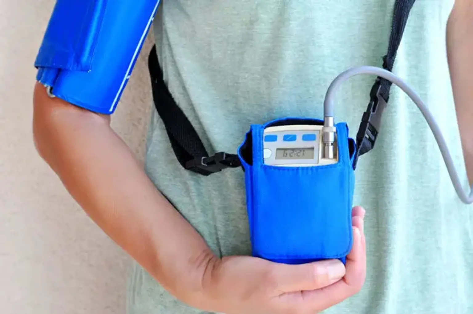
The procedure involves wearing a small, portable device called a Holter monitor, which is attached to the chest with electrodes. These electrodes measure the electrical signals produced by the heart. Over the 24-hour period, the monitor records the data, providing detailed insights into the heart’s rhythm and detecting irregularities that may not show up in a typical ECG.
Pre-Surgical Assessment and Safety Protocols
Before undergoing a Holter monitoring test, patients typically meet with a cardiologist to assess their heart health. The test is safe, with minimal risks involved. However, patients should avoid activities that might interfere with the monitor’s readings, such as heavy exercise or getting the electrodes wet. The procedure is non-invasive, and patients can resume normal activities while the test is being performed.
The Benefits of 24-Hour Holter Monitoring
Early Detection of Heart Disease and Cardiac Arrhythmias
Holter monitoring is incredibly effective in detecting irregular heartbeats, which can indicate conditions like arrhythmias or heart disease. These conditions are sometimes missed by standard ECG tests, but the 24-hour monitoring provides a broader view of the heart’s behavior, increasing diagnostic accuracy.
Non-Invasive Nature and Long-Term Heart Monitoring
Unlike other heart tests, the Holter monitor is non-invasive and allows for continuous monitoring without requiring hospitalization. This makes it particularly useful for patients with heart conditions that need to be observed over an extended period, offering detailed insights into heart rhythms, especially in cases where symptoms are intermittent.
Benefits for Patients in Korea
In Korea, Holter monitoring is widely used due to the country’s advanced healthcare system. Many patients turn to Korean hospitals for this test, which offers cutting-edge technology and high success rates in detecting heart conditions early. With Korea’s growing reputation for medical tourism, patients from around the world travel there for precise and reliable heart health monitoring.
How Accurate is the 24-Hour Holter Monitoring Test?
Effectiveness of Holter Monitoring in Diagnosing Heart Conditions
Holter monitors provide highly accurate data over a 24-hour period, making them one of the most reliable methods for diagnosing heart arrhythmias and other irregularities. Unlike traditional ECG tests, which only provide a snapshot of heart activity at one moment, the Holter monitor captures a complete picture of how the heart functions over time.
Specific Use Cases in Korean Healthcare
Korean hospitals are equipped with advanced cardiac monitoring technology, allowing them to perform precise Holter monitoring. The test is especially valuable for diagnosing conditions like atrial fibrillation, which may go undetected without continuous monitoring. Thanks to this technology, Korean healthcare providers offer some of the most accurate heart disease diagnoses globally.
How the 24-Hour Holter Monitoring Test is Performed
Detailed Procedure: What to Expect Before, During, and After the Test
Before the test, a cardiologist will explain how the Holter monitor works and what to expect. Electrodes are placed on your chest, connected to the Holter monitor, which is usually worn on a belt or shoulder strap. The device records the heart's electrical signals continuously for 24 hours. During the test, patients can go about their daily activities, but they should avoid intense exercise or getting the electrodes wet.
Recovery and Aftercare
Once the 24-hour monitoring period is complete, the electrodes are removed, and the device is returned to the medical team for analysis. The results are carefully examined by a cardiologist to identify any irregularities in the heart's rhythm. In most cases, there’s no need for recovery time, and patients can resume normal activities right after the test. Follow-up appointments will discuss the results and any necessary treatments.
The Costs and Availability of 24-Hour Holter Monitoring in Korea
Holter Monitoring Cost in Korea
The cost of a 24-hour Holter monitoring test in Korea can vary depending on the healthcare facility and whether the patient is local or an international visitor. Typically, it ranges from a few hundred to over a thousand dollars. While the procedure may seem costly, it provides invaluable information for diagnosing and managing heart conditions, potentially saving money by avoiding more invasive tests or treatments later.
Availability of Holter Monitoring at Korean Hospitals
Korea is home to world-class hospitals with advanced medical technology, including the 24-hour Holter monitoring test. Major medical centers, especially in cities like Seoul, offer this procedure. Korean hospitals are renowned for their high-quality care and cutting-edge diagnostics, making the country a top destination for medical tourism.
Understanding Heart Disease and How Holter Monitoring Helps
Heart Disease Detection with Holter Monitoring
Heart disease can develop silently, with few visible symptoms until it reaches a critical point. Holter monitoring offers early detection of conditions like arrhythmias, which are irregular heartbeats that can lead to more severe health problems if left untreated. By continuously monitoring the heart's electrical activity, the test can identify issues that would otherwise go unnoticed, allowing for early intervention and treatment.
Heart Health Monitoring Over Time
For patients with chronic heart conditions or those at risk, continuous heart monitoring is crucial. Holter monitoring allows doctors to observe the heart's rhythm over an extended period, providing insights into how the heart functions day-to-day. This long-term monitoring can help adjust treatments as needed, ensuring better management of heart health.
Holter Monitoring and Korean Medical Technology
Innovative Cardiac Monitoring Technology in Korea
Korea is a leader in adopting and advancing medical technology, particularly in the field of cardiology. The 24-hour Holter monitoring test is a prime example of this. Korean hospitals utilize state-of-the-art ECG Holter devices, ensuring precise heart rhythm analysis. These advancements allow Korean healthcare providers to offer the best possible care for heart disease patients, helping them manage conditions more effectively.
Popularity of the 24-Hour Holter Monitoring Test in Korea
Holter monitoring is widely used in Korea, where high standards of healthcare are paired with innovative technology. The test is particularly popular for diagnosing heart arrhythmias and assessing overall heart health. As the country becomes a hub for medical tourism, international patients seek out Korean hospitals for their expertise in providing reliable and accurate heart health diagnostics, including 24-hour Holter monitoring.
Risks and Limitations of the 24-Hour Holter Monitoring Test
Understanding the Risks
The 24-hour Holter monitoring test is generally safe with minimal risks. However, some patients may experience mild skin irritation from the electrodes attached to the chest. In rare cases, the adhesive may cause allergic reactions or discomfort. The monitor is lightweight and portable, but some patients may find it slightly cumbersome to wear for an extended period.
Limitations of the Test
While the Holter monitor provides a detailed snapshot of the heart’s activity over 24 hours, it does have limitations. For instance, if a patient’s symptoms occur outside the monitoring period or are sporadic, the test may not detect the problem. Additionally, the test primarily focuses on heart rhythm and does not provide insights into other heart conditions, like structural abnormalities or heart disease related to blockages. As a result, it’s often used in conjunction with other tests for a comprehensive diagnosis.
The Global Popularity of Holter Monitoring in Cardiology
International Appeal and Popularity of the 24-Hour Holter Test
Holter monitoring has become a go-to diagnostic tool worldwide, especially in cardiology. Its non-invasive nature and ability to detect hidden heart issues make it a valuable option for healthcare providers. Many patients from around the world seek out Holter monitoring tests in advanced medical centers, particularly in Korea, where the procedure is widely available and highly regarded.
Success Stories from Korean Hospitals
In Korea, the use of 24-hour Holter monitors has significantly advanced the diagnosis and management of heart conditions. Patients who had suffered from undiagnosed arrhythmias were successfully treated after Holter monitoring revealed their heart irregularities. Success stories from Korean hospitals are not only a testament to the test’s effectiveness but also show how the procedure has helped improve patient outcomes by catching heart issues early.
Comparing the 24-Hour Holter Monitoring Test with Traditional ECG Tests
Key Differences Between Holter Monitoring and Standard ECG
Unlike a traditional ECG, which only records the heart's electrical activity at a single point in time, the 24-hour Holter test continuously monitors the heart’s rhythms for 24 hours. This extended observation is crucial for detecting arrhythmias that might not occur during a brief ECG test. Holter monitoring is ideal for patients experiencing intermittent symptoms or those with suspected arrhythmias that are not constant.
When to Choose One Over the Other
A traditional ECG is suitable for a quick check-up or emergency situation, but for long-term monitoring or diagnosing irregular heartbeats that occur unpredictably, Holter monitoring is the preferred option. Doctors typically recommend Holter monitoring when a more comprehensive view of heart activity is needed, particularly in patients with symptoms like dizziness, fainting, or irregular heartbeats.
Holter Monitoring for Arrhythmia Diagnosis in Korea
Specific Applications of the Test for Arrhythmia
Holter monitoring plays a crucial role in diagnosing arrhythmias, which are abnormal heart rhythms. Arrhythmias, such as atrial fibrillation, can sometimes go unnoticed during brief ECG tests because they may occur intermittently. The 24-hour Holter test, however, offers the opportunity to capture these irregular rhythms, making it an essential tool in the management of heart disease in Korea.
Importance of Early Diagnosis in Managing Cardiac Arrhythmia
Early detection of arrhythmias through Holter monitoring can prevent severe complications, including stroke or heart failure. In Korea, where heart disease is a significant health concern, the ability to monitor and diagnose arrhythmias early allows for timely interventions, improving patient outcomes and reducing the risk of long-term damage to the heart.
The Role of Holter Monitoring in Managing Cardiac Arrhythmias
Treatment Options for Diagnosed Arrhythmias
Once an arrhythmia is diagnosed through 24-hour Holter monitoring, a variety of treatment options may be considered based on the severity and type of arrhythmia. Common treatments include medications like antiarrhythmic drugs, which help regulate the heart's rhythm. In more severe cases, procedures like catheter ablation may be recommended to target and treat abnormal electrical pathways in the heart. For patients with atrial fibrillation, anticoagulant therapy might be prescribed to reduce the risk of stroke. Holter monitoring allows doctors to tailor treatments based on the specific irregularities detected in the heart’s rhythm.
Patient Case Studies from Korea
In Korea, many patients have benefited from early arrhythmia diagnosis through Holter monitoring. For example, a patient who had experienced dizziness and shortness of breath was diagnosed with atrial fibrillation after a 24-hour Holter test. As a result, the patient received timely treatment, which included medication and lifestyle adjustments, leading to a significant improvement in their heart health. This type of early detection and intervention is one of the key advantages of using Holter monitoring in Korea's advanced healthcare system.
Patient Experience and Expectations with 24-Hour Holter Monitoring in Korea
What to Expect During the Monitoring Period
The 24-hour Holter monitoring test is simple and doesn’t require hospitalization, making it convenient for patients. When undergoing the test, patients are fitted with the Holter monitor and are instructed to go about their daily activities, while avoiding actions like showering or heavy exercise that might affect the electrodes. Though it’s common to feel slightly aware of the device, most patients find the test comfortable and unobtrusive. The monitor records heart data throughout the day and night, which is then analyzed by doctors to identify any irregular heart rhythms.
Patient Testimonials from Korean Medical Centers
Many patients who have undergone Holter monitoring in Korea share positive experiences, particularly regarding the non-invasive nature of the procedure and the comfort of wearing the portable monitor. One patient, a foreign traveler to Korea for heart health evaluation, appreciated the seamless process and quick results provided by the local hospital. This patient had suffered from irregular heartbeats that were missed by previous ECG tests but were detected through Holter monitoring, allowing for appropriate treatment. Testimonials like this show the confidence patients have in Korea's ability to provide accurate and efficient heart care.
Frequently Asked Questions (FAQs) about the 24-Hour Holter Monitoring Test
What is the Difference Between Holter Monitoring and Other Tests?
Holter monitoring offers continuous heart rate monitoring over 24 hours, unlike traditional ECGs that only provide a snapshot of the heart’s activity at a specific moment. This continuous observation makes Holter monitoring especially useful for detecting arrhythmias or other irregularities that may not be present during a brief ECG test. Holter monitoring is particularly valuable for patients who experience intermittent symptoms like dizziness or palpitations.
How Long Does the Test Last?
The Holter monitoring test lasts for 24 hours. During this period, the patient wears the Holter monitor, which records heart activity. After the 24-hour period, the device is removed, and the recorded data is analyzed by a cardiologist. While the monitoring period is short, it provides valuable insights into heart health over an extended time.
Is the Holter Monitor Comfortable to Wear?
Most patients find the Holter monitor comfortable to wear. It is a small, portable device that attaches to the chest using electrodes and is typically worn on a belt or shoulder strap. While some patients may experience mild skin irritation from the adhesive, the device is generally lightweight and non-intrusive, allowing patients to go about their normal activities with minimal discomfort.
Conclusion
The Growing Importance of 24-Hour Holter Monitoring for Heart Health
The 24-hour Holter monitoring test is an essential tool for diagnosing and managing heart conditions. By providing continuous monitoring, it allows for the detection of arrhythmias and other heart issues that may not be visible during a standard ECG test. This diagnostic tool is particularly valuable for early detection of heart disease, enabling doctors to offer timely interventions.
Why Choose Korean Medical Centers for Holter Monitoring?
Korea’s healthcare system is known for its advanced technology and high standards of care. The 24-hour Holter monitoring test is widely available in top hospitals throughout the country, and many international patients seek treatment in Korea for its world-class medical services. With skilled cardiologists and cutting-edge medical equipment, Korean hospitals provide accurate diagnoses and effective treatments, making Korea a leading destination for heart health monitoring.
Holter monitoring in Korea represents not only medical excellence but also patient-centered care, ensuring that heart conditions are detected early and treated promptly, improving the quality of life for patients both locally and internationally.