Adrenal tumors
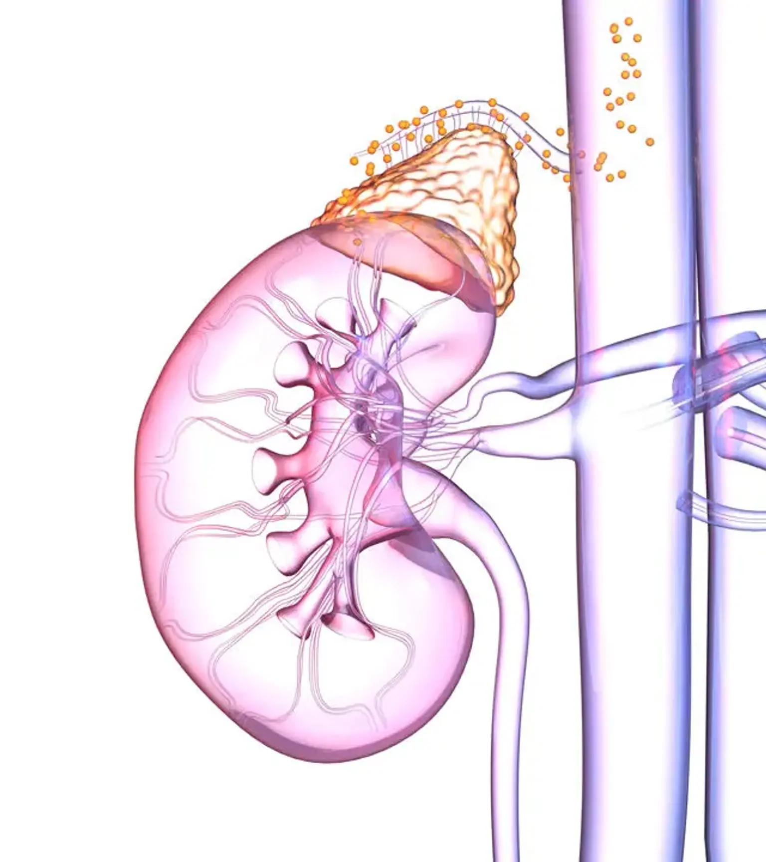
The adrenal gland is an endocrine organ that serves two purposes. The outer adrenal cortex releases steroid hormones such as cortisol and mineralocorticoids (aldosterone and the androgen), as well as glucocorticoids like cortisol. The glucocorticoids are involved in the metabolism of carbohydrates, proteins, and fats. Mineralocorticoids are necessary for maintaining sodium (Na) and potassium (K) balances as well as fluid homeostasis.
Survival requires glucocorticoids and mineralocorticoids. Catecholamines are produced by the inner adrenal medulla (dopamine, epinephrine, and norepinephrine).
Adrenal mass is a common finding in the adrenal gland. While most lesions are benign, when a patient comes with hypercortisolism, adrenocortical cancer should be considered. Occasionally, adrenal masses are discovered during a normal examination.
These lesions can be classified as either functional (hormone-secreting) or silent (non-hormone-secreting) (either benign or malignant). Less than 1% of these tumors are malignant in total.
Epidemiology of Adrenal Tumors
According to the National Institute of Health's Office of Rare Diseases Research, there are less than 200000 occurrences of adrenocortical carcinoma in the United States, with a global incidence of 0.5 to 2 per million and 0.72 per million in the US alone.
Adrenocortical carcinoma is responsible for 0.2 percent of all fatalities in the United States each year. It appears in adults in their fourth and fifth decades of life; nevertheless, it also appears in children under the age of five. Women are more likely to be affected than males (female to male ratio is between 2.5 and 3 to 1).
Cigarette smoking and the use of oral contraceptives were shown to be associated with the development of adrenocortical cancer (ACC) in a 1996 analysis of risk factors.
Pathophysiology of Adrenal Tumors
Several molecular alterations, including inactivation of tumor suppressor genes and activation of a variety of distinct oncogenes, DNA mutations, and epigenetic modifications, have been implicated in the development of adrenocortical carcinoma in recent decades. Large-scale alterations in gene expression, chromosomal abnormalities such as chromosomal gains, losses, and heterozygozity, DNA methylation, and dysregulation of microRNA (miRNA) lead to overexpression of these sequences occur at the genome level.
Initially, hereditary disorders that predispose to ACC allowed signaling pathways involved in the pathophysiology of adrenal tumors to be identified. Specifically, the Tp53 pathway and IGF-II overexpression. In 50 percent to 80 percent of infants with ACC, germline abnormalities in the Tp53 pathway are the underlying genetic etiology.
The incidence of germline TP53 mutations in adult ACC ranges from 3% to 7% in the general population. However, more than half of adult patients with ACC have somatic TP53 mutations in tumor tissue or loss of heterozygozity at the TP53 gene, and TP53-mutated tumors are linked with an aggressive phenotype.
Histopathology of Adrenal Tumors
Large size with irregular borders, invasion of surrounding tissues, and the existence of distant metastases are all characteristics of adrenocortical cancer. The color of the tumor's surface varies from brown to orange to yellow, depending on the amount of lipid in the cells.
The presence of necrosis is usually always evident. Importantly, ACC is characterized by tumor invasion of the capsule, extra-adrenal soft tissue, or direct invasion of lymphatic channels/blood vessels. However, because these markers are very generic and are commonly lacking in early illness, it can be difficult to distinguish ACC from benign adrenal tumors.
A nine-criteria histopathologic categorization scheme for adrenocortical tumors is based on the following:
- Nuclear grade (III or IV)
- High mitotic rate (more than five mitoses per 50 high-power field
- Presence of atypical mitoses
- Percentage of clear cells (less than or equal to 25% of tumor)
- Diffuse architecture
- Microscopic necrosis
- Venous invasion
- Sinusoidal invasion
- Capsular invasion
- The presence of more than three or more features is consistent with adrenocortical carcinoma.
Despite its extensive usage, poorly differentiated ACC can still be mistaken for renal cell carcinoma, small cell lung carcinoma, melanoma, or hepatocellular carcinoma. To distinguish cortical carcinoma from adenoma, numerous diagnostic methods were presented in the previous WHO edition.
The current WHO classification acknowledges certain histological variations in addition to traditional adrenocortical cancer. Oncocytic, myxoid, and sarcomatoid carcinomas are the three types, in order of decreasing frequency. Sarcomatoid carcinomas and myxoid carcinomas of the adrenal gland are uncommon.
The stroma of myxoid carcinoma contains a lot of extracellular mucin. The lack of cortical differentiation is visible in sarcomatoid cancer. A common cortical carcinoma zone may be present in the cancer (biphasic). When the usual cortical carcinoma zone is monophasic, sarcomatoid carcinoma must be distinguished from retroperitoneal sarcoma affecting the adrenal gland.
The major cell type in adrenal oncocytic carcinoma is an oncocyte, which is a cell with dense, granular cytoplasm associated with the accumulation of mitochondria and endoplasmic reticulum. Adrenocortical carcinomas can be classified as low-grade or high-grade depending on their mitotic rates.
Causes of Adrenal Tumors
Adrenocortical carcinomas can develop on their own or as part of a family cancer syndrome. Although the molecular pathways causing carcinogenesis are well recognized in many inherited diseases, the molecular pathophysiology of sporadic ACC is less well defined. Furthermore, the majority of ACC occurrences appear to be episodic.
Although adrenocortical hyperplasia appears to be a polyclonal process, the majority of adrenocortical tumors, whether benign or malignant, are monoclonal lesions, indicating that genetic mutations at particular genomic loci may play a role in adrenal carcinogenesis.
A multistep tumor progression of tumorigenesis has been proposed from genes that are known to be associated with hereditary tumor syndromes described below:
Li-Fraumeni syndrome:
It is a cancer disease caused by a germline mutation in the TP53 gene on chromosome 17p13. Breast cancer, sarcomas, brain tumors, leukemia, and adrenocortical carcinoma are all possible in patients. Variants of this condition may arise in children with sporadic ACC and germline p53 mutations who do not have a typical Li-Fraumeni syndrome background.
Familial adenomatous polyposis (FAP) or Gardner syndrome:
Which is a mutation of the adenomatous polyposis coli gene (APC) found on chromosome 5q21, is characterized by numerous colonic polyps and a higher risk of developing early colon carcinomas.
FAP is also linked to pigmented retinal lesions, desmoids tumors, osteomas, thyroid adenoma/carcinomas, and a variety of other malignant tumors. Adrenocortical tumors, including nonfunctional nodular hyperplasia, cortisol-producing adenomas (CPA), and ACC, affect 7 to 13% of FAP patients.
Beckwith-Wiedemann syndrome (BWS):
It is distinguished by genetic and epigenetic events involving the cyclin-dependent kinase inhibitor and insulin-growth-factor II (IGF-II), resulting in overgrowth disorders, visceromegaly (macroglossia, hemihyperplasia), malformations (wall defect, umbilical hernia), and predisposition to embryonal malignancies.
ACC is part of the BWS tumor spectrum, which also includes Wilms tumor, hepatoblastoma, rhabdomyosarcoma, and neuroblastoma, with a 7.5 percent overall risk of tumor formation in children; the majority of tumors arise in the first 8 to 10 years of life.
Multiple endocrine neoplasia type 1:
It has an aberrant genetic locus at 11q13, where a defective MEN1 gene manufactures a faulty tumor suppressor protein called the Menin protein. The patient is predisposed to parathyroid, pituitary, endocrine pancreatic, and adrenocortical malignancies, particularly adrenocortical carcinoma, due to the aberrant function of this protein.
Types of Adrenal Tumors
Adrenal cancer is divided into two classes in the Fourth Edition of the World Health Organization (WHO) classification of endocrine cancers, which was released in 2017. Tumors of the adrenal cortex are classified as the first category, whereas tumors of the adrenal medulla and extra-adrenal paraganglia are classified as the second.
The importance of genetics in adrenal cortical carcinoma and pheochromocytoma/paraganglioma, as well as the inclusion of a few uncommon malignancies of the adrenal cortex and medulla, is a fundamental aspect of the WHO classification.
Tumors of adrenal cortex
Adrenocortical adenoma:
Adrenocortical adenomas are exceedingly prevalent benign malignancies of the adrenal cortex (present in 1-10 percent of persons at autopsy). They are not the same as adrenocortical "nodules," which are not true neoplasms.
Adrenocortical adenomas are uncommon in persons under the age of 30 and occur equally in both sexes. These neoplasms have a dual clinical relevance. First, due to the increased use of CT scans and magnetic resonance imaging in a range of medical contexts, they have been found as accidental findings with increasing frequency in recent years. This can necessitate costly extra testing and invasive treatments to rule out the likelihood of an early adrenocortical cancer.
Second, a small percentage of adrenocortical adenomas (about 15%) are "functional," meaning they produce glucocorticoids, mineralcorticoids, and/or sex steroids, resulting in endocrine disorders such as Cushing's syndrome, Conn's syndrome (hyperaldosteronism), female virilization, or male feminization. Functional adrenocortical adenomas can be removed surgically.
Adrenocortical carcinoma:
Adrenocortical carcinoma (ACC) is a rare, aggressive malignancy of the adrenal cortical cells that can affect both children and adults. Many ACCs are "functional," generating steroid hormones and causing endocrine dysfunction similar to that found in many adrenocortical adenomas.
Most adrenocortical carcinomas are not identified until they have become fairly big due to their placement deep in the retroperitoneum. They typically invade major veins such as the renal vein and inferior vena cava, as well as metastasizing to the lungs and other organs via the lymphatics and blood.
Tumors of the adrenal medulla
The adrenal medulla is located in the center of each adrenal gland and is made up of neuroendocrine (chromaffin) cells that manufacture and release epinephrine (adrenaline) into the circulation in response to sympathetic nervous system activity. The two most common cancers that develop from the adrenal medulla are neuroblastoma and pheochromocytoma. Both tumors can develop from non-adrenal locations, notably the sympathetic chain's paraganglia.
Neuroblastoma:
Neuroblastoma is an aggressive malignancy of immature neuroblastic cells (neuronal progenitors) that is one of the most prevalent pediatric malignancies, with a median age of two years at diagnosis. Adrenal neuroblastoma usually manifests as a quickly growing abdominal tumor.
Despite the fact that the tumor has frequently migrated to distant regions of the body at the time of diagnosis, this cancer is unique in that many instances are extremely treatable when the spread is restricted to the liver, skin, and/or bone marrow (stage IVS). Ganglioneuroblastoma and ganglioneuroma are related but less malignant tumors made of more developed brain cells.
Neuroblastic tumors frequently create high quantities of catecholamine hormone metabolites such as vanillylmandelic acid (VMA) and homovanillic acid, and they can cause severe watery diarrhea by producing vasoactive intestinal peptide.
Pheochromocytoma:
Pheochromocytoma is a tumor that is made up of cells that resemble the chromaffin cells of the adult adrenal medulla. Pheochromocytomas can be spontaneous or linked with a hereditary cancer syndrome, such as multiple endocrine neoplasia (MEN) types IIA and IIB, neurofibromatosis type I, or von Hippel-Lindau syndrome.
Only 10% of adrenal pheochromocytomas are malignant, with the remainder being benign tumors. The tendency of pheochromocytomas to produce substantial levels of the catecholamine hormones epinephrine (adrenaline) and norepinephrine is the most clinically significant trait.
Incidentalomas:
Adrenal incidentaloma is a tumor detected by chance in the absence of clinical signs or suspicion. It is a common unexpected result found by computed tomography (CT), magnetic resonance imaging (MRI), or ultrasonography.
Signs and Symptoms of Adrenal Tumors
Adrenocortical carcinoma can manifest itself in three ways. Approximately one-third of patients arrive with symptoms of hormonal excess, another third with non-specific symptoms, and the other third is found coincidentally when imaging exams are performed for other medical issues.
Plethora (rounded face), diabetes mellitus, osteoporosis, purple striae on the abdomen wall, muscular weakness, truncal obesity, and muscle atrophy are all symptoms of hypercortisolism (Cushing syndrome). Excess glucocorticoids can cause hypokalemia and hypertension. Malignant adrenal tumors typically manifest with hypercortisolism, quickly increasing muscular weakness, and severe muscle weakness.
Male pattern baldness, virilization, hirsutism, and menstrual irregularities are all symptoms of hyperandrogenism. Excess estrogen production affects 1 to 3% of patients. When men overproduce estrogen, they develop a feminization syndrome, which includes impotence, reduced libido, testicular shrinkage, and gynecomastia.
Approximately half of the patients who report with hormonal excess also have hypercortisolism and hyperandrogenism. Males with isolated hyperandrogenism are frequently misdiagnosed.
As a result of autonomous aldosterone secretion, hypertension and hyperkalemia can ensue. Children are more likely than adults to develop adrenocortical carcinomas (90 percent of cases). The majority of these tumors produce androgens, causing early puberty and virilization.
Although paraneoplastic symptoms are uncommon, tumor-related, IGF-2-mediated hypoglycemia known as Anderson syndrome is prevalent. Leukocytosis, polycythemia, and hyperreninemic hyperaldosteronism can all be caused by tumor chemokines. Deep venous thrombosis and pulmonary embolism are further symptoms of ACC.
Local tumor development, stomach or flank discomfort, early satiety, and abdominal fullness are nonspecific symptoms. At the time of diagnosis, the tumor is typically 10 to 13 cm in size. A small percentage of patients have tumors that are smaller than 6 cm in size.
Nonfunctional tumors are often bigger than functional tumors at presentation, and a considerable percentage are detected accidentally during abdominal or thoracic imaging for other reasons. Some individuals with nonfunctional tumors are asymptomatic until they develop signs or symptoms of metastatic disease (e.g., jaundice, bone pain). The most frequent metastasis locations are the lung (40 % to 80 %), liver (40 % to 90 %), and bone (40 % to 90 %) (5 %to 20 %).
How Adrenal Tumors can be Diagnosed?
A basic metabolic panel (electrolytes, glucose, BUN, liver function test) and a complete blood count are required for patients. A magnetic resonance imaging (MRI) or computed tomography (CT) scan of the pelvis and abdomen, as well as a CT scan of the chest, should be performed as part of the staging process. Another clinical suspicion should lead to the consideration of another imaging test (e.g., bone scan for skeletal metastasis).
Biochemical evaluation
Cortisol, mineralocorticoids, androgens, and oestrogens are all measured in the blood and urine as part of the biochemical examination.
The European Network for the Study of Adrenal Tumors recommends a pre-operative hormonal workup to rule out a probable ACC. Basal cortisol, ACTH, DHEAS, 17-hydroxyprogesterone, testosterone, androstenedione, and estradiol, as well as a dexamethasone suppression test and urine free cortisol, are all recommended. Recently, it appears that certain ACC, previously thought to be non-secreting, may produce some urinary steroid metabolites.
Hormone levels in the blood or hormonal symptoms in individuals with adrenocortical carcinoma have no relationship with tumor growth.
When no steroid hormone production is obvious, it is advisable to rule out a pheochromocytoma in addition to steroidal hormone measures. Pheochromocytoma is diagnosed by detecting the levels of metanephrine and normetanephrine in the urine or plasma during a 24-hour period.
Imaging procedure
An adrenal incidentaloma is a previously undetected adrenal tumor identified during cross-sectional imaging for another cause. Adrenocortical carcinomas often have a diameter of greater than 6 cm. The size of the tumor raises the possibility that it is malignant. Tumors bigger than 4 cm have a sensitivity of 97% and a specificity of 52%, whereas tumors larger than 6 cm have a sensitivity of 91% and a specificity of 80%.
However, a small percentage of minor lesions (less than 4 cm in diameter) may be malignant. Benign adenomas, on the other hand, account for 65% of masses smaller than 4 cm and 18% of masses larger than 6 cm.Because to internal bleeding, necrosis, and calcification, adrenocortical cancer has a variable appearance with frequent heterogeneous enhancement. Imaging may potentially identify lung, lymph node, or liver metastases. Extension into the renal vein and/or inferior vena cava, as well as neighboring organs, is possible.
Before surgery, all patients with ACC must have a chest CT scan to detect pulmonary metastases. At the moment, no one imaging approach can classify a localized adrenal mass as ACC. If there is a suspicion of ACC, an abdominal CT scan is required. In a case with an adrenal mass with a density that is >10UH, the contrast media washout can be used to discriminate the benign adrenal lesions from the ACC.
Positron emission tomography (PET) with fluorodeoxyglucose (FDG) and metomidate (MTO) are useful to confirm the diagnosis of a malignant lesion or establish a tumor of adrenocortical origin. ACC typically presents as a heterogeneous and large mass with intense FDG uptake greater than the liver background.
How Adrenal Tumors can be Treated?
Because of its many endocrine activities, adrenocortical carcinoma presents a unique treatment problem. There are no general standards because of the rarity of adrenocortical carcinomas and the limited clinical series.
Expert consensus opinion acquired from medical institutes that specialize in the treatment of ACC influences current practice. At the moment, the most practicable strategy to ACC is total tumor excision. The goal of adjuvant therapy is to reduce the incidence of recurrence.
The natural history of recurrence following surgery is unknown, although even with total resection, the rate of local recurrence ranges from 19 to 34 percent, depending on tumor stage. As a result, adjuvant therapy following surgery, which involves the use of mitotane and tumoral irradiation, is widespread.
Advanced ACC treatment must be regarded palliative. This therapy encompasses both enhancing the patient's quality of life by required measures (e.g., pain control, avoidance of fractures caused by bone metastases, and sufficient control of hormonal symptoms) and reducing the negative effects of antineoplastic treatments.
Postoperative and Rehabilitation Care
The handling of follow-up is not well-standardized. However, because adrenocortical carcinomas are aggressive tumors, patients should be followed up on every three months following therapy. When the patient enjoys a recurrence-free period of 2 to 3 years, the follow-up period may be extended to every six months for a total of five years.
After five years of disease-free survival, monitoring can be performed every 1 to 2 years, because, while rare, some individuals may experience a late recurrence. Patients should have a hormone test, a full imaging work-up, including a chest and abdomen CT scan, and a thorough physical examination.
Prognosis of Adrenal Tumors
The prognosis of adrenocortical carcinoma was previously quite poor. In the past, an average overall survival in patients with adrenocortical carcinoma was 14.5 months with a 5-year mortality rate of about 75 to 90%.
Median survival for metastatic disease (stage IV) at the time of diagnosis is reported to be less than one year. Of those who had undergone treatment, about 30 to 60% survived 5-years. Furthermore, it is worth noting that only about 30% of ACC cases are curable at early stages, which further implies an overall poor prognosis of ACC.
Conclusion
The phrase "adrenal tumor" refers to benign and malignant mass lesions of the adrenal gland, including primary adrenal tumors and extra-adrenal metastases. Adrenal tumors have grown more common as 'incidentalomas' as imaging techniques have become more widely used.
The discovery of an adrenal tumor presents two concerns: Is the mass cancerous? Is the mass hormonally active? Whereas malignancy is assessed using particular imaging features (imaging phenotype), hormonal secretion is assessed using a focused clinical examination and certain biochemical assays.
A 4 cm adrenal tumor with evident benign characteristics on imaging and a normal hormonal workup does not necessitate therapy. If cancer is suspected, more diagnostic investigations and/or adrenalectomy are recommended. Surgery is typically regarded the therapy of choice for hormonally active tumors; however, the decision for surgery must be customized for aldosterone-secreting tumors and cortisol-secreting tumors with only moderate cushing's syndrome.
In patients with big tumors (> 4 cm), as well as those with inconclusive malignancy or hormonal activity, the choice for future care must be taken on an individual basis. In adrenal tumors less than 6 cm in size and without local invasion of neighboring tissues, a less invasive surgical technique may be explored.
It is possible to use both laparoscopic (transabdominal) and retroperitoneoscopic methods. The surgical result is determined on the surgeon's level of experience. In the examination and treatment of adrenal tumors, extensive multidisciplinary teamwork is required.