Aging of veins
Various physiological mechanisms are deteriorating with aging. Vascular aging is connected with changes in the mechanical and structural features of the vascular wall, which results in decreased arterial and venous elasticity and compliance.
The change in the mechanical and structural characteristics of the arterial and venous wall is the most evident feature of vascular ageing. Vessels contain inherent and functional qualities that are affected by stress and hormonal changes throughout their lives. Modifiable cardiovascular risk factors such as hypertension, obesity, smoking, and lifestyle, as well as non-modifiable risk factors such as age, genetics, and family history, alter these features.
One of the most significant cardiovascular risk factors is aging. Genetic, mechanical, and hemodynamic variables all play a role in vascular aging. The latter is supported by the fact that age-related alterations in capillaries and veins, which are less susceptible to changes in plasticity and blood pressure, are less prominent or even non-existent.
In large resistance vessels, age-related morphologic changes include intima-media thickening and increased matrix material deposition, ultimately leading to decreased compliance. Vascular aging is characterized mostly by decreased endothelial-dependent vasorelaxation. The expression of endothelial nitric oxide synthase, which produces vasodilatatory nitric oxide, increases with age.
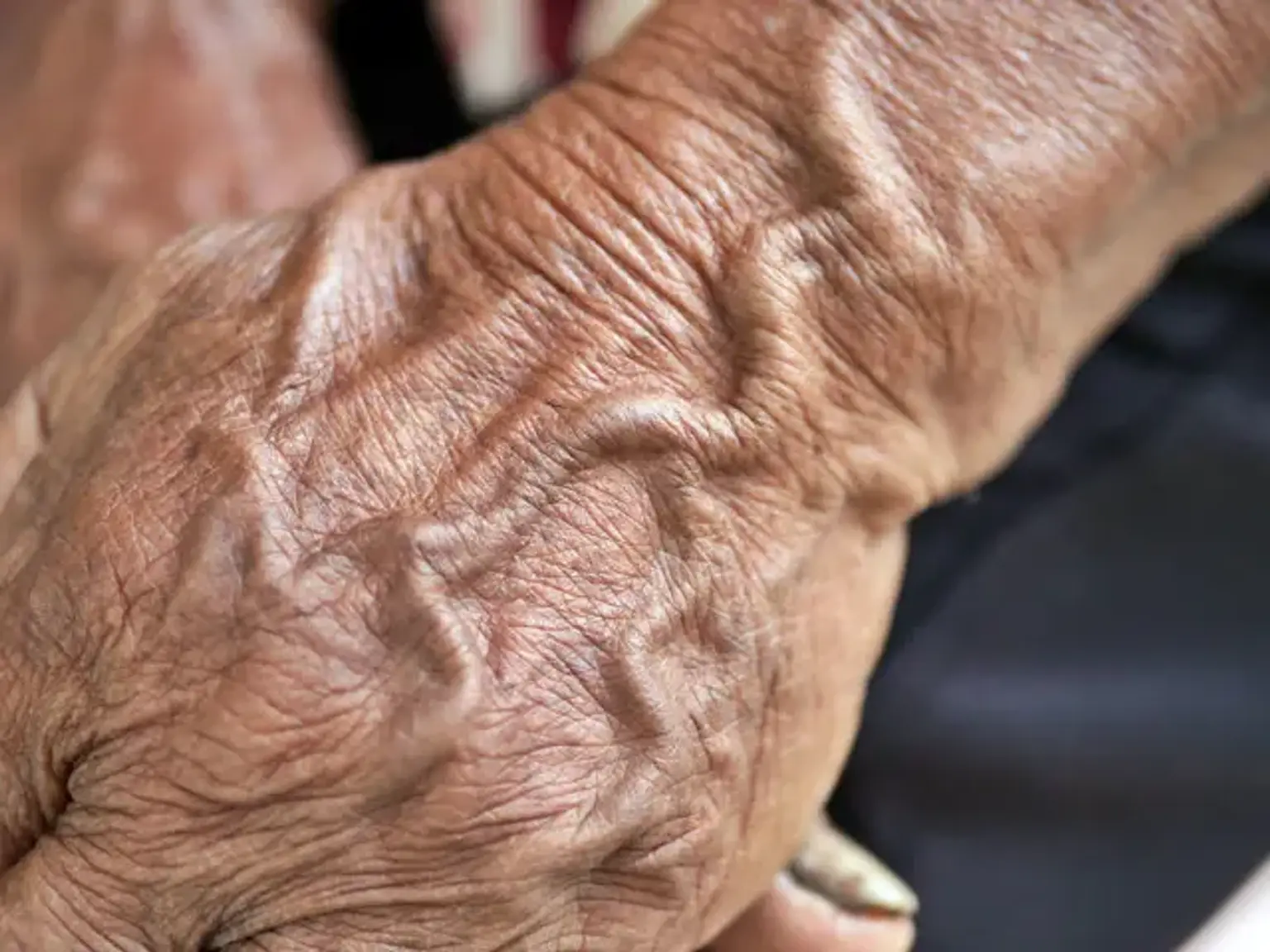
Why do hand veins bulge?
Variations in blood pressure and skin elasticity can cause veins to bulge and become more visible over time. As previously stated, hand veins are a good predictor of age. This is because the most prevalent cause of noticeable, bulging hand veins is aging. As people age, their skin thins, causing veins to become more visible. Furthermore, increased blood pressure might cause them to swell.
Bulging, visible hand veins aren't usually a cause for concern, but. Before identifying the appropriate treatment for bulging hand veins, the underlying cause should be determined:
- Aging: Is the most common cause of bulging veins, but it’s not the only cause. Although prominent hand veins are healthy veins that return blood to the heart, some may become bulgy and protrude at the back of the hand with age due to many factors such as loss of skin and vein wall elasticity, loss of subcutaneous fat and water content of the tissues.
- Varicose veins: Even though varicose veins are typically associated with legs, they can develop in other areas of your body, including hands.
- Low body weight: If you’re underweight or have low body fat, your veins may be more visible.
- Hot weather or high temperatures: These can cause prominent veins due to expanding blood vessels.
- Exercise: Increased blood pressure from working out as well as lifting weights can cause more visible veins.
- Phlebitis: Infections and/or vein trauma can cause inflammation and swelling.
- Familial: large, visible veins may also run in your family.
How to get rid of bulging hand veins?
Hand veins are treated with painless sclerotherapy in a 15-20-minute operation. Following the injections, the patient will be asked to wear compression gloves for 12 hours before returning to regular activities.
Sclerotherapy for hand veins is a very safe operation. For more than 30 years, there have been no issues with hand injections. For the first 24 hours after treatment, there is minimal black and blue and occasionally mild edema. Sclerotherapy is the gold standard for treating hand veins. In rare circumstances, a mini-phlebectomy may be performed. The outcome is great, with long-term effects.
If age spots are evident after the veins have been treated, they may be treated with a laser or another method. Injections of fillers or growth factors might be used if the affected hands are bony and thin.
The cost for hand injections is $1,000 for the first treatment and $750 for any treatment after that.
What causes veins in the face?
Dilated facial veins, also known as broken blood vessels or spider veins, arise when blood vessels dilate or swell just beneath the skin's surface. This produces little, reddish lines that spread out into a web-like pattern. They can appear anywhere on the body, although the face and legs are the most commonly affected.
While ruptured blood vessels are generally benign, they can become a nuisance if they make you feel self-conscious. Some people are more prone to developing spider veins than others. Broken blood vessels can happen to anybody, including children, at any age. The risk factors depend on the particular cause which summed up below:
- Heredity and genetics: For unknown reasons, spider veins tend to run in families. Individual risk factors also increase with age.
- Pregnancy: An increase in estrogen hormones during pregnancy can lead to broken blood vessels. Pregnancy-related spider veins heal on their own after delivery. Skin changes are common in pregnancy.
- Rosacea: This common skin condition leads to excessive redness and flushing. With erythemato-telangiectatic rosacea, broken blood vessels are common.
- Sun exposure: Excessive sun exposure can enlarge the blood vessels. when exposed to sun, the top layer of skin may peel and temporarily make some of the blood vessels more noticeable.
- Weather changes: Hot weather increases blood vessel dilation.
- Environmental or chemical irritants.
- Alcohol consumption: Moderate or occasional alcohol consumption can cause the skin to flush due to the enlargement of blood vessels. Binge drinking and heavy alcohol use can eventually lead to spider veins.
- Facial injuries: Minor to significant injuries can lead to bruising. With bruises on the face, broken blood vessels may also be noticeable.
- Vomiting or sneezing: Sudden, extreme pressure in the face vasculature from a violent sneeze or a vomiting spell can break the blood vessels in the skin.
Basic classifications of facial veins
facial veins are catogorized based on their size and depth, as well as where they are found – i.e. the size of the vein, the depth of the vein – either inside or under the dermis, and the anatomical part of the face impacted. Fortunately, there is a strong relationship between anatomical position and the kind of vein present at each place. As a result, when we characterize veins by anatomical region on the face, we normally mention the type of vein that is likely to be detected.
Before delving into the many classifications of each type of vein, keep in mind that the venous system is a network of small veins that drain into bigger veins. It is commonly referred to as 'branching,' yet it is the inverse of branching. Thousands of small veins (called venules) drain venous blood from capillaries, and these veins connect to bigger veins that are diverse and unnamed. The venous blood drains from them into the bigger identified veins, such as the temporal or facial vein, and then to the jugular vein, superior vena cava, and right heart. When evaluating a vein that resembles a branching tree, keep in mind that blood flow is from peripheral to central and that it is essentially a collection of tributaries draining to a central vein.
Because the most superficial veins will connect and drain into somewhat deeper veins, and these deeper veins may do the same, one kind of issue vein may be related with another type of problem vein of different size and depth. As a result, there is no clearly defined classification of problem veins to be treated. Problem veins that require treatment may intersect two or more types of face veins.
Telangiectasia (‘spider’ or ‘thread’ veins):
Telangiectasia is characterized by exceedingly thin, superficial veins. If the blood is bright red, it is generally in tiny arteries before the capillaries, but if the blood is blue or purple, it is usually in veins after the capillary network.
The veins themselves are colorless and seem white when they are empty. The color of veins is solely due to the blood within the vein. The thicker the vein wall and the deeper the vein, the more the color shifts from blue to green until, when the vein is deep enough, there is no color observed in the subdermal veins.
Reticular veins:
Reticular veins are veins that appear as a green line through the skin but don't bulge. This is the same term as reticular veins, which can be found in the legs or elsewhere in the body. When it comes to prospective therapies, the fact that they look to be green is critical.
Subdermal veins:
When veins are both large and deep enough, they can cause the skin above them to expand, producing the appearance of a varicose vein. Of course, a varicose vein in the face is not the same as a varicose vein in the legs. Varicose veins develop in the legs when the valves fail and blood collects in the vein owing to gravity. Blood flows with gravity back to the heart through veins in the face.
However, these veins can still inflate, generating aesthetically unattractive bulges, especially when inciting actions such as smiling, talking, straining, or leaning forward are done. The ''Whiteley-Smith provocation sign'' for forehead veins is a novel sign that has never been documented to our knowledge before to this publication. After manual cheek compression, subdermal veins of the forehead developed in this novel indication.
This simulates the dilatation of the same veins seen with the other provocative maneuvers mentioned above. Because these veins are so deep into the dermis, the color of the blood cannot be seen through such a thick layer of vein wall and skin.
Is the presence of dilated facial veins preventable?
While not all facial veins can be avoided, there are certain precautions you may take. Again, this is challenging since some factors, such as heredity, may make prevention impossible. However, the most popular preventative actions are listed below:
- Avoid extreme heat: High temperatures and hot weather can cause blood vessels to dilate. If at all possible, avoid spas, saunas, hot tubs, and excessively hot weather.
- Limit sun exposure: Avoid the sun as much as possible, especially during peak daytime hours. To protect your skin from the sun, use sunscreen, a wide hat, and layers of dark clothes.
Treatment of dilated facial veins
Fortunately, face veins are easier to diagnose and treat than leg veins for cosmetic practitioners. Unlike leg veins, these veins do not require venous duplex ultrasonography to search for underlying valve dysfunction. Treatment options for facial veins may nearly always be determined just by visual examination.
Before treatment, appropriate diagnosis should be made. This is fairly simple and requires only a visual examination of the affected area. There are a wide variety of vein treatments, but some of the most effective treatments include the following:
- Sclerotherapy: This is the most common treatment. During the procedure, the specialist will use an injectable solution to address facial veins. The solution is injected directly into the vein to collapse and dissolve it.
The utility of sclerotherapy in face veins is debatable. Sclerotherapy is used to treat tiny veins on the legs and body, and it has also been used to treat bigger veins. A brief online search will reveal that several practitioners provide sclerotherapy for face veins. Sclerotherapy, on the other hand, "may be employed on bigger blue telangiectasia but may be compounded by unintended injection into arterioles," according to specialists in the area. This can result in skin necrosis or, in rare cases, blindness in one eye.
Furthermore, the most often used detergent sclerosant in the UK, sodium tetradecyl sulphate (STS), is not permitted for use in facial veins, and the nursing and midwifery council (NMC) has previously ruled in one case where STS was used and a problem occurred.
Practitioners in the United Kingdom can use items for unlicensed indications, but they must be able to demonstrate that no licensed drug or procedure can be used instead. Given that there are other procedures for treating all types of face veins that do not include the hazards associated with sclerotherapy, it is difficult to see how practitioners can justify utilizing sclerotherapy for facial veins. Although problems are uncommon, they can be serious when they do occur.
- Laser vein treatment: This utilizes advanced laser technology to treat facial veins. The laser targets the affected area and reduces the appearance of the facial vein.
Because a laser emits a specific wavelength, the proper laser may be selected to target any specific pigment, such as oxyhaemoglobin or haemoglobin. When a pigment or chemical is targeted by laser, it is referred to as a 'chromophore.' Different wavelengths of light are absorbed or reflected by chromophores. When it absorbs enough energy, it may be disturbed or heated up. As a result, for vascular lesions such thread veins on the face, a wavelength that is easily absorbed by oxyhaemoglobin or deoxygenated haemoglobin is utilized.
Other lasers that can be employed are the KTP (532nm – green light) and ND:YAG (1064nm – microwave) lasers. Because KTP does not penetrate very far, it is only utilized on extremely superficial veins that are bright red or blue. It warms the haemoglobin, causing heat to be transferred to the vein wall and damaging it.
The ND:YAG, whose wavelength is exactly twice that of the KTP, likewise reacts with haemoglobin, heating it. This longer wavelength, on the other hand, penetrates deeper into the skin, enabling for the treatment of bigger green veins that lay deeper beneath the skin. It is effective to at least 1.5mm below the skin's surface.
- Intense pulsed light (IPL): IPL devices use pulses of white light to create incredibly intense and well-controlled bursts of light. This white light contains some infrared and ultraviolet wavelengths ranging from 400nm to 1200nm. A configurable number of pulses of varying lengths may be selected, with a predetermined amount of rest in between each pulse. The intensity of the white light is such that it will heat tissue.
Different hand components then place various filters in the path of the white light before it reaches the skin. A yellow filter is used for red vein treatments. This filter permits a relatively small range of light wavelengths, approximately 500nm, to pass through it (yellow light). Because the absorption peaks for oxyhaemoglobin (found mostly in red veins) and deoxygenated haemoglobin (found primarily in blue veins) are 418nm and 542nm, respectively, this is perfect for treating face telangiectasia.
- Phlebectomy: Facial vein phlebectomy is a technically demanding procedure that takes a lot of experience. In our experience, it is particularly effective for bigger bulging veins in the periorbital, temporal, and frontal areas. Veins in the center of the forehead, in particular, are frequently quite big, subdermal, and hence colorless.
These are too deep and big for electrocautery, IPL, or transdermal laser, and sclerotherapy is not a viable option because to the high flow rates possibly passing through them into the orbital veins.
Facial phlebectomy should not be performed by anyone who does not have a thorough understanding of the anatomy of the area, has received some training from an experienced trainer, and performs the procedure on a regular enough basis to ensure they get through the learning curve and maintain their experience.
Conclusion
Various physiological pathways deteriorate as we age. Vascular aging is associated with changes in the mechanical and structural characteristics of the vascular wall, resulting in diminished arterial and venous elasticity and compliance.
Facial and hand vein therapy is a typical cosmetic procedure. As a general rule, the smaller and more superficial the vein, the easier and more frequently it is treated.
An aesthetic practitioner will be able to know which veins they are comfortable treating and which veins they should refer to practitioners who specialize in the more difficult and less common facial veins if they have a good understanding of the different veins that can be encountered and the different methods of treatment that are available.
Finally, as with the majority of cosmetic problems, there is no medical need to treat every dilated vein simply because the patient demands it. Practitioners should maintain high index of suspicion for any lesion that is described as a facial vein but does not appear normal, and they should not feel obligated to treat every patient who comes in for a consultation. Atypical vascular lesions should be referred for a second evaluation to relevant specialists.