Aneurysm repair
An aortic aneurysm is a dilation (enlargement) of the aorta that is more than 1.5 times its usual size. Except when ruptured, they normally do not cause any symptoms. Abdominal, back, or leg discomfort may occur on occasion.
The prevalence of abdominal aortic aneurysm (AAA) has been shown to range between 2 and 12 %, with around 8% of males over the age of 65 being affected. AAA causes around 15,000 deaths per year in the United States and 6,000 to 8,000 deaths per year in the United Kingdom and Ireland. Between 2001 and 2006, about 230,000 AAA surgical repairs on Medicare patients were done in the United States.
An intracranial aneurysm, also known as a brain aneurysm, is a cerebrovascular condition that produces a localized dilatation or ballooning of a cerebral artery or vein.
Aneurysms in the posterior circulation (basilar, vertebral, and posterior connecting arteries) are more likely to rupture. Although basilar artery aneurysms account for approximately 3–5% of all cerebral aneurysms, they are the most prevalent aneurysms in the posterior circulation.
Although they can arise in any blood channel, aneurysms of the Circle of Willis in the brain, aortic aneurysms affecting the thoracic aorta, and abdominal aortic aneurysms are particularly dangerous instances. Following a cardiac attack, aneurysms can form in the heart itself, including both ventricular and atrial septal aneurysms. Congenital atrial septal aneurysms are an uncommon cardiac abnormality.
Epidemiology of Aortic and Cerebral aneurysms
Aortic aneurysms killed around 152,000 people in 2013, up from 100,000 in 1990.
The prevalence of intracranial aneurysm is roughly 1–5% (10 million to 12 million people in the United States), and the incidence is approximately 1 per 10,000 people each year (approximately 27,000), with 30- to 60-year-olds being the age group most affected. Intracranial aneurysms are more common in women (3 to 2) and are infrequently encountered in pediatric populations.
Classification of Arterial aneurysm
Aneurysms are classified by type, morphology, or location:
True and false aneurysms:
A true aneurysm is one that affects all three layers of an artery's wall (intima, media and adventitia). True aneurysms include atherosclerotic, syphilitic, and congenital aneurysms, as well as ventricular aneurysms caused by transmural myocardial infarctions (aneurysms that involve all layers of the attenuated wall of the heart are also considered true aneurysms).
A fake aneurysm, also known as a pseudoaneurysm, is a collection of blood that has leaked fully out of an artery or vein but has been restrained near to the vessel by surrounding tissue. This blood-filled space will ultimately thrombose sufficiently to close the leak or explode out of the surrounding tissue.
Pseudoaneurysms can be induced by trauma that punctures the artery, such as knife and bullet wounds, or by percutaneous surgical operations such as coronary angiography or arterial grafting, or by the use of an artery for injection.
Morphology:
Aneurysms are also characterized based on their macroscopic forms and sizes, which are classed as saccular or fusiform. The form of an aneurysm does not correspond to a particular condition. The size of the base or neck can help determine the likelihood of endovascular coiling.
Saccular aneurysms, also known as "berry" aneurysms, are spherical in shape and include only a piece of the artery wall; they typically vary in diameter from 5 to 20 cm (2.0 to 7.9 in) and are frequently filled, either partially or completely, by a thrombus.
Saccular aneurysms feature a "neck" that links the aneurysm to the aneurysm's primary artery, a wider, spherical portion known as the dome.
Fusiform aneurysms (also known as spindle-shaped aneurysms) vary in width and length; their diameters can reach up to 20 cm (7.9 in). They usually include large parts of the ascending and transverse aortic arch, abdominal aorta, or, less commonly, iliac arteries.
Location:
Aneurysms can also be classified by their location:
- Arterial and venous, with arterial being more common.
- The heart, including coronary artery aneurysms, ventricular aneurysms, aneurysm of sinus of Valsalva, and aneurysms following cardiac surgery.
- The aorta, namely aortic aneurysms including thoracic aortic aneurysms and abdominal aortic aneurysms.
- The brain, including cerebral aneurysms, berry aneurysms, and Charcot–Bouchard aneurysms.
- The legs, including the popliteal arteries.
- The kidney, including renal artery aneurysm and intraparechymal aneurysms.
- Capillaries, specifically capillary aneurysms.
- The Large vessels such as external and internal jugular veins.
- Cerebral aneurysms, also known as intracranial or brain aneurysms, most usually form in the anterior cerebral artery, which is part of the Willis circle. This can result in serious strokes and death. The internal carotid artery is the second most prevalent location of brain aneurysm occurrence.
Size:
The size and symptomatology of abdominal aortic aneurysms are often used to classify them. An aneurysm is often defined as an outer aortic diameter more than 3 cm (normal aortic diameter is about 2 cm), or greater than 50% of the normal diameter of a healthy individual of the same sex and age. The aneurysm is deemed large if its outer diameter exceeds 5.5 cm.
Abdominal aorta size classification:
- Ectatic or mild dilatation: >2.0 cm and <3.0 cm.
- Moderate: 3.0–5.0 cm.
- Large or severe: >5.0 or 5.5 cm.
Signs and Symptoms of Arterial aneurysm
Aneurysm presentation may range from life-threatening complications of hypovolemic shock to being found incidentally on X-ray. Symptoms will differ by the site of the aneurysm and can include:
Cerebral aneurysm:
When an aneurysm pushes on a structure in the brain, symptoms might emerge. Symptoms will vary depending on whether or not an aneurysm has ruptured. Until the aneurysm ruptures, there may be no symptoms at all. The following symptoms may arise in symptomatic patient with cerebral aneurysm:
- Fatigue
- Loss of perception
- Loss of balance
- Speech problems
- Double vision
- Severe headaches
- Loss of vision
- Double vision
- Neck pain or stiffness
- Pain above or behind the eyes
Abdominal and thoracic aortic aneurysm:
Abdominal aneurysms are normally asymptomatic; however, they might induce lower back pain or lower limb ischemia in rare circumstances.
The vast majority of aneurysms are asymptomatic. However, if the abdominal aorta grows and/or ruptures, the aneurysm can become painful, causing pulsing feelings in the belly as well as discomfort in the chest, lower back, legs, or scrotum.
The patient may present initially with unfavourable complications include rupture, peripheral embolization, acute aortic occlusion, and aortocaval or aortoduodenal fistulae. Physical examination reveals a palpable and pulsatile abdominal mass. Bruises might occur as a result of renal or visceral arterial stenosis.
Severe discomfort in the lower back, flank, abdomen, or groin may be evidence of a ruptured AAA. It is possible to feel a mass that pulses in time with the heartbeat. The bleeding can cause hypovolemic shock, which is characterized by low blood pressure and a rapid heart rate. This may result in a short passing out. AAA rupture can be fatal in up to 90% of cases.
Between 65 and 75 percent of patients die before arriving at the hospital, and up to 90 percent die before reaching the operation room. The bleeding might be retroperitoneal or abdominal in nature. Aortic rupture can also result in a link between the aorta and the colon or the inferior vena cava. Flank ecchymosis, commonly known as Grey Turner's sign, is a symptom of retroperitoneal hemorrhage.
Renal aneurysm:
- Flank pain and tenderness
- Hypertension
- Hematuria
- Signs of hypovolemic shock
What are the Causes Aortic aneurysm?
The precise reasons of the degenerative process are unknown. There are, nevertheless, certain ideas and risk factors that can be identified:
- Tobacco use: Approximately 90% of those who acquire a AAA have smoked at some point in their life.
- Alcohol and hypertension: Prolonged alcohol consumption causes inflammation, and the hypertensive consequences of abdominal edema, which leads to hemorrhoids, esophageal varices, and other disorders, is also thought to be a long-term cause of AAA.
- Genetic influences: The impact of hereditary variables is significant. AAA is four to six times more prevalent among known patients' male siblings, with a risk of 20–30%. The high family incidence rate is mainly noticeable in men. There are several possibilities concerning the specific genetic condition that may produce a greater prevalence of AAA among male members of afflicted families.
Some hypothesized that alpha 1-antitrypsin deficiency was important, whereas other studies supported the concept of X-linked mutation, which would explain the decreased prevalence in heterozygous females. Other genetic cause ideas have also been proposed. AAA has also been linked to connective tissue disorders such as Marfan syndrome and Ehlers-Danlos syndrome. Relapsing polychondritis and pseudoxanthoma elasticum can both lead to abdominal aortic aneurysm.
- Atherosclerosis: Because the walls of the AAA typically have an atherosclerotic burden, the AAA was long thought to be caused by atherosclerosis. This idea, however, cannot be utilized to explain the initial fault and the progression of occlusion observed during the procedure.
What are the Causes of Cerebral aneurysm?
Intracranial aneurysms can develop as a result of acquired disorders or hereditary abnormalities. The development of brain aneurysms is linked to hypertension, smoking, alcohol, infection, and obesity. Cocaine usage has also been linked to the occurrence of cerebral aneurysms.
How Cerebral or Aortic aneurysm Diagnosed?
A physical exam, abdominal ultrasound, or CT scan are commonly used to detect an abdominal aortic aneurysm. When an aneurysm's walls are calcified, plain abdomen radiographs may show the contour of the aneurysm. However, in less than half of all aneurysms, the outline will be apparent on X-ray.
Ultrasonography is used to detect aneurysms and determine the size of those that are present. Furthermore, free peritoneal fluid can be identified. It is non-invasive and sensitive, although its use may be limited by the presence of intestinal gas or obesity. A CT scan has a nearly 100% sensitivity for aneurysms and is also valuable in preoperative planning, outlining the anatomy and the prospect of endovascular surgery.
Intracranial aneurysms can be identified radiologically via magnetic resonance or CT angiography if they are suspected. However, these approaches have low sensitivity for diagnosing small aneurysms and are frequently unable to differentiate them from infundibular dilations without a proper angiography.
The evaluation of whether an aneurysm has ruptured is crucial for diagnosis. Lumbar puncture (LP) is the gold standard procedure for diagnosing aneurysm rupture (subarachnoid hemorrhage). Following an LP, the CSF is examined for RBC count and the presence or absence of xanthochromia.
Aortic aneurysm Management
Asymptomatic AAA therapy options include conservative care, observation with the goal of future repair, and immediate repair. An AAA can be repaired in two ways: open aneurysm repair and endovascular aneurysm repair (EVAR). If the aneurysm expands more than 1 cm each year or is larger than 5.5 cm, surgery is usually indicated. Aneurysm repair is also recommended for symptomatic aneurysms. The total survival rate ten years following open AAA repair was 59 %.
Conservative:
Conservative therapy is recommended in individuals whose repair poses a high mortality rate and in patients where repair is unlikely to extend life expectancy. The conservative treatment's mainstay is smoking cessation.
Surveillance is recommended for small asymptomatic aneurysms (less than 5.5 cm in diameter) if the risk of repair outweighs the danger of rupture. The danger of rupture increases as the diameter of a AAA increases. Surveillance until an aneurysm reaches a diameter of 5.5 cm has not been found to be more dangerous than early intervention.
Medical therapy:
There is no medical therapy that has been shown to be beneficial in slowing the development or rupture rate of asymptomatic AAAs. Blood pressure and lipids, on the other hand, should be managed as normal.
Surgery:
The repair threshold varies significantly from person to person, depending on the balance of risks and benefits when weighing repair vs continuing observation. The size of an individual's native aorta, as well as the existence of comorbidities that enhance operational risk or lower life expectancy, may impact this. However, evidence typically does not support repair if the size is smaller than 5.5 cm.
- Open repair:
Open repair is suggested as an elective operation in young individuals, as well as in developing or big, symptomatic or ruptured aneurysms. During the surgery, the aorta must be pinched off, denying blood to the abdominal organs and parts of the spinal cord; this might result in a variety of consequences.
Because it is necessary to complete the critical component of the procedure as quickly as possible, the incision is often created big enough to allow for the quickest healing. It takes a long time to recover after open AAA surgery. A few days in intensive care, a week in the hospital overall, and a few months before full recovery are the bare minimums.
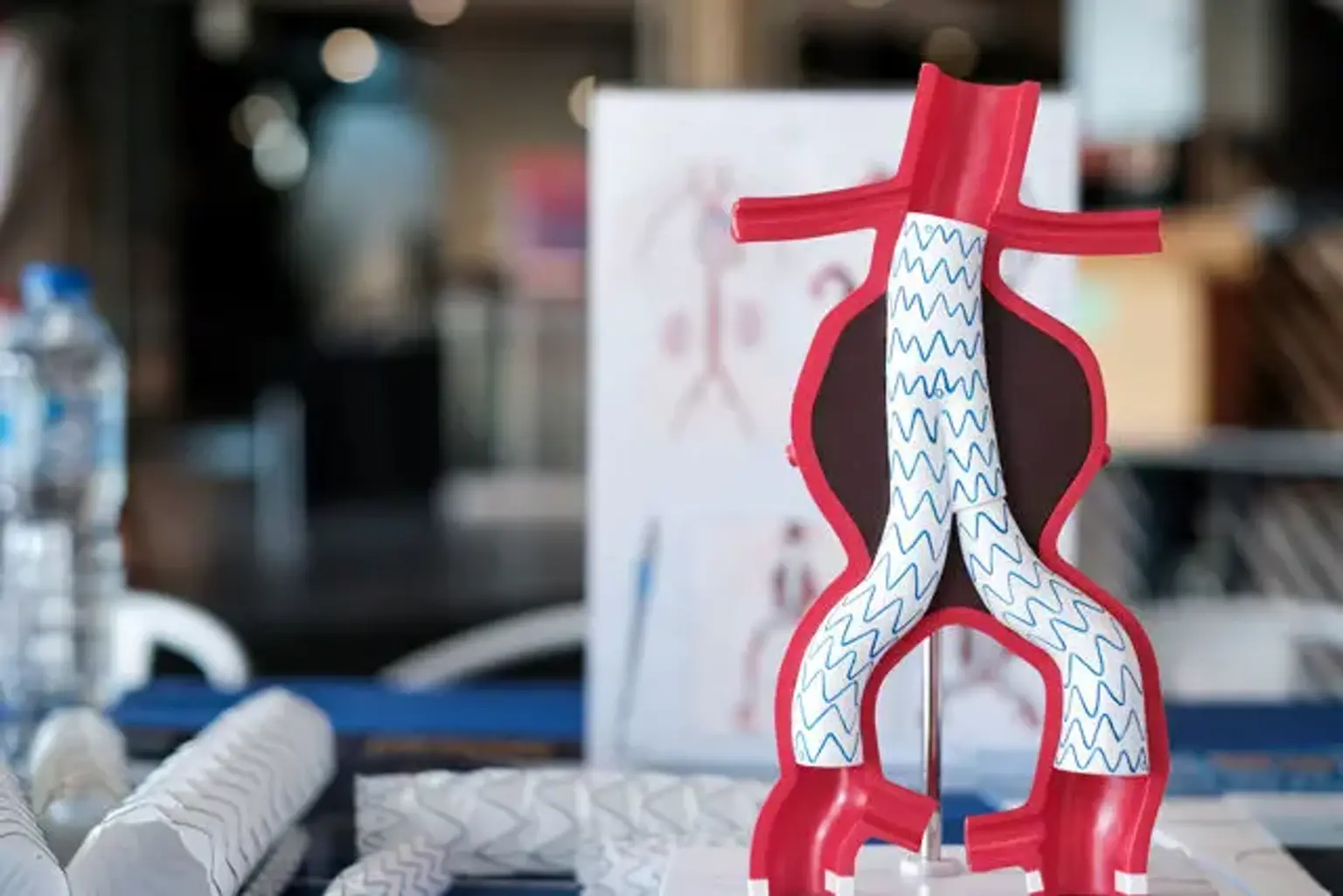
Endovascular repair:
Endovascular repair initially became practicable in the 1990s, and while it is now a well-established alternative to open surgery, its purpose remains unclear. It is typically used in elderly, high-risk patients or those who are unable to undergo open repair. However, depending on the form of the aneurysm, endovascular repair is only possible for a subset of AAAs.
The key advantages of this procedure over open repair include lower peri-operative mortality, less time in critical care, less time in the hospital overall, and a faster return to regular activity. The disadvantages of endovascular repair include the need for more frequent continuing hospital assessments and a higher likelihood of further surgeries being necessary.
According to the most recent research, the EVAR treatment does not improve overall survival or health-related quality of life when compared to open surgery, despite the fact that aneurysm-related mortality is reduced.
Management of Cerebral aneurysm
Individuals with a ruptured cerebral aneurysm should receive emergency treatment that includes restoring deteriorating respiration and lowering intracranial pressure. There are currently two options for securing intracranial aneurysms: surgical clipping or endovascular coiling. To occlude the burst aneurysm and limit the risk of recurrent hemorrhage, surgical clipping or endovascular coiling is often performed within the first 24 hours after bleeding if feasible.
Surgical clipping:
Aneurysms can be treated by using a specifically constructed clip to clip the aneurysm's base. While craniotomies are normally used for this, a novel endoscopic endonasal technique is being tested. Walter Dandy of the Johns Hopkins Hospital pioneered surgical cutting in 1937. A catheter angiography or CTA can be done after clipping to ensure complete clipping.
Endovascular coiling:
The insertion of platinum coils into the aneurysm is referred to as endovascular coiling. A catheter is placed into a blood vessel, most often the femoral artery, and then carried through blood arteries into the cerebral circulation and the aneurysm. Coils are either inserted into the aneurysm or discharged into the bloodstream prior to the aneurysm.
When the coils are deposited into the aneurysm, they expand and cause a thrombotic response. If successful, this stops additional aneurysm hemorrhage. In the event of broad-based aneurysms, a stent may be inserted into the parent artery first to act as a scaffold for the coils.
Cerebral bypass surgery
When a patient has a blood vessel aneurysm or a tumor at the base of the skull wrapped around a blood vessel, surgeons replace the problematic vascular with an artery from another part of the body.
Conclusion:
True aneurysms are abnormal arterial dilatation caused by a compromised vessel wall. False aneurysms, on the other hand, are external hematomas with a sustained connection to a leaking artery. The symptoms are usually determined by the location and size of the aneurysm. There are surgical and endovascular treatment options available, with the decision depending on the type of aneurysm and whether or not symptoms or consequences are present.
Aortic aneurysm (AAA) is a localized dilatation of the abdominal aorta that is more than 1.5 times its usual diameter. AAAs are characterized as either suprarenal or infrarenal aneurysms based on their location. Men of advanced age are more likely to develop them; smoking and hypertension are other key risk factors.
If a dissection or aneurysm rupture happens, the prognosis is significantly worse. AAA rupture is characterized by a rapid onset of acute ripping back or abdominal pain, a painful pulsatile mass, and hypovolemic shock, and it should be treated with emergency surgery. To rule out a AAA, all males between the ages of 65 and 75 with a history of smoking should be checked with an ultrasound once.
AAAs are typically asymptomatic and therefore discovered by chance. AAAs can cause lower back discomfort, a pulsatile abdominal mass, and a bruit on auscultation. The best primary and confirmatory diagnostic for diagnosing AAAs and determining their extent is abdominal ultrasonography.
Small aneurysms should be treated with observation, close monitoring, and a decrease in cardiovascular risk factors, but larger (> 5.5 cm) or rapidly growing aneurysms require surgery. Surgical therapy consists of open aneurysm excision with graft implantation or, increasingly, endovascular stent insertion.