Introduction
Angiomyoma, also known as angioleiomyoma, is a benign smooth muscle tumor that arises from vascular smooth muscle cells. Although it is considered benign, this soft tissue tumor can cause discomfort and other symptoms, particularly due to its location. Angiomyomas are a part of a larger family of soft tissue tumors, which include other vascular and smooth muscle growths. These tumors are often found in the subcutaneous tissues and are most commonly located in the extremities, especially the lower legs.
Although the tumor itself is not cancerous, its presence can lead to pain and swelling, especially when the vascular elements contract. Angiomyomas are generally slow-growing, and many cases remain undiagnosed until they cause noticeable symptoms or complications. Understanding the symptoms, diagnosis, and treatment options is essential for individuals who may be dealing with this condition.
Epidemiology and Risk Factors
Angiomyomas are relatively rare, accounting for a small percentage of soft tissue tumors. They most commonly affect adults between the ages of 40 and 60, with a slight predominance in females (F:M ratio of 1.7:1). The tumors are often found in the lower extremities (50-70% of cases), although they can also appear on the head, trunk, and upper limbs.
The exact cause of angioleiomyomas is not well understood, but there are several factors believed to contribute to their development. Some researchers suggest that minor trauma to the area, venous stasis, or hormonal influences—especially estrogen—could play a role in their formation. This might explain why the condition is more commonly found in individuals who are middle-aged and why it might be more frequent in women.
Clinical Presentation
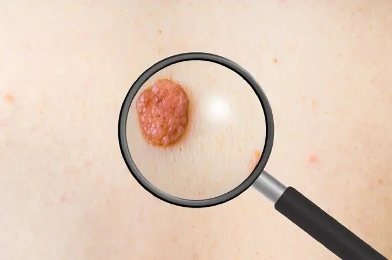
The hallmark of an angioleiomyoma is a painful, slow-growing, solitary nodule. These tumors often develop under the skin (subcutaneous tissue) and are most noticeable on the lower extremities. The pain associated with angioleiomyomas is thought to be caused by ischemia (reduced blood flow) within the vascular elements of the tumor, which results in localized tissue pressure and nerve irritation. However, it’s important to note that pain is not always present, and not all individuals with an angioleiomyoma experience discomfort.
Patients typically report the appearance of a firm, well-demarcated lump, which may gradually increase in size. Despite its slow growth, the tumor can cause significant concern due to its location and potential for discomfort. In some cases, multiple lesions may develop, though this is less common.
The clinical challenge with diagnosing angioleiomyomas is their similarity to other types of tumors, such as glomus tumors, neuromas, or angiolipomas. These conditions can present with similar symptoms and appearances, so distinguishing between them may require additional tests, including imaging and biopsy.
Diagnostic Challenges
Diagnosing an angioleiomyoma can be tricky, as the symptoms and appearance can resemble other soft tissue tumors. A complete diagnosis often requires histopathological examination, meaning that a biopsy is typically necessary to confirm the presence of the tumor. Clinicians may suspect angioleiomyoma based on clinical examination and the typical presentation of pain and a subcutaneous nodule, but definitive diagnosis often requires the analysis of tissue samples under a microscope.
In terms of imaging, ultrasound and MRI can provide valuable information. On ultrasound, an angioleiomyoma typically appears as a well-defined, homogeneous mass with increased vascularity. MRI scans show the characteristic features of a well-circumscribed mass, which may appear hyperintense on T1-weighted images and have a heterogeneous signal on T2-weighted scans. Contrast-enhanced imaging further aids in visualizing the blood vessels within the tumor.
Because of the overlap in symptoms with other conditions, diagnosis can sometimes be delayed. However, once the tumor is identified, treatment can begin promptly. The key to diagnosing angioleiomyoma is recognizing the potential for benign smooth muscle tumors in clinical settings where pain and subcutaneous masses are present.
Radiologic Features
Radiologic imaging plays a key role in diagnosing angioleiomyomas. While a physical exam and biopsy are essential for confirming the diagnosis, imaging studies like ultrasound and MRI help visualize the tumor’s characteristics and guide treatment.
On ultrasound, angioleiomyomas typically appear as well-defined, oval-shaped masses with a homogeneous texture. These tumors often have increased vascularity, which can be noted by color Doppler ultrasound, helping to differentiate them from other soft tissue lesions.
An MRI scan is especially useful in assessing deeper tumors. The mass usually shows up as a well-circumscribed lesion. On T1-weighted MRI, the tumor appears hyperintense, while T2-weighted images show a heterogeneous signal, reflecting the mixed tissue composition of muscle fibers and blood vessels. Contrast-enhanced MRI can further define the tumor's vascular structures, making it a valuable tool for pre-surgical planning.
Although imaging is crucial, the final diagnosis still requires histological confirmation, typically via biopsy.
Histopathological Features
The hallmark of an angioleiomyoma is its histopathological features, which consist of a mixture of smooth muscle fibers and blood vessels. Under the microscope, the tumor typically presents as a well-defined lesion with smooth muscle bundles and vascular structures. The vascular elements may be dilated, and the smooth muscle cells are arranged in a regular, non-atypical pattern.
A biopsy sample typically shows two main components: smooth muscle tissue, which accounts for the tumor's leiomyoma aspect, and vascular tissue, which gives the tumor its angio- (blood vessel) component. This unique combination of smooth muscle and vascular structures helps differentiate angioleiomyoma from other types of soft tissue tumors.
Histopathological examination remains the gold standard for diagnosis, especially when the tumor’s clinical presentation is unclear. It also helps rule out other conditions, like angiolipomas or glomus tumors, which may have overlapping symptoms but distinct microscopic features.
Treatment Options for Angiomyoma
The primary treatment for angioleiomyoma is surgical excision. Given that these tumors are benign, complete removal is generally curative. Surgery involves carefully excising the mass along with a small margin of surrounding healthy tissue to ensure the tumor is entirely removed and reduce the risk of recurrence. The procedure is typically straightforward, though its complexity may increase depending on the tumor's size and location.
For smaller lesions that cause minimal symptoms, conservative management might be considered, especially if the tumor is asymptomatic or growing very slowly. However, if the lesion causes discomfort or interferes with function, surgery is recommended.
In rare cases, particularly for tumors that are located in challenging areas or have a complex structure, reconstructive surgery may be necessary after excision to repair any damage to surrounding tissue. Non-surgical treatments are rarely used but could include corticosteroid injections for reducing inflammation and pain, though they do not target the root cause—the tumor itself.
Surgical Considerations and Challenges
Surgical removal of an angioleiomyoma is generally safe, but it does come with certain considerations. The main challenge during surgery is ensuring clear margins, as incomplete excision may result in recurrence. Surgeons aim to remove the entire tumor while preserving as much surrounding healthy tissue as possible. Tumors located near vital structures, such as nerves or blood vessels, may pose additional challenges.
Another consideration is postoperative care. Depending on the location and size of the tumor, patients may need time for wound healing and physical therapy to restore function, particularly if the tumor was in an area like the foot or hand. Most patients recover quickly after excision, but follow-up appointments are necessary to monitor for any signs of recurrence.
Post-Operative Care and Recovery
After surgery, recovery from an angiomyoma excision is usually straightforward. Most patients experience minimal pain, which can be managed with over-the-counter medications like acetaminophen or ibuprofen. Swelling and bruising are common around the surgical site but typically subside within a few days.
Patients are often advised to rest and avoid strenuous activity for a few weeks to allow the surgical site to heal properly. If the tumor was located in a joint or weight-bearing area, physical therapy may be recommended to restore full function and mobility. It is also essential to follow wound care instructions to prevent infection and promote healing.
Follow-up visits are crucial to monitor for signs of recurrence. While complete excision usually results in a permanent cure, there is a small chance the tumor could grow back if not fully removed. Recurrence rates are low, but patients should stay vigilant, especially if symptoms reappear.
Prognosis and Long-Term Outlook
The prognosis for individuals with angioleiomyoma is generally very good, especially after successful surgical excision. Most patients experience complete resolution of symptoms with no recurrence. Studies show that with proper surgical removal, the cure rate for angioleiomyoma is high, with a recurrence rate of less than 10% when the tumor is completely excised.
For those with larger tumors or multiple lesions, the risk of recurrence may be slightly higher, but overall, the condition does not pose a long-term threat to health. Angioleiomyomas are benign, meaning they do not spread to other parts of the body, and they do not transform into cancer.
It’s important to note that while surgery is highly effective, patients should remain vigilant for any new symptoms or changes in the surgical site, particularly in the first few months after treatment.
Global Popularity and Trends
Angioleiomyomas are relatively rare tumors, but they are well-documented in the medical community. Their global prevalence is not easily measured, as the tumors are often misdiagnosed or go unnoticed until they cause symptoms. However, they are more commonly reported in developed countries where diagnostic imaging and surgical options are readily available.
Recent advances in medical imaging and surgical techniques have made the diagnosis and treatment of angioleiomyomas more efficient. MRI and ultrasound have improved preoperative planning, while minimally invasive surgical methods have helped reduce recovery time and surgical risks.
Globally, the treatment trends for angioleiomyomas largely follow the standard approach of surgical excision, with varying access to resources depending on the country. In more resource-rich nations, patients benefit from advanced imaging and surgical options, while in lower-income regions, diagnosis may be delayed due to limited access to diagnostic tools. Nonetheless, the overall prognosis remains favorable worldwide.
Frequently Asked Questions (FAQs)
What are the symptoms of angioleiomyoma?
Angioleiomyomas typically present as a painful, slow-growing nodule under the skin, often on the lower limbs. The pain is usually due to reduced blood flow within the tumor, though not all patients experience discomfort. The tumor may feel firm and well-defined upon physical examination.
Is surgery always required for treatment?
Surgery is the primary treatment for angioleiomyoma and is generally recommended if the tumor causes pain or grows significantly. Small, asymptomatic tumors may be monitored without surgery, but excision is the preferred option for symptomatic or larger lesions.
Can angioleiomyoma recur after surgery?
While the recurrence rate is low, recurrence can occur if the tumor is not completely removed during surgery. This is why surgeons aim to excise the tumor with clear margins to reduce the risk of recurrence.
Is angioleiomyoma a cancerous condition?
No, angioleiomyomas are benign (non-cancerous) tumors. They do not spread to other parts of the body and do not become malignant. However, if left untreated, they can cause pain or discomfort, which is why timely treatment is recommended.
Potential Complications and Risks
Though rare, complications can occur after angiomyoma excision. The most common issues include infection, bleeding, and poor wound healing, particularly in larger or deeper tumors. Scarring is another possible concern, especially when the tumor is located in visible or cosmetically sensitive areas like the face or hands.
In some cases, the surgical site may develop numbness or nerve damage, especially if the tumor was near a nerve. This is usually temporary, but there may be long-term changes in sensation in the affected area. If the excision was incomplete, the tumor could recur, necessitating additional surgery.
Despite these risks, with proper care and surgical expertise, the chance of complications is minimal, and most patients recover fully without long-term issues.
Prevention of Angioleiomyoma Recurrence
While angioleiomyomas are typically benign and treated effectively with surgical excision, there are steps that can help minimize the chances of recurrence. The key factor in preventing recurrence is ensuring that the tumor is completely excised with clear margins, meaning the entire tumor and a small margin of surrounding tissue are removed during surgery. This reduces the likelihood of any remaining tumor cells that could regrow.
Post-surgical follow-up appointments are essential for monitoring the site of the excision, particularly in the first year after surgery. Recurrence rates are generally low when the tumor is adequately removed, but patients should remain vigilant for any signs of regrowth, such as pain or the reappearance of a lump.
For individuals with multiple tumors or those who have had a recurrence in the past, regular imaging and clinical exams may be recommended to ensure that no new lesions are developing. Early detection and treatment are key to preventing further complications.
Costs and Accessibility
The cost of diagnosis and treatment for angioleiomyoma can vary depending on geographical location, healthcare system, and the complexity of the case. In developed countries, the primary cost is related to imaging (ultrasound or MRI) and surgical procedures, which can range from a few hundred to a few thousand dollars, depending on whether the surgery is outpatient or requires an overnight hospital stay. Postoperative care and follow-up visits also contribute to overall costs.
In regions with limited healthcare resources, access to advanced imaging and surgical facilities may be restricted, which could lead to delayed diagnoses or less comprehensive treatment. However, the fundamental treatment of surgical excision remains relatively simple, and many healthcare systems offer assistance programs or more affordable care options.
For individuals seeking treatment abroad, the cost of surgery may be lower in some countries, but it’s important to weigh the quality of care, potential complications, and long-term follow-up requirements before choosing medical tourism for treatment.
Current Research and Advancements
Although angioleiomyomas are benign and relatively well-understood, there are ongoing studies focusing on improving the diagnosis and treatment of this condition. New imaging techniques, including advanced MRI sequences and contrast agents, are helping to better visualize vascular components and predict surgical outcomes. These advances allow for more precise excisions and reduced complications.
Researchers are also investigating minimally invasive procedures, such as laser removal or radiofrequency ablation, as potential alternatives to traditional surgery. These treatments aim to reduce recovery time and minimize scarring, but more studies are needed to establish their effectiveness in treating angioleiomyomas.
Additionally, advancements in genetic research are exploring the molecular pathways that might lead to the development of angioleiomyomas. Understanding these pathways could lead to the development of targeted therapies that could prevent the growth of new tumors or even shrink existing ones without the need for surgery. While these treatments are still in the experimental phase, they represent an exciting area of future research.
Patient Resources and Support
For patients diagnosed with angioleiomyoma, it can be helpful to connect with support groups or patient advocacy organizations that focus on benign tumors or soft tissue tumors. These groups can provide valuable resources on treatment options, connect individuals with healthcare providers who specialize in tumor excision, and offer emotional support.
In addition to professional support, online forums and community groups can offer a space for patients to share their experiences with others who have undergone similar treatments. Patients can ask questions, share tips for managing recovery, and find reassurance from others who have successfully navigated the treatment process.
Patients should always ensure that any resources they rely on are credible and endorsed by medical professionals. Consulting with a healthcare provider is the best way to ensure that the information they receive is accurate and applicable to their specific situation.
Conclusion
Angioleiomyoma is a benign tumor that can cause significant discomfort, but with proper diagnosis and surgical excision, most patients experience a full recovery. The outlook for individuals with this condition is generally positive, with low recurrence rates when the tumor is completely removed. Imaging, biopsy, and surgical techniques have improved significantly, providing effective solutions for managing this rare tumor.
While the condition is not life-threatening, it can significantly affect a person’s quality of life if left untreated. Early detection, patient education, and access to advanced healthcare options are crucial in managing angioleiomyomas effectively. Research continues to advance the understanding of these tumors, and new treatments may emerge in the future, offering even better outcomes for patients.
For anyone experiencing symptoms like painful lumps or subcutaneous masses, it’s important to seek medical attention. With proper care and follow-up, most people can return to their normal activities without the burden of an angioleiomyoma.