Introduction
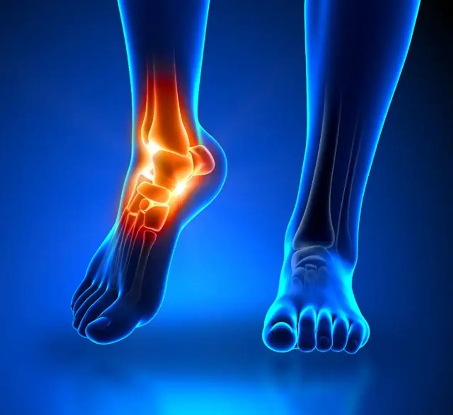
Ankle fusion surgery, also known as arthrodesis, is a procedure used to treat severe ankle joint arthritis or injuries that haven’t responded to other treatments. When the ankle joint becomes damaged from conditions like arthritis, trauma, or deformities, fusion surgery helps reduce pain and restore function by permanently joining the bones of the ankle joint.
This procedure has gained popularity worldwide as an effective solution for those who can no longer tolerate the discomfort or immobility caused by damaged ankle joints. Ankle fusion can significantly improve quality of life by eliminating the pain that comes with joint movement.
Indications for Ankle Fusion Surgery
Ankle fusion surgery is typically considered when other treatment options—such as medication, physical therapy, or joint injections—no longer provide relief from pain and stiffness. The most common reasons for ankle arthrodesis include:
End-stage arthritis: This is the most frequent reason for ankle fusion surgery. When the cartilage in the ankle joint is severely worn down, arthritis leads to constant pain and limited mobility.
Severe ankle fractures: In cases where the bones are irreparably damaged and cannot be properly reconstructed, fusion provides a stable and functional alternative.
Ankle deformities: Congenital deformities or those resulting from previous injuries can also lead to the need for fusion to restore proper alignment and prevent further joint degradation.
Techniques Used in Ankle Fusion Surgery
Ankle fusion can be performed through different surgical approaches, depending on the patient’s condition and the surgeon’s preference:
Open Ankle Fusion: The most traditional technique, where the surgeon makes an incision to directly access the joint and perform the fusion.
Arthroscopic Ankle Fusion: A minimally invasive option, where smaller incisions are made and a camera is used to guide the surgery. This technique is usually preferred for certain cases as it results in less soft tissue damage and a quicker recovery.
The surgeon may also use bone grafting to enhance healing, especially in cases where the bone quality is poor. Internal or external fixation techniques (plates, screws, rods, or external frames) are used to hold the bones together while they fuse.
What is Ankle Fusion Surgery?
Ankle fusion surgery involves fusing the bones of the ankle joint together, usually by using screws, plates, or rods to ensure the bones heal in a fixed position. This prevents the ankle joint from moving and eliminates the pain associated with damaged cartilage and bones rubbing against each other.
While this surgery eliminates the joint's natural movement, it can restore stability, which is crucial for walking and standing. It is especially beneficial for patients with advanced arthritis, severe fractures, or joint deformities that cannot be treated through less invasive methods like injections or physical therapy.
Benefits of Ankle Fusion Surgery
Ankle fusion surgery offers several key benefits, especially for patients suffering from chronic ankle pain due to arthritis or severe injuries. The primary benefit is pain relief, which can drastically improve a patient's quality of life. By eliminating joint movement, the friction that causes pain is also removed.
Other benefits include:
Improved stability: After fusion, the ankle becomes more stable, allowing patients to walk more confidently without the constant fear of instability.
Restoration of mobility: While the joint no longer moves, the patient can regain a functional level of mobility for everyday activities, including walking and standing.
Long-term results: With proper care, the fusion can last for many years, making it an enduring solution for those with severe ankle arthritis or deformities.
However, it’s important to note that while the procedure alleviates pain, it comes at the cost of losing natural ankle joint motion. This limitation may affect certain high-impact activities like running or jumping.
Ankle Fusion Surgery for Special Populations
Certain groups may face additional challenges with ankle fusion surgery:
Elderly patients: Older adults often have slower healing times, but surgery can still provide relief from debilitating pain, improving overall function and quality of life.
Diabetic patients: People with diabetes may face increased risks of infection or delayed healing. However, with proper management, they can still benefit from the procedure.
Patients with previous infections or poor bone quality: For these individuals, additional considerations like bone grafting and more intensive post-op care may be needed to ensure a successful fusion.
Preparing for Ankle Fusion Surgery
Proper preparation for ankle fusion surgery can significantly enhance the recovery process. Here are a few key steps involved in preparing for the procedure:
Imaging and Medical Evaluation: Before surgery, the surgeon will perform tests like X-rays, CT scans, or MRIs to assess the severity of the joint damage. These images help the surgeon decide on the best approach for the fusion.
Health Optimization: Patients should work on improving their overall health before the surgery. This includes stopping smoking (since smoking can hinder healing), managing weight, and ensuring optimal blood sugar levels for diabetic patients.
Home and Post-Surgery Care: Post-surgery, patients will need to rest with their foot elevated for several weeks. Preparing the home for recovery, like ensuring easy access to food, setting up mobility aids (e.g., crutches), and planning for help with daily activities, can make the healing process more comfortable.
Pre-Surgery Consultation: During the consultation, the surgeon will discuss the surgery in detail, explain anesthesia options, and provide instructions for post-operative care, including wound care and medications.
Walking and Mobility Post-Surgery
After ankle fusion surgery, mobility will be limited for a period of time as the bones need to heal. Immediately post-surgery, patients will need crutches or a walker to keep weight off the fused joint.
Timeline for walking: It typically takes 6 to 12 weeks before patients can begin bearing weight on the ankle, depending on healing speed. Full recovery may take 6 months to a year.
Physical therapy: Once the bones have fused, physical therapy is crucial to regain strength and mobility in the foot and surrounding muscles.
Though full ankle motion is lost, patients usually regain enough stability to walk without pain.
Return to Normal Activities After Ankle Fusion
While high-impact activities like running and jumping are generally avoided post-surgery, most patients can return to low-impact activities:
Daily activities: Walking, driving (once cleared by a doctor), and most light activities are possible after recovery.
Sports and exercise: After healing, patients may return to low-impact sports such as swimming or cycling. However, sports involving twisting or running, like basketball, are often not recommended long-term.
The extent to which normal activities can resume depends on the individual’s healing process and how well they adapt to the limitations of the fused joint.
What Happens During Ankle Fusion Surgery?
Ankle fusion surgery typically lasts between 1 to 2 hours, depending on the complexity of the case. Here’s a general overview of what happens:
Anesthesia: The patient will be given general anesthesia or a regional block to numb the area. This ensures the procedure is painless.
Surgical Approach: The surgeon will make an incision over the ankle joint and clean out any damaged tissue or cartilage. In some cases, arthroscopic tools may be used for a less invasive approach.
Bone Fusion: The bones of the ankle joint are aligned and fixed using plates, screws, or rods. Bone grafting may be added to help promote healing if necessary.
Wound Closure: Once the bones are properly fused and stabilized, the surgeon will close the incision with sutures and apply a dressing or brace to protect the area.
After the procedure, the patient will be taken to a recovery room where they will be monitored as the anesthesia wears off. Patients can expect to stay in the hospital for a couple of days before being discharged to recover at home.
Cost of Ankle Fusion Surgery
The cost of ankle fusion surgery can vary widely based on location, healthcare provider, and whether insurance covers the procedure. On average, the cost ranges from $10,000 to $30,000 in the U.S., but this figure can be lower in other countries or with insurance coverage.
Insurance coverage: Many health insurance plans will cover ankle fusion surgery, especially if it is deemed medically necessary due to arthritis or severe trauma.
Out-of-pocket costs: For patients without insurance, the procedure may be costly, but some clinics offer payment plans or financial assistance programs.
It’s important to consult with both the surgeon and insurance provider to fully understand the costs involved.
Ankle Fusion Surgery: Success Rates and Outcomes
Ankle fusion surgery has a high success rate, with most patients experiencing significant pain relief and improved mobility. Success rates range from 80% to 90% for patients who undergo the procedure due to arthritis or traumatic injuries.
Pain relief: The primary benefit of ankle fusion is the elimination of pain caused by damaged cartilage. Most patients report significant improvements in pain management, which leads to a better quality of life.
Long-term outcomes: In the long term, ankle fusion can provide a stable and functional ankle, though the loss of natural movement can affect activities that require a flexible joint.
Success largely depends on the surgeon’s skill, the patient's adherence to post-surgery care, and overall health.
Alternative Treatments to Ankle Fusion Surgery
Before opting for ankle fusion, patients may consider other non-surgical treatments that can alleviate pain and improve function, including:
Physical therapy: Targeted exercises to strengthen the muscles surrounding the ankle and improve flexibility.
Injections: Steroid injections or hyaluronic acid injections can temporarily reduce pain and swelling in the joint.
Bracing: A custom ankle brace can provide support and stability for people with mild arthritis or instability.
Ankle replacement: For some patients, a total ankle replacement may be a better option, especially if the goal is to preserve joint movement.
Ankle fusion is usually recommended only after these conservative treatments fail to provide relief.
Can You Play Sports After Ankle Fusion Surgery?
After recovery, patients can return to some physical activities, but sports that require running or jumping may be off-limits:
Low-impact activities: Swimming, cycling, and walking are typically safe and often encouraged for improving overall health and mobility.
High-impact sports: Activities that involve high stress on the ankle, such as running or playing basketball, are generally not advised. The fused joint can’t handle the twisting and force of such movements.
Though sports may be limited, many patients find that they can still enjoy an active lifestyle without the pain they once had.
The Psychological Impact of Ankle Fusion Surgery
Chronic ankle pain can have a significant emotional toll. The surgery not only improves physical function but also helps address the psychological challenges associated with living with pain:
Relief from constant pain: Many patients experience a major improvement in mental health after surgery due to the relief from persistent pain.
Anxiety and recovery: The fear of post-surgery complications and the long recovery process can cause anxiety, but patients often find peace of mind as they see progress in healing.
Addressing the emotional and psychological impact is an essential part of the recovery process, with support from family, friends, and healthcare providers.
Risks and Complications of Ankle Fusion Surgery
Like all surgeries, ankle fusion carries some risks. Understanding these risks can help patients make informed decisions:
Infection: As with any surgical procedure, there is a risk of infection, especially since the surgery involves the use of implants (plates, screws).
Nonunion or delayed healing: In some cases, the bones may fail to fuse properly, resulting in prolonged pain and the need for further procedures.
Adjacent joint arthritis: After fusion, the nearby joints (like the subtalar joint) may be subjected to extra stress, potentially leading to arthritis in those areas over time.
Nerve injury: Although rare, there is a risk of nerve damage, which could result in loss of sensation or movement.
Certain factors like smoking, obesity, and poor circulation can increase the likelihood of complications, which is why pre-surgical assessments and lifestyle changes are essential for optimizing the chances of a successful outcome.
Recovery Timeline After Ankle Fusion Surgery
The recovery process after ankle fusion surgery varies from person to person, but patients can generally expect a few key milestones during the healing process:
Initial recovery (0-6 weeks): During this phase, patients are usually non-weight-bearing and must use crutches or a walker. The ankle is typically in a cast or a boot to immobilize it.
Partial weight-bearing (6-12 weeks): After about 6 weeks, once the surgeon confirms initial healing, patients can start putting limited weight on the ankle with the aid of a walking boot.
Full weight-bearing (3-6 months): By 3 months, most patients can walk without crutches. However, the fusion process may continue for up to 6 months or longer.
Final recovery (6 months to 1 year): Full recovery, including the complete fusion of the bones, typically takes about 6 to 12 months. During this time, physical therapy helps regain strength and mobility in the foot and leg muscles.
Adherence to post-surgery care and regular check-ups are essential for a successful recovery.
Ankle Fusion vs. Ankle Replacement: Which is Right for You?
Ankle fusion and ankle replacement are both options for patients with severe arthritis or joint damage, but they differ significantly in their approach:
Ankle Fusion (Arthrodesis): This procedure involves permanently joining the bones of the ankle joint, which eliminates pain but also reduces mobility. It’s often recommended for patients with advanced arthritis or significant joint deformities.
Ankle Replacement: Also known as total ankle arthroplasty, this procedure involves replacing the damaged joint with a prosthetic. It preserves more natural movement in the ankle but may not be suitable for everyone, particularly those with poor bone quality.
The choice between the two depends on factors like the patient’s age, activity level, and overall health. Ankle fusion is often preferred for younger patients or those with severe deformities, while ankle replacement may be a better option for older patients or those who want to maintain some joint movement.
How to Choose the Right Surgeon for Ankle Fusion Surgery
Choosing an experienced and qualified surgeon is crucial to achieving a successful outcome from ankle fusion surgery. Consider the following factors when selecting a surgeon:
Specialization: Ensure the surgeon specializes in foot and ankle surgery. They should have experience in performing ankle fusions specifically.
Reputation and reviews: Look for reviews from previous patients to understand the surgeon’s track record. Recommendations from your primary care doctor or other specialists can also help.
Consultation: During your consultation, ask about the surgeon’s approach to the procedure, success rates, and the recovery process. A good surgeon will clearly explain the potential risks and benefits and answer all your questions.
The surgeon’s expertise can make a significant difference in the outcome of the surgery and the patient’s overall experience.
Conclusion
Ankle fusion surgery is an effective solution for people suffering from severe arthritis or joint injuries that have failed to respond to other treatments. While it eliminates pain and provides stability, it does come with the trade-off of losing natural joint movement.
Before deciding on ankle fusion, it’s important to consider your activity level, health, and long-term goals. Consulting with a specialized surgeon and weighing all treatment options—including non-surgical methods and ankle replacement—will help ensure you make an informed decision.
With proper preparation and recovery, ankle fusion can significantly improve quality of life, allowing many patients to regain mobility and freedom from pain.