Anterior Cervical Microdiscectomy
Overview
An intervertebral disc is a cushion that sits between each bone in the spinal column. These discs keep the bones from grinding together and serve as shock absorbers during falls, exercise, and daily activities.
These discs can be injured, resulting in discomfort ranging from mild to severe. An ACDF surgery can be performed on any of the seven cervical bones' discs.
Anterior cervical discectomy is a surgical procedure used to alleviate or remove persistent discomfort in the neck and back caused by a disc problem.
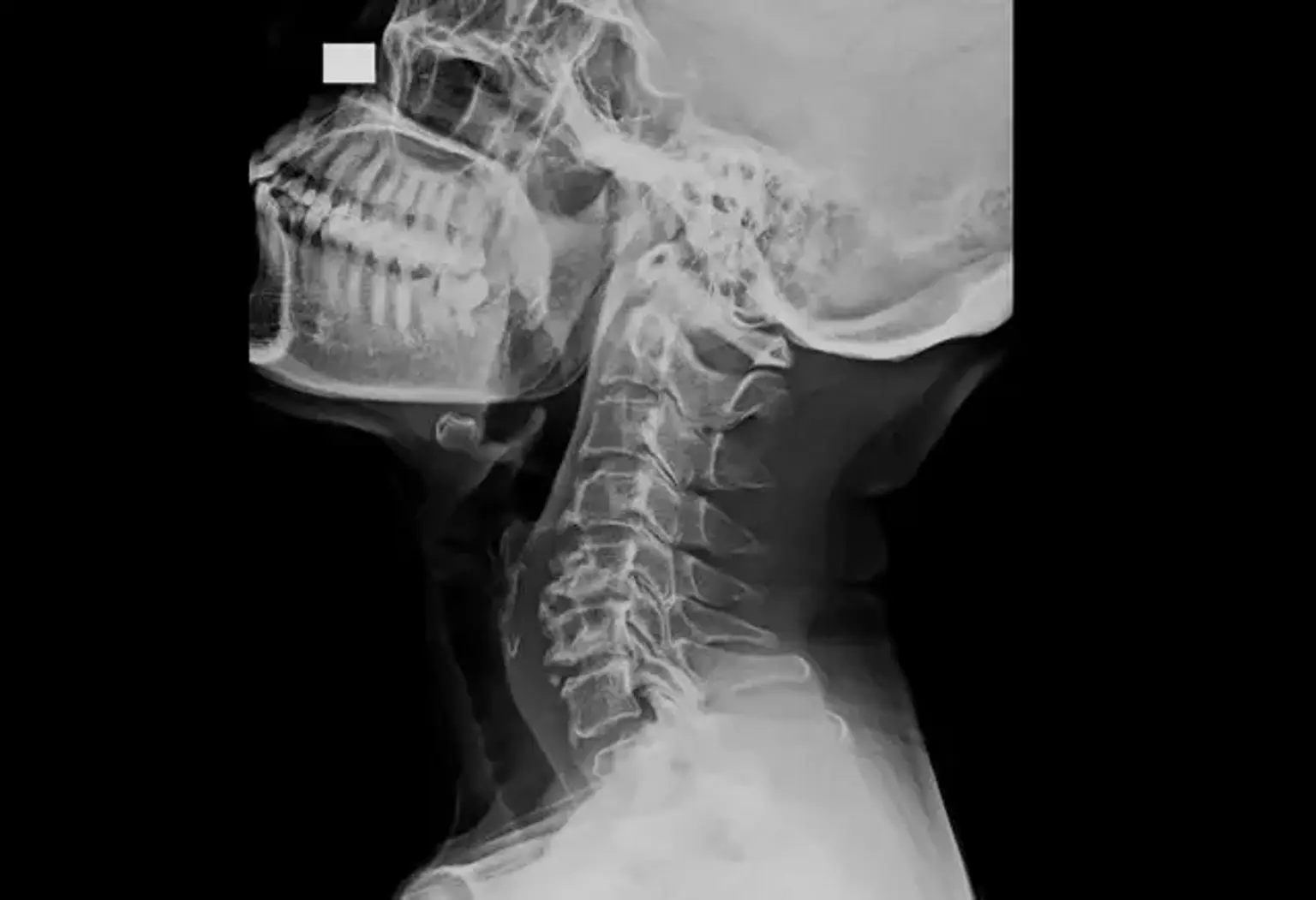
The Cervical Spine
The spinal canal and intervertebral foramina are bone tunnels in the spine that house the spinal cord and spinal nerves (nerve roots). They safeguard the nerves and spinal cord by allowing them to travel in a safe manner. However, when the size of these tunnels is reduced, there is less space for the spinal nerves and/or spinal cord, putting pressure on these structures.
The cervical spine runs from the base of the skull to the base of the neck and bears the weight of the head. The spinal cord connects the brain to the cervical spine and controls the operation of the body's organs and limbs. Soft pads or discs are located between each of the cervical spine's seven vertebrae, acting as shock absorbers and allowing for head bending and movement. Each disc is composed of two parts: a softcore known as the nucleus and a tough outer band known as the annulus.
Cervical Pain
Millions of individuals are bothered by neck and arm discomfort. A rupture or herniation of one or more cervical discs is a typical cause of cervical discomfort. When the annulus of the disc rips, the soft nucleus squeezes out. As a result, pressure is exerted on the nerve root or spinal cord, causing discomfort in the neck, shoulders, arms, and, in rare cases, the hands. Cervical disc herniations can occur as a result of age, normal wear and tear, or unexpected stress, such as an accident.
The majority of cases of cervical discomfort do not require surgery and are managed non-surgically using medicines, physical therapy, and/or bracing. However, if patients continue to endure substantial pain and weakness, surgery may be required.
What are disc osteophytes?
Intervertebral discs are located between each vertebra in the spine. They serve as shock absorbers while also enabling proper mobility of the bones in your neck. Each disc has a fibrous outer ring (annulus fibrosis) and a soft jelly-like center (nucleus pulposis)
The annulus, which joins each spinal bone, is the hardest component of the disc. The disc's soft and juicy core acts as the primary shock absorber. An annular tear occurs when the annulus fibrosis tears, which is frequently the initial step in the process of disc prolapse. An annular tear can induce neck discomfort as well as arm pain.
A cervical disc prolapse (or herniation) happens when the nucleus pulposis slips out of its normal place and bulges into the spinal canal, occasionally pressing on nerves or the spinal cord.
Degenerative disc disease causes the discs or cushion pads between your vertebrae to decrease, causing wear and herniation. Your spine may also include osteoarthritic regions. Because of strain on the spinal nerves and/or spinal cord, this degeneration and osteoarthritis can produce pain, numbness, tingling, and weakness.
Osteophytes are aberrant bone spurs that grow as part of the degenerative process or after a lengthy period of disc prolapse. This additional bone growth can result in spinal stenosis and intervertebral foraminal stenosis, which can compress the spinal cord and/or spinal nerves.
Anterior cervical discectomy and fusion definition
Anterior cervical discectomy and fusion (ACDF) is a surgical procedure used to remove a herniated or degenerative disc from the neck. The bones are fused together after the surgeon removes the injured disc.
The procedure is called anterior because the surgeon enters the disc at the front of the neck rather than the rear.
The surgeon can access the spinal column more easily through the throat since going through the back of the neck might injure the neck muscles and the spinal column. The surgeon next pushes the tissue within the neck and throat aside to get access to the spine, where any damaged discs are removed.
The procedure normally comprises the fusing of at least two bones to guarantee that the spine is aligned and to prevent the bones of the spine from rubbing against one another. The surgeon replaces the disc at this stage in the surgery.
There are a few options for disc replacement:
- Bone graft: A bone graft occurs when the surgeon connects bone to the location where the disc is to be replaced. The bone might originate from elsewhere in the person's body or from a bone bank.
- Bone graft substitute: This method, like a bone transplant, employs human-manufactured materials containing shavings from the patient's bones.
- Arthroplasty: This is the procedure in which the surgeon replaces the disc with an artificial disc.
Once the new disc is in place, the surgeon attaches the bones with a titanium plate and screws. When a bone transplant is used in surgery, the bones will ultimately grow together. Until then, the plate and screws give support.
An X-ray machine assists the surgeon in ensuring that the new disc is in the correct position. Following the surgery, the surgeon repositions the tissue of the neck and throat and closes the incision with stitches.
Uses
Normally, the discs of the spinal column allow for pleasant mobility. However, aging, traumas, and other degenerative disorders, such as arthritis, can cause disc injury.
Discs might thin, dry out, swell and bulge, resulting in insufficient cushioning. Degeneration occurs when discs get damaged. Discs can also expand or split open, a condition known as herniation.
Muscle stiffness and soreness are sometimes caused by pain. It can also spread to other parts of the body, resulting in headaches, back discomfort, and shoulder pain.
When conservative methods (pain medicines, nerve sheath injections, physical therapy, neck collars, etc.) have failed or the degree of spinal compression is severe, surgery is typically suggested. Surgery may be the best first-line therapy option in situations of substantial instability or neurological issues.
What do you need to tell the doctor before surgery?
It is necessary that you notify your surgeon if you:
- Have clotting or bleeding problems?
- Have you ever encountered blood clots in your legs or lungs (DVT or deep venous thrombosis)? (pulmonary emboli)
- Are you using aspirin, warfarin, or other anticoagulants, or anything else (including herbal supplements) that might cause your blood to thin?
- Have you had high blood pressure?
- Do you have any allergies?
- Do you have any other health issues?
What do you need to do before surgery?
It is essential that you stop smoking before surgery, and you should not smoke for at least 12 months afterward (it is preferable that you cease permanently). Smoking impedes the fusion process and results in worse post-surgery outcomes.
If you are fairly overweight, it is recommended that you begin a healthy weight loss program before undergoing surgery. Please with your primary care physician and a neurosurgeon about this.
We usually prescribe prehabilitation with one of our exercise physiologists prior to surgery. This is done to get you in the greatest possible form for surgery and to prepare you for post-operative rehabilitation. To avoid unnecessary bleeding before or after surgery, you must cease taking aspirin and any other antiplatelet (blood-thinning) drugs or substances, including herbal remedies, at least two weeks before your operation
If you typically use warfarin or other anticoagulants, you may be admitted to the hospital three or four days before surgery. At that point, your warfarin will be stopped (it takes a few days to wear out) and you may be started on shorter-acting anti-clotting medications for a few days. These can then be discontinued about a day before surgery. If you are taking other anticoagulants, your preparation may differ from this, and your neurosurgeon and perioperative physician will advise you.
Ideally, you should take a Zinc pill every day for one month before surgery and for three months thereafter. This should aid in the healing of wounds.
How is ACDF performed?
To put you to sleep, a general anesthetic will be delivered. An endotracheal tube will be placed, and intravenous antibiotics and steroids will be administered (to prevent infection and post-operative nausea). Throughout the procedure, calf compression devices will be utilized to reduce the danger of blood clots in your legs.
Your skin will be cleansed with an antiseptic solution, and a local anesthetic will be administered.
The incision across the front of your neck is around 2-2.5cm long. It is generally horizontal and can be performed on either the left or right side of the neck. The tiny muscle just beneath the skin has broken. The dissection is then carried out along the natural planes of the neck, between the food pipe and windpipe on one side and the carotid artery (a key blood vessel leading to the brain) on the other.
The front of the spine's thin layer of fibrous tissue ('fascia') is peeled away from the disc space. To ensure that the proper disc is being operated on, a needle is put into the disc space and an x-ray is taken.
The disc is subsequently removed (discectomy) by first severing the outer annulus fibrosis (the fibrous ring that surrounds the disc) and removing the nucleus pulposus (the soft inner core of the disc). To help invisibility of the canal and nerves, the dissection is conducted using a microscope or special surgical magnifying glasses.
A combination of specific equipment is used to remove the disc. Adjacent bone is frequently removed with a tiny drill to recontour the disc space for eventual fusion, to enable safe access to the spinal canal, and to eliminate excess bone growth ('osteophytes') towards the rear of the disc space.
A ligament (the 'posterior longitudinal ligament') right in front of the spinal cord is gently removed to provide access to the spinal canal and the removal of any disc debris that may have extruded through the ligament.
In certain cases, instrumentation (screws with or without a plate) will be utilized to increase spine stability.
Another X-ray is taken to ensure proper cage, plate, and screw location, as well as cervical spine alignment.
Dissolving sutures are used to seal the wound. In certain situations, a wound drain may be needed for up to 24 hours after surgery.
What are the possible implications of surgery?
The majority of patients are hospitalized on the same day as their operation, however, some are admitted the day before. Patients hospitalized the day before surgery include those who reside in rural areas, interstate, or abroad; have severe medical problems or use blood-thinning drugs or anticoagulants; require more investigations before surgery, or are first on the operating list for the day. Before your admittance, you will be given instructions on when to cease eating and drinking.
Typically, you will be hospitalized for 1-2 days following your procedure. You will be instructed on any physical restrictions that will apply following surgery, as well as how to care for your incision.
During surgery, X-rays of your neck will be obtained to ensure that the right spinal level is fused and to optimize the location of cages, screws, and plates. It is crucial that you notify us if you are pregnant or suspect you might be pregnant, as X-rays can be dangerous to an unborn child.
There is a lot of variation amongst people when it comes to the result of surgery and how long it takes to recover. Physical constraints, as well as your return to work and resumed leisure activities, will be explained to you. You should not drive a car or operate heavy machinery unless your neurosurgeon has cleared you to do so.
Because anesthesia might momentarily cloud your mind, you should not sign or witness legal documents until they have been examined by your GP post-operatively.
A crucial aspect of spinal fusion is that by fusing a level of the spine, the levels directly above and below the fusion are subjected to somewhat increased stress. This raises the risk of degeneration at these levels and, as a result, the likelihood that you may require more surgery in the future. The risk of this is estimated to be 3% or less every year. You should speak with your neurosurgeon about this more.
The limitation of mobility in the neck caused by cervical spine fusion is most noticeable while bending your neck forwards and backward. This loss of mobility is generally not perceptible in a one-level fusion (if at all). Following a two-level fusion, there is normally a slight but noticeable loss of mobility, with a more severe loss of movement following a three- or four-level fusion.
What happens After Surgery?
Patients will have some pain following surgery, particularly at the incision site. Pain medicines are typically administered to assist manage the pain. Moist heat and periodic repositioning, as directed by a physician, can also give some comfort. While tingling or numbness are normal and should subside with time, they should be reported to a doctor. Within a few hours of surgery, the majority of patients are up and moving around. In fact, this is advised in order to maintain regular circulation and avoid blood clots.
However, most patients must remain in the hospital for many days before being discharged, gradually increasing the amount of time they are up and walking. Prior to release, the doctor will give the patient-specific instructions on activities that can be undertaken and those that should be avoided.
Patients are frequently urged to engage in regular low-impact exercise. Walking, gradually increasing the distance each day, is the best form of exercise following this sort of surgery. Some discomfort is natural, but pain signals that it is time to slow down and relax.
Arm discomfort is generally the symptom that improves the most consistently following surgery. Neck pain and headaches frequently improve, although they do not always (very occasionally they can be worse). Weakness is frequently the second symptom to improve.
However, your strength may not return to normal totally. Strength usually improves over the course of several weeks or months. Because the nerve fibers carrying feeling are weaker and more prone to pressure, surgery may or may not relieve numbness or pins & needles. It might take up to a year to recover from numbness.
The likelihood of gaining a major benefit from surgery is determined by a number of factors. Your neurosurgeon will give you an idea of your chances of success in your unique instance.
What are the specific risk of an ACDF surgery?
In general, surgery is quite safe, with significant consequences being unusual. The likelihood of a mild problem is less than 3 or 4%, while the likelihood of a serious complication is less than 1 or 2%. Over 90% of patients should be able to complete their procedure without difficulties. Complication rates vary from surgeon to surgeon, so getting a second opinion before undergoing surgery is a good idea. Other potential issues include:
- Ongoing pain
- Paralysis/weakness/numbness
- Functional impairment (clumsiness, poor fine motor skills, and coordination)
- Problems with walking and balance
What is the cost of the surgery?
Private patients who have surgery will almost always have some out-of-pocket payments. A quote for surgery will be sent, but it is merely an estimate. The ultimate fee may vary depending on the operation performed, surgical results, technical concerns, and so forth.
Before proceeding with surgery, you should fully understand the costs involved, and you should share any concerns with your physician or the Precision Brain Spine and Pain Centre administration staff.
What are the alternatives to an ACDF surgery?
- Pain medications. A variety of drugs may be beneficial in the treatment of pain. These include opioid and non-opioid analgesics, membrane stabilizers, and anticonvulsants, as well as Pregabalin. In some cases, specialized medical therapies such as Ketamine infusions may be necessary.
- Nerve sheath injections. Under CT scan guidance, a local anesthetic may be given into the skin of the neck around the compressed nerve. This is sometimes referred to as a 'foraminal block.' Patients typically profit significantly from this therapy, and surgery can sometimes be postponed or avoided. Unfortunately, the advantage of this operation is generally just brief, and it wears off after a few days, weeks, or even months. This treatment is also a great diagnostic tool, particularly when the MRI scan indicates that numerous nerves are compressed and your neurosurgeon wants to know which nerve is causing your problems.
- Physical therapies. These include physiotherapy, osteopathy, hydrotherapy, and massage.
- Activity modification. Simply changing your employment and leisure activities to minimize heavy lifting and repetitive neck or arm movements can sometimes speed up the healing process.
- Other surgical approaches. Foraminotomy, posterior cervical decompression (laminectomy) with or without fusion, and artificial disc replacement are among the procedures available (also known as disc arthroplasty). You should talk to your neurosurgeon about these options, as well as their possible risks and advantages.
Conclusion
An anterior cervical discectomy (decompression) and fusion (ACDF) is a surgical procedure performed via the front of the neck to relieve pressure on the spinal cord and/or nerves while also stabilizing the spine. Because a discectomy is a type of surgical decompression, the technique is also known as anterior cervical decompression.
Whether or not the fusion procedure is used, the ACD approach produces good results. In terms of disc height and neural foramen height, fusion with a semirigid plate provides an advantage at the operating level in the early postoperative period. However, semirigid anterior plates, by definition, do not prevent sinking, thus the benefit provided by this treatment is not long-lasting.
In contrast, using a fusion process with a semirigid plate system results in much less disc height narrowing as compared to a simple ACD procedure.