Understanding Cruciate Ligament Injuries
The anterior cruciate ligament (ACL) is one of the four major ligaments in the knee. It connects the thigh bone (femur) to the shin bone (tibia), providing stability to the knee joint. ACL injuries are common, particularly among athletes who engage in high-impact sports like soccer, basketball, or skiing.
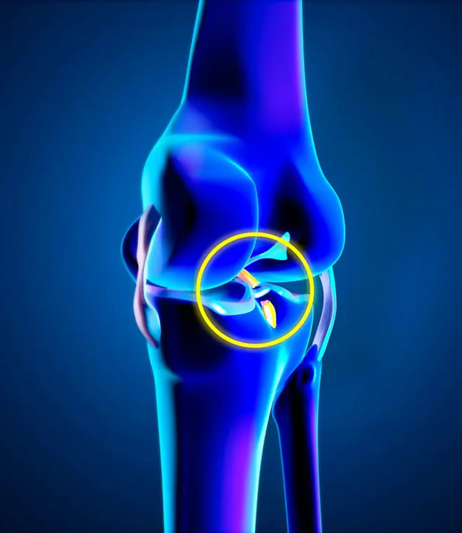
An ACL tear typically occurs during sudden, forceful movements, such as pivoting, jumping, or landing awkwardly. Common symptoms include a popping sound at the time of injury, swelling, knee instability, and difficulty bearing weight. If conservative treatments like physical therapy and bracing do not relieve symptoms or restore knee function, surgery may be recommended to reconstruct the torn ligament.
Types of Grafts Used in ACL Reconstruction
The choice of graft for ACL reconstruction depends on factors such as the patient’s age, activity level, and the surgeon’s preference. The three main types of grafts are:
Autografts: The most common option, where the surgeon uses tissue from the patient’s own body. Typically, this involves using the patellar tendon (from the knee), hamstring tendons, or quadriceps tendon. The benefit is a lower risk of rejection, but it requires harvesting tissue from another part of the body, which may add to recovery time.
Allografts: These grafts come from a deceased donor. They are a good option for older patients or those who don’t want an additional incision site. However, there is a slight risk of disease transmission, though this is minimized through screening and sterilization.
Synthetic Grafts: Though less common, some surgeons use synthetic materials for ACL reconstruction. These offer the advantage of not requiring additional tissue removal but may have a higher failure rate compared to biological grafts.
Each type of graft has its advantages and potential drawbacks, and the decision is made based on individual patient needs and surgical goals.
Surgical Techniques for ACL Reconstruction
There are several techniques available for ACL reconstruction, but the most common approach is arthroscopic surgery, a minimally invasive method that uses small incisions and a camera to guide the surgeon. This technique allows for faster healing, reduced scarring, and less pain post-operation.
Traditional ACL Reconstruction involves making larger incisions for better visibility and access to the knee, though this method is less commonly used today due to the benefits of arthroscopy.
Robotic-assisted ACL Reconstruction is an emerging technique that uses computer guidance to improve the accuracy of graft placement and alignment. This method promises a more precise surgery with potentially faster recovery times.
While all of these techniques aim to restore knee function, the choice of method will depend on the surgeon’s expertise, the patient’s specific condition, and available technology.
What is Cruciate Ligament Reconstruction Surgery?
Cruciate ligament reconstruction surgery is a procedure designed to repair a torn ACL, helping restore knee stability and function. The surgery typically involves replacing the torn ligament with a graft, which may come from the patient’s own tissue (autograft), a donor (allograft), or synthetic material.
In the most common form of ACL reconstruction, the surgeon makes small incisions around the knee and uses a camera (arthroscope) to guide the procedure. This minimally invasive technique allows for quicker recovery, less scarring, and reduced risk of complications compared to traditional open surgery. The graft is inserted through tunnels drilled in the bones, and over time, it integrates with the bone and tissue, gradually becoming a functional ligament.
Risks and Complications of ACL Reconstruction
Like any surgery, ACL reconstruction carries some risks. Common complications include:
Infection: Though rare, infection can occur at the surgical site.
Blood clots: A potential risk after any surgery, blood clots can be dangerous if they travel to the lungs.
Nerve damage: The surgery may inadvertently affect nearby nerves, leading to temporary or permanent numbness.
Graft failure or re-injury: Sometimes, the graft doesn’t heal properly, or the patient may re-tear the ACL, particularly if they return to intense activity too soon.
To minimize these risks, it’s essential to follow post-surgery care instructions, take medications as prescribed, and attend regular follow-up appointments.
Expected Results
Most patients experience significant improvement in knee stability and function after ACL reconstruction surgery. The goal is to restore mobility, eliminate pain, and enable patients to return to their daily activities and sports.
Full recovery typically takes about 9 to 12 months, with the first few months focusing on basic mobility and strength. By the 6-month mark, most patients can return to low-impact activities. High-impact sports, however, may require longer recovery and careful monitoring.
With proper rehabilitation, ACL reconstruction has a high success rate, allowing athletes and active individuals to return to their pre-injury performance levels.
The Role of Physical Therapy in Recovery
Physical therapy is crucial for recovery after ACL reconstruction surgery. It helps rebuild strength, flexibility, and stability in the knee joint. The rehab process typically begins with gentle exercises to reduce swelling and improve mobility, gradually progressing to more intense strength training and functional exercises.
Rehabilitation is typically divided into phases, with the initial focus on regaining range of motion and reducing pain. Later stages involve strengthening the muscles around the knee, particularly the quadriceps and hamstrings, which provide essential support. Compliance with physical therapy is vital for achieving optimal outcomes and reducing the risk of re-injury.
Pre-Surgery Considerations
Before undergoing ACL reconstruction, a thorough evaluation is essential. This includes imaging tests like MRI to assess the extent of the injury and pre-surgery assessments to ensure you’re healthy enough for surgery.
Choosing a qualified surgeon with experience in ACL procedures is crucial for a successful outcome. The surgeon will discuss the best graft option and surgical technique based on your age, activity level, and injury severity.
In the weeks leading up to surgery, patients may be advised to strengthen the muscles around the knee through physical therapy, as this can improve recovery outcomes. It's also important to discuss any medications you take and follow the surgeon’s instructions to ensure the safest procedure possible.
Rehabilitation and Return to Sport
Rehabilitation is an ongoing process after ACL reconstruction and can take several months. Initially, physical therapy focuses on reducing swelling, restoring range of motion, and re-establishing muscle function. Strengthening exercises, especially for the quadriceps and hamstrings, are critical to support the knee joint.
As you approach the 3- to 6-month mark, the focus of rehab shifts toward functional exercises that mimic the movements involved in your daily activities or sport. This includes jumping, running, and lateral movements.
For athletes, the general guideline is to avoid high-impact activities until at least 9 months post-surgery to minimize the risk of re-injury. Full clearance for return to sport is given once the knee is strong enough to handle the stresses of competitive activity, typically after a comprehensive rehabilitation program and testing by your healthcare provider.
Managing Expectations
It's important to set realistic expectations when it comes to ACL surgery recovery. While the majority of patients see significant improvement, a full recovery to pre-injury levels can take time, especially for athletes. Expect to gradually regain strength and stability over several months.
Knee function post-surgery varies between individuals, and some may experience lingering discomfort or mild instability. Factors like age, overall health, and adherence to rehabilitation protocols play a role in the final outcome. While most patients achieve excellent results, it's important to manage your expectations and understand that some residual issues, like mild discomfort during certain movements, may persist.
Post-Surgery Care
After ACL reconstruction surgery, post-operative care plays a vital role in ensuring a successful recovery. The first 48 hours typically focus on managing pain and swelling. Ice packs, elevation, and prescribed pain medications are commonly used to keep swelling in check.
For the first few weeks, you may need to wear a knee brace or crutches to limit movement and protect the healing graft. Gradually, you’ll be introduced to basic exercises aimed at regaining knee mobility and strength. These exercises often start with gentle range-of-motion movements and progress to strengthening routines as healing progresses.
You should avoid putting excessive weight on the knee until your doctor approves it, and regular follow-ups are necessary to monitor the graft's healing progress. If you experience unusual pain, fever, or swelling, consult your healthcare provider immediately to rule out any complications.
The Global Popularity of ACL Surgery
ACL reconstruction surgery is widely performed around the world, with increasing demand among athletes and active individuals. Sports injuries, particularly those occurring in football, basketball, and soccer, have contributed to the rise in ACL surgeries. In countries like the United States, where sports are a significant part of the culture, ACL reconstruction has become a common procedure for both professional athletes and recreational sports enthusiasts.
Technological advancements in surgery and rehabilitation have made ACL reconstruction more effective and accessible, leading to better outcomes and faster recovery times. As awareness of ACL injuries grows, more people are seeking surgical options to restore knee function and prevent long-term instability, making it one of the most successful and popular orthopedic surgeries globally.
Cost of ACL Surgery
The cost of ACL reconstruction surgery can vary widely depending on the country, healthcare system, and the type of facility where the procedure is performed. In the United States, for example, the total cost can range from $10,000 to $30,000, including hospital fees, surgeon’s fees, and anesthesia. Additional expenses, such as physical therapy, medications, and follow-up visits, can further increase the overall cost.
Insurance coverage plays a significant role in reducing out-of-pocket expenses for many patients. In many cases, health insurance plans will cover a substantial portion of the surgery, provided the procedure is deemed medically necessary. It’s important to verify insurance coverage and clarify what is included, as some policies may have restrictions on the type of graft used or require prior approval.
For patients without insurance or those in countries with less access to healthcare, exploring financing options or medical tourism to regions with lower costs can be alternative solutions.
Potential Alternatives to ACL Surgery
While ACL reconstruction surgery is often necessary for athletes or individuals with significant knee instability, there are non-surgical treatments available for those who may not be good candidates for surgery.
Physical therapy: This is the first line of treatment for many individuals with an ACL injury, especially if the tear is partial or if the patient is not involved in high-impact sports. Therapy focuses on strengthening the muscles around the knee to improve stability.
Bracing: Some patients use knee braces to protect the ACL and provide additional stability during physical activities. This can be particularly useful for people who wish to continue with low-impact exercises.
Injections (PRP or stem cell therapy): Platelet-rich plasma (PRP) or stem cell injections are gaining popularity as a non-surgical option to promote healing and repair damaged tissues. However, these treatments are still considered experimental in many cases and may not be effective for everyone.
While non-surgical methods may offer relief and improve function for some, ACL surgery remains the most effective option for most patients, particularly athletes or those seeking to return to high-intensity physical activities.
Innovations in ACL Reconstruction
In recent years, technological advancements have significantly improved ACL reconstruction surgery. Some of the most promising innovations include:
Robotic-assisted surgery: This advanced technology allows surgeons to precisely plan and execute the procedure, improving graft placement and alignment. The use of robotics can reduce human error, minimize damage to surrounding tissues, and potentially shorten recovery times.
3D printing: Surgeons are now experimenting with 3D-printed models of a patient’s knee to better understand the injury and plan the best approach for surgery. This customization can improve surgical accuracy and outcomes.
Biological grafts and regenerative medicine: Research into biological grafts, such as stem cell therapies, is progressing rapidly. These techniques aim to regenerate tissue more effectively and reduce the risk of graft rejection or failure, offering promising alternatives to traditional surgical methods.
These innovations are making ACL surgery less invasive, more precise, and potentially leading to faster recovery times, making the procedure even more successful and appealing for patients worldwide.
Long-Term Effects of ACL Reconstruction
While ACL reconstruction surgery is generally very effective in restoring knee function, it does have long-term implications that patients should consider.
Osteoarthritis risk: One of the most significant long-term concerns after ACL reconstruction is the increased risk of developing osteoarthritis in the affected knee. Even with successful surgery, the knee joint may undergo degenerative changes over time, especially in younger patients or those who return to high-impact sports.
Graft durability: The success of the graft depends on factors like the type of graft used, the patient’s rehabilitation process, and how well they protect the knee post-surgery. Though most grafts will function well for many years, there is always a risk of graft failure, particularly in the first few years following surgery.
Preventive care: To mitigate these risks, it’s crucial to maintain a healthy weight, engage in regular low-impact exercises, and avoid repetitive stress on the knee joint. Additionally, using proper techniques when engaging in physical activity can help protect the knee from future injuries.
While ACL reconstruction can significantly improve knee stability and quality of life, long-term care and attention to joint health are important to ensure the best possible outcome over the years.
Psychological Impact of ACL Injury and Surgery
An ACL injury and the subsequent need for surgery can have a profound psychological impact on patients, especially athletes who are used to being active. The recovery process, which can take months, often leads to frustration, anxiety, and even depression, particularly if the individual is unable to participate in the sports or activities they love.
For many, the uncertainty about the success of the surgery and fear of re-injury during rehabilitation can add to the emotional burden. It is important for patients to acknowledge these feelings and seek psychological support or counseling if necessary. Engaging in support groups, working with sports psychologists, and setting small, achievable goals can help maintain mental health during recovery.
Additionally, family and friend support plays a crucial role in the emotional well-being of patients, offering encouragement and reassurance during the long recovery process.
Return to Activity After ACL Reconstruction
A common concern for individuals undergoing ACL reconstruction surgery is when they can safely return to their favorite activities, particularly sports. The timeline for returning to full activity varies by individual but generally follows a progression of recovery:
Weeks 0-6: Focus on reducing pain, swelling, and improving knee range of motion. Limited activity is allowed.
Weeks 6-12: Introduction of strengthening exercises, with a gradual return to low-impact activities like cycling or swimming.
Months 3-6: Increased focus on functional exercises, including jogging and agility drills, under the supervision of a physical therapist.
Months 6-12: Athletes can begin sport-specific exercises. Full return to high-intensity or contact sports is typically allowed after 9-12 months, once strength, stability, and confidence have been restored.
It's essential not to rush back into sports or strenuous activities before your knee is fully prepared. Prematurely returning to high-impact exercise increases the risk of re-injury. Most athletes and active individuals can successfully return to their previous level of performance with proper rehabilitation.
Impact of Age on ACL Surgery Outcomes
Age plays an important role in the success of ACL reconstruction and recovery. Younger, more active individuals, especially those under 30, typically have faster recovery times and better long-term outcomes following ACL surgery. Younger patients generally heal more quickly due to higher levels of natural healing and stronger tissue regeneration.
In contrast, older patients may face a longer recovery time and an increased risk of complications such as graft failure or the development of arthritis. Additionally, older adults may not be as physically active post-surgery, which can impact their rehabilitation process and final recovery.
While ACL reconstruction can still be successful for older patients, they should carefully consider their goals for surgery, as well as the potential long-term implications, including the risk of developing knee osteoarthritis later in life.
Conclusion
ACL reconstruction surgery is a life-changing procedure for individuals dealing with a torn or damaged anterior cruciate ligament, offering the opportunity to regain knee stability, reduce pain, and return to physical activity. While the road to recovery can be long and challenging, advancements in surgical techniques, rehabilitation, and post-surgery care have significantly improved outcomes for patients worldwide.
From the importance of choosing a qualified surgeon to adhering to a comprehensive rehabilitation program, the success of ACL reconstruction depends heavily on preparation, commitment, and realistic expectations. Though risks and complications exist, most patients can look forward to a positive outcome, particularly if they follow recovery protocols and seek psychological support if needed.
Ultimately, ACL surgery is not just about physical healing; it’s about restoring confidence, improving quality of life, and enabling individuals to return to the activities that define their lifestyle. With ongoing innovations and a holistic approach to care, the future for ACL surgery is bright, offering even better results and faster recoveries for patients around the globe.