Aortic valve stenosis
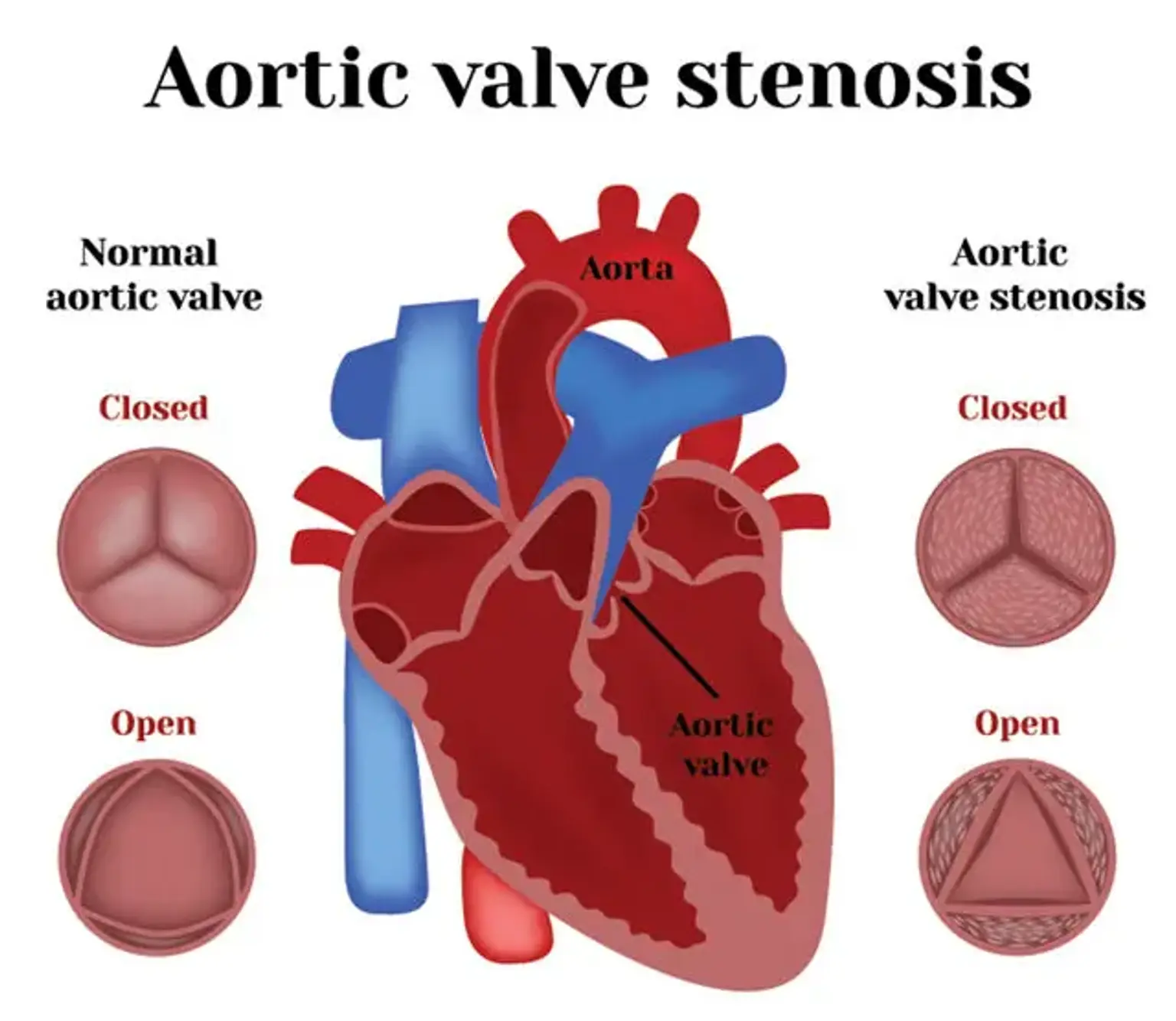
Aortic valve stenosis is the most prevalent and dangerous valve disease concern. The aortic valve aperture narrows due to aortic stenosis. As a result, it reduces blood flow from the left ventricle towards the aorta and can influence the left atrium's pressure.
Some people develop aortic valve stenosis as a result of a congenital heart abnormality known as a bicuspid aortic valve. However, the condition is usually caused by aging since calcium or scarring destroys the valve and restricts blood flow.
Signs and Symptoms of Aortic Valve Stenosis
The severity of aortic valve stenosis varies from mild to chronic. When the valve severely narrows down, signs and symptoms begin to appear. Aortic valve stenosis might go unnoticed for many years in some persons.
Generally, the signs and symptoms of aortic valve stenosis that are likely to occur include;
- An irregular heart sound or heart murmur that can be detected using a stethoscope
- With activity, you may get chest pain (angina) or tightness.
- You may feel dizzy or even faint in an exertion
- Shortness of breath, particularly after a vigorous exercise
- Fatigue, especially when there is a lot of activity.
- A fluttering, rapid heart pulse (palpitations)
- Not consuming enough food, especially in minors who have aortic valve stenosis
- Inadequate weight gain, mostly in children with aortic valve stenosis
Heart failure can also occur due to aortic valve stenosis. Shortness of breath, fatigue, and swelling ankles and feet are all signs and symptoms of heart failure.
Causes of Aortic Valve Stenosis
The four valves of the heart maintain blood circulation the right way. The mitral valve, pulmonary valve, tricuspid valve, and aortic valve are among these valves. During every heart pulse, flaps (leaflets or cusps) open and close on each valve. These valves don't always open or close appropriately. As a result, blood flow slows or gets obstructed when the valve does not entirely open or close.
The aortic valve between the lower left heart chamber (left ventricle) and the aorta doesn’t open fully in aortic valve stenosis. The passageway for blood from the heart towards the aorta narrows (stenosis). The heart has to work harder to pump sufficient blood into the aorta and the other parts of the body when the opening of the aortic valve narrows.
The left ventricle can thicken and grow as a result of the heart's additional work. The tension can eventually weaken the heart muscle, which can cause cardiac failure and other severe complications.
The common causes of aortic valve stenosis are;
Accumulation of calcium on the valve: Calcium is a mineral that can be present in the bloodstream. Calcium deposits on the heart valves can form as blood runs over the aortic valve repeatedly (aortic valve calcification).
A congenital heart defect: Sometimes, children are born with only two cusps on their aortic valve (bicuspid aortic valve) rather than three (tricuspid aortic valve). Aortic valves can have one (unicuspid) or four (quadricuspid) cusps on rare occasions.
Rheumatic fever: One of the most common causes of heart valve issues is rheumatic fever. It can affect several regions of the body, including the brain, joints, heart, and skin. Adults and children with or who have had strep throat are likely to develop rheumatic fever.
Risk Factors of Aortic Valve Stenosis
Some of the factors that can contribute to developing aortic valve stenosis are;
- Old age
- Previous history of infections affecting the heart
- Some heart problems that are present at birth, including bicuspid aortic valve
- Chronic kidney condition
- Cardiovascular risk factors, including high cholesterol, diabetes, and high blood pressure
- Chest radiation therapy history
Complications of Aortic Valve Stenosis
Sometimes, aortic valve stenosis is associated with several complications such as;
- Stroke
- Heart failure
- Bleeding
- Formation of blood clots
- Arrhythmias
- Heart infections (endocarditis)
Aortic Valve Stenosis Diagnosis
The doctor or cardiologist will start by examining your signs and symptoms, evaluate your medical history, and perform a physical examination to determine if you have aortic valve stenosis. He or she will use a stethoscope to listen to your heart to identify whether you have a heart murmur that could indicate an aortic valve problem.
Cardiologists can conduct various tests and procedures to rule out or verify aortic valve stenosis. They might as well help determine the severity and underlying cause of the condition. These diagnostic tests and procedures include;
Chest x-ray: Your doctor can use a chest x-ray to see if your heart is enlarged due to aortic valve stenosis. Swelling of the aorta and calcium deposits on the aortic valve is also a possible symptom that can be detected with this technique.
Echocardiogram: This method utilizes sound waves to generate pictures of the heart in motion in this exam. An ultrasonic beam is sent through your chest to your heart by firmly pressing a device (transducer) against the skin. The transducer takes a record of the sound wave echoes in the heart. These echoes are then converted into images that your doctor may see on a monitor.
Computerized tomography (CT) scan of the heart: A cardiac CT scan is a type of x-ray that combines numerous x-ray pictures to provide a more comprehensive cross-sectional image. A cardiac CT scan can help determine the size of the aorta and examine the aortic valve more precisely.
MRI of the heart: A cardiac MRI creates comprehensive pictures of the heart using strong magnetic fields and radio waves. This test can help figure out how serious the disease is and how big the aorta is.
An electrocardiogram (ECG or EKG): This is a painless procedure that uses small sensors (electrodes) connected to the arms, chest, and, in certain cases, legs to diagnose and record the electrical activity of the heart. The technique can also detect cardiac illness, enlarged chambers, and irregular heart pulses.
Cardiac catheterization: This procedure is not commonly used when it comes to aortic valve stenosis diagnosis. However, it might be recommended if other diagnostic tests fail to diagnose or identify the severity of the illness. It may help ensure that the arteries that supply blood to the heart muscle (coronary arteries) don’t get obstructed before aortic valve surgery.
Aortic Valve Stenosis Treatment
There is no particular aortic valve stenosis treatment. This is because once the condition develops, it cannot be reversed. Instead, the physician can recommend medication to manage the symptoms of the disorder. This can also treat the underlying health problems causing the problem.
Medications:
Medicine cannot cure aortic valve stenosis. Nonetheless, the doctor may recommend a few drugs to help manage the symptoms or relieve the strain on your heart. The following are some examples of effective medications:
Antibiotics: Antibiotics are useful in preventing rheumatic fever from progressing and resulting in heart damage.
High blood pressure drugs: Calcium channel blockers, or beta-blockers, can help you lower and maintain the normal blood pressure level.
Blood thinners: These are drugs that thin the blood. Coumadin or other blood thinners may thus be helpful for aortic valve stenosis patients.
Anti-arrhythmics: These medications are sometimes used to control the heart's rhythm.
Surgery and other procedures:
For other cases, the healthcare provider can suggest surgery or a procedure to replace and repair the damaged valve. Valvuloplasty is a minimally invasive treatment for valve repair. This treatment can be done with a catheter, which is a thin, flexible tube that is less invasive compared to traditional surgery.
This procedure involves inserting a long, tiny catheter with a small balloon at the tip into an artery, normally in the groin. The surgeon will direct the tube and inflates the balloon in the heart. The balloon and catheter are then removed once the valve has been opened. This is a minimally invasive treatment. Hence, the recovery time is less than that of open-heart surgery.
The surgeon may recommend replacing the damaged valve. This will necessitates performing open-heart surgery. The procedure can involve inserting a mechanical valve or the valve obtained from a cow or pig. Valves from human cadavers are occasionally used. The recovery time for open-heart surgery is substantially longer.
Managing Aortic Valve Stenosis Symptoms
Aortic valve stenosis is not always a congenital abnormality. This means that you were not born with it, but you may acquire it later in life. In such case, you can take the following healthy lifestyle and aortic valve stenosis diet measures to help ease your heart’s burden;
- Consume a healthful, low-saturated-fat diet.
- Exercise frequently.
- Maintain a healthy body mass index (BMI).
- Avoid smoking
- Consult the doctor if you have unusual health conditions
- To avoid rheumatic fever, see your doctor if you have a severe sore throat.
- Dental infections can move via the circulation and cause harm to the heart valves and muscles; hence, practicing proper dental hygiene is crucial.
Conclusion
Aortic valve stenosis, also known as aortic stenosis, is a narrowing of the aortic valve in the heart. The valve does not fully open, reducing or blocking blood flow from the heart to the body's main artery (aorta) and other parts.
The severity of the disorder determines the treatment. The valve might require repair or sometimes replacement, which will necessitate surgery. Severe stenosis of the aortic valve can be fatal if left untreated.