Introduction
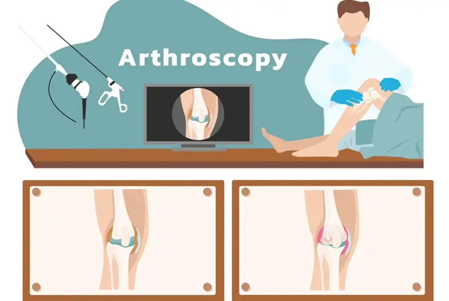
Arthroscopy is a minimally invasive surgical procedure used to examine, diagnose, and treat joint problems. Unlike traditional open surgeries, which involve large incisions, arthroscopy uses small cuts, typically no larger than half an inch, to insert a small camera (arthroscope) and tiny surgical instruments into the joint. This allows surgeons to view and treat joint conditions with greater precision and less damage to surrounding tissues.
Arthroscopy is commonly performed on joints like the knee, shoulder, ankle, and elbow, and has become a preferred method due to its quicker recovery times and reduced risk of complications. Whether it's repairing damaged cartilage or removing inflamed tissue, arthroscopy offers an effective solution with minimal discomfort for patients.
The Evolution of Arthroscopy
The history of arthroscopy dates back to the early 20th century, with significant advancements over the decades. Originally, it was a diagnostic tool, allowing doctors to view the inside of a joint for the first time using a simple lens and light. However, over time, technological improvements—such as the development of fiber optics, high-definition cameras, and miniature surgical instruments—have turned arthroscopy into a highly effective treatment method.
In the past, joint surgeries required large incisions, resulting in longer recovery times, increased pain, and higher risks of infection. Today, arthroscopic surgery is a preferred method for many conditions, offering a minimally invasive approach that results in fewer complications, faster healing, and less scarring.
How Arthroscopy Works
During an arthroscopic procedure, the surgeon first makes small incisions near the affected joint. A sterile saline solution is often injected to expand the joint space, making it easier to view and treat the problem. An arthroscope, a thin, flexible tube with a camera, is then inserted through one incision, allowing the surgeon to view the inside of the joint on a monitor. Through another incision, specialized surgical tools can be introduced to repair or remove damaged tissue.
This technique offers a high level of precision, as the camera provides a clear view of the joint's interior. Surgeons can diagnose problems like torn ligaments, damaged cartilage, and joint inflammation without having to perform a large open surgery. The small size of the incisions also reduces trauma to the surrounding tissues, leading to less post-surgical pain and a faster recovery.
Common Conditions Treated with Arthroscopy
Arthroscopy is used to treat a wide range of joint issues. The most common conditions include:
Meniscal Tears: A tear in the cartilage of the knee, which can cause pain and swelling. Arthroscopy allows surgeons to trim or repair the damaged cartilage.
ACL Injuries: Arthroscopic surgery can help reconstruct a torn anterior cruciate ligament (ACL), commonly injured during sports.
Rotator Cuff Tears: The rotator cuff, a group of muscles and tendons in the shoulder, can tear during repetitive motions or trauma. Arthroscopy can repair these tears with minimal disruption to the shoulder joint.
Cartilage Damage: Arthroscopy is often used to treat chondromalacia (softening of cartilage), as well as to remove damaged cartilage from the knee or shoulder.
In addition, arthroscopy can be used for synovitis (inflammation of the joint lining), joint infections, and even the removal of loose bodies (fragments of bone or cartilage floating in the joint). These minimally invasive techniques provide relief with less downtime and faster healing.
What to Expect During an Arthroscopic Procedure
An arthroscopic procedure typically begins with the patient being placed under anesthesia, which could range from local anesthesia (numbing just the affected area) to general anesthesia (where the patient is asleep during the surgery), depending on the joint and the complexity of the procedure.
Once the patient is prepared, the surgeon makes one or more small incisions around the affected joint. Through these incisions, the arthroscope—a small, tube-like instrument equipped with a camera and light—is inserted. The surgeon then carefully inspects the joint, using the real-time images displayed on a monitor.
If any issues are found, such as damaged tissue or cartilage, the surgeon can use specialized instruments inserted through separate incisions to repair or remove the damaged areas. These procedures are often quicker and more precise than traditional open surgery.
Other Joints Treated with Arthroscopy
Although the knee and shoulder are the most commonly treated joints, arthroscopic surgery can be performed on a variety of other joints, including the ankle, hip, elbow, and wrist.
Ankle: Arthroscopy is frequently used for treating ligament injuries, joint inflammation, and cartilage damage in the ankle. It allows surgeons to access hard-to-reach areas with minimal disruption, making it an ideal choice for patients with chronic ankle pain or instability.
Hip: In the hip joint, arthroscopy is used for treating conditions like femoroacetabular impingement (a condition where bone spurs form and cause damage to cartilage) or to repair labral tears. It can offer significant pain relief and improved mobility with a much shorter recovery time than traditional hip surgery.
Elbow and Wrist: For the elbow, arthroscopy can be used to remove damaged tissue, repair torn ligaments, or treat conditions like tennis elbow. In the wrist, it can help with treating issues like cartilage tears, arthritis, or wrist impingement.
Each joint benefits from the minimally invasive nature of arthroscopy, offering patients a faster and less painful recovery.
Global Popularity and Advancements in Arthroscopy
Over the years, arthroscopy has gained significant popularity worldwide, becoming the go-to method for joint surgeries. The procedure’s minimally invasive nature and quicker recovery times have made it a top choice for both patients and healthcare providers. It is now commonly performed across the globe, especially in countries with advanced healthcare systems.
Technological advancements have further improved the success of arthroscopic procedures. High-definition cameras and robot-assisted surgery have enabled surgeons to perform more precise and efficient procedures. New tools and techniques have expanded the range of conditions treatable through arthroscopy, making it even more versatile.
As arthroscopy continues to evolve, it holds promise for treating even more complex joint issues with even less risk and better outcomes, making it a key advancement in modern orthopedic surgery.
Benefits of Arthroscopy Over Traditional Surgery
One of the biggest advantages of arthroscopic surgery is its minimally invasive nature, which provides multiple benefits over traditional open surgeries:
Less Trauma: The small incisions used in arthroscopy result in less damage to surrounding tissues. This leads to less bleeding, reduced pain, and a faster recovery.
Shorter Recovery Time: Because arthroscopic surgery involves less tissue disruption, most patients experience a quicker return to normal activities compared to traditional surgery. Many arthroscopic procedures are done on an outpatient basis, allowing patients to go home the same day.
Reduced Risk of Infection and Complications: Smaller incisions mean a lower chance of infection. The precision of the procedure also reduces the risk of complications like nerve or blood vessel damage, which are more common in traditional open surgeries.
Less Scarring: The small incisions leave less scarring compared to large surgical cuts, which is an important consideration for many patients, especially those concerned with cosmetic outcomes.
Improved Precision and Better Outcomes: Surgeons can use high-definition cameras and specialized tools to view and treat the joint with great precision, often leading to more successful outcomes and faster healing.
These benefits have made arthroscopy a go-to choice for surgeons and patients alike, offering a less invasive, more effective way to address joint issues.
Post-Operative Pain Management
After an arthroscopic procedure, pain management is an essential part of recovery. Most patients experience minimal discomfort due to the small incisions and reduced tissue damage. Pain typically lasts only a few days to a week, and over-the-counter pain relievers, such as acetaminophen or ibuprofen, are often sufficient.
In some cases, local anesthesia is used during the procedure to numb the area, which can help reduce post-surgery pain. For more complex surgeries, stronger pain medications may be prescribed, but the goal is to avoid narcotics to reduce the risk of dependency. Ice packs and anti-inflammatory medications can also be helpful in managing swelling and pain.
The Impact of Arthroscopy on Joint Health
One of the major benefits of arthroscopic surgery is its ability to preserve joint function. By repairing or removing damaged tissue, arthroscopy can prevent further degeneration and delay the need for more invasive procedures, like joint replacement.
Because the procedure is less disruptive to the surrounding tissues, it can also reduce the likelihood of post-surgical complications. This preservation of joint health allows patients to enjoy improved mobility and reduced pain, even in cases of arthritis or cartilage damage.
For athletes or active individuals, arthroscopy offers a way to repair joint damage without interrupting their active lifestyle for extended periods, helping to maintain long-term joint health.
How to Prepare for Arthroscopic Surgery
Proper preparation before arthroscopic surgery can help ensure a smooth procedure and faster recovery. Here are some general steps to follow:
Pre-Surgery Consultation: During this visit, the surgeon will review your medical history, perform a physical exam, and discuss the procedure. Any medications you’re currently taking, especially blood thinners, will need to be addressed.
Follow Instructions on Fasting: You may be asked to avoid eating or drinking for a certain period before the surgery, especially if general anesthesia is used.
Prepare Your Home for Recovery: Ensure your home is prepared for your post-surgery recovery. Arrange for assistance if necessary, and make sure you have access to medications, ice packs, and comfortable resting spaces.
Wear Comfortable Clothing: On the day of surgery, wear loose, comfortable clothing, as the procedure typically involves a small incision near the joint.
Risks and Complications of Arthroscopy
Although arthroscopy is generally a safe procedure, as with any surgery, there are potential risks. Some of the common complications include infection, bleeding, or nerve damage. However, because the procedure involves small incisions, the risk of these issues is significantly lower than with traditional open surgery.
Other risks include blood clots, especially in the lower legs, or stiffness in the joint following surgery. These can be managed with proper care and rehabilitation, and following the surgeon’s instructions carefully can greatly reduce the chances of complications.
The key to a successful outcome lies in choosing a skilled surgeon with experience in arthroscopic techniques.
Arthroscopy for Arthritis and Degenerative Diseases
Arthroscopy can also be a useful tool for managing osteoarthritis and other degenerative joint diseases. In cases of arthritis, the procedure can remove damaged cartilage or clean out debris in the joint, helping to alleviate pain and improve function.
While arthroscopic surgery can’t cure arthritis, it can offer significant relief for patients experiencing joint pain due to cartilage loss or inflammation. For some patients, it may also delay the need for joint replacement surgery, offering a less invasive alternative with a faster recovery time.
In cases of rheumatoid arthritis, arthroscopy can be used to remove inflamed tissue from the joint lining, providing pain relief and improving movement.
Arthroscopy for Knee and Shoulder Injuries
One of the most common uses of arthroscopy is for treating injuries in the knee and shoulder joints, which are prone to wear and tear, especially in active individuals.
Knee Injuries: Arthroscopy is often used to repair meniscal tears, treat ACL injuries, or smooth rough surfaces of the cartilage in cases of osteoarthritis. The procedure can address these issues without requiring large incisions, allowing for a quicker recovery and minimal scarring.
Shoulder Injuries: For the shoulder, arthroscopic surgery is commonly used to repair rotator cuff tears, treat shoulder impingement, or remove damaged cartilage. Arthroscopy allows surgeons to repair the shoulder without the need for a large incision, which can significantly reduce recovery time and post-operative pain.
These procedures are highly effective, especially for athletes or anyone involved in physically demanding activities, as they can help restore joint function while minimizing the disruption to surrounding tissues.
Return to Normal Activities
One of the major advantages of arthroscopic surgery is the quick recovery time, allowing patients to return to their normal activities faster than with traditional surgery. Most patients can return to light activities within a few weeks, although high-impact activities like running or sports may take longer—often 2 to 3 months, depending on the type of joint involved.
For athletes or active individuals, arthroscopy is a game-changer, as it allows them to get back to their routine sooner, while also reducing the risk of further injury. However, it’s important to follow the doctor’s recommendations and avoid overloading the joint too soon to prevent re-injury.
Cost of Arthroscopy and Insurance Coverage
The cost of arthroscopic surgery can vary significantly depending on the type of procedure, location, and whether the surgery is done in a hospital or outpatient facility. On average, patients may pay anywhere from $3,000 to $10,000 or more for arthroscopy, depending on factors like insurance coverage and geographical location.
Most insurance plans, including Medicare and private health insurance, typically cover arthroscopic surgery as it is a recognized and effective treatment for joint issues. Patients should check with their insurance providers to confirm coverage details and any out-of-pocket costs.
For those without insurance, some hospitals or surgery centers may offer financing options or payment plans to help manage the cost.
Rehabilitation After Arthroscopy
Rehabilitation is a critical part of recovery following arthroscopic surgery. The rehabilitation plan typically includes physical therapy to restore strength, mobility, and flexibility to the affected joint. The length and intensity of rehab depend on the type of surgery performed and the patient’s overall health.
For instance, after knee arthroscopy, patients may start with gentle range-of-motion exercises to prevent stiffness. As the healing progresses, strengthening exercises are introduced to rebuild muscle strength and improve joint function. Full recovery can take anywhere from a few weeks to several months, depending on the complexity of the surgery and the individual’s commitment to rehabilitation.
Adhering to the prescribed rehab program is crucial to ensuring the best long-term outcome, preventing complications like joint stiffness or weakness.
Choosing the Right Surgeon for Arthroscopy
Selecting the right surgeon is crucial for the success of arthroscopic surgery. It's important to choose a board-certified orthopedic surgeon with extensive experience in performing arthroscopic procedures. An experienced surgeon will not only have the technical skills required but also be able to provide personalized care and answer any questions you may have.
Before undergoing surgery, consider asking the surgeon about their experience with the specific procedure you're having, the types of technologies they use, and their track record of patient outcomes. A good surgeon will be transparent, explain the risks and benefits clearly, and make you feel comfortable with your decision.
The Future of Arthroscopy: Innovations on the Horizon
The field of arthroscopic surgery continues to evolve with advancements in medical technology. One exciting development is the use of robot-assisted surgery, which enhances precision and allows for even smaller incisions. This technology helps surgeons plan and execute procedures with greater accuracy, which can lead to improved outcomes and quicker recoveries.
Additionally, there is growing research in regenerative medicine for joint repair. Procedures such as stem cell injections and platelet-rich plasma (PRP) therapy are being explored to complement arthroscopy for better healing and tissue regeneration. These innovations offer the potential to further reduce the need for invasive surgeries and promote faster recovery and longer-lasting joint health.
Conclusion
Arthroscopy has revolutionized the way joint problems are treated, offering a minimally invasive approach that provides numerous benefits over traditional surgery. With smaller incisions, reduced recovery times, and fewer risks of complications, arthroscopy has become the preferred choice for treating a wide variety of joint issues, from sports injuries to arthritis.
Whether you are an athlete, an active individual, or someone dealing with the natural wear and tear of aging, arthroscopic surgery can help you regain mobility and alleviate pain, often with quicker results and less post-operative discomfort.
As advancements in technology continue to evolve, arthroscopy will only become more precise and effective, making it an increasingly valuable tool in modern orthopedics. If you’re considering this procedure, selecting an experienced surgeon and adhering to post-surgery care can greatly influence the success of the treatment and your recovery process.
Ultimately, arthroscopy offers hope for millions, providing a safe, effective, and less invasive option for joint repairs that enhances quality of life and encourages a swift return to daily activities.