Artificial bladder
Overview
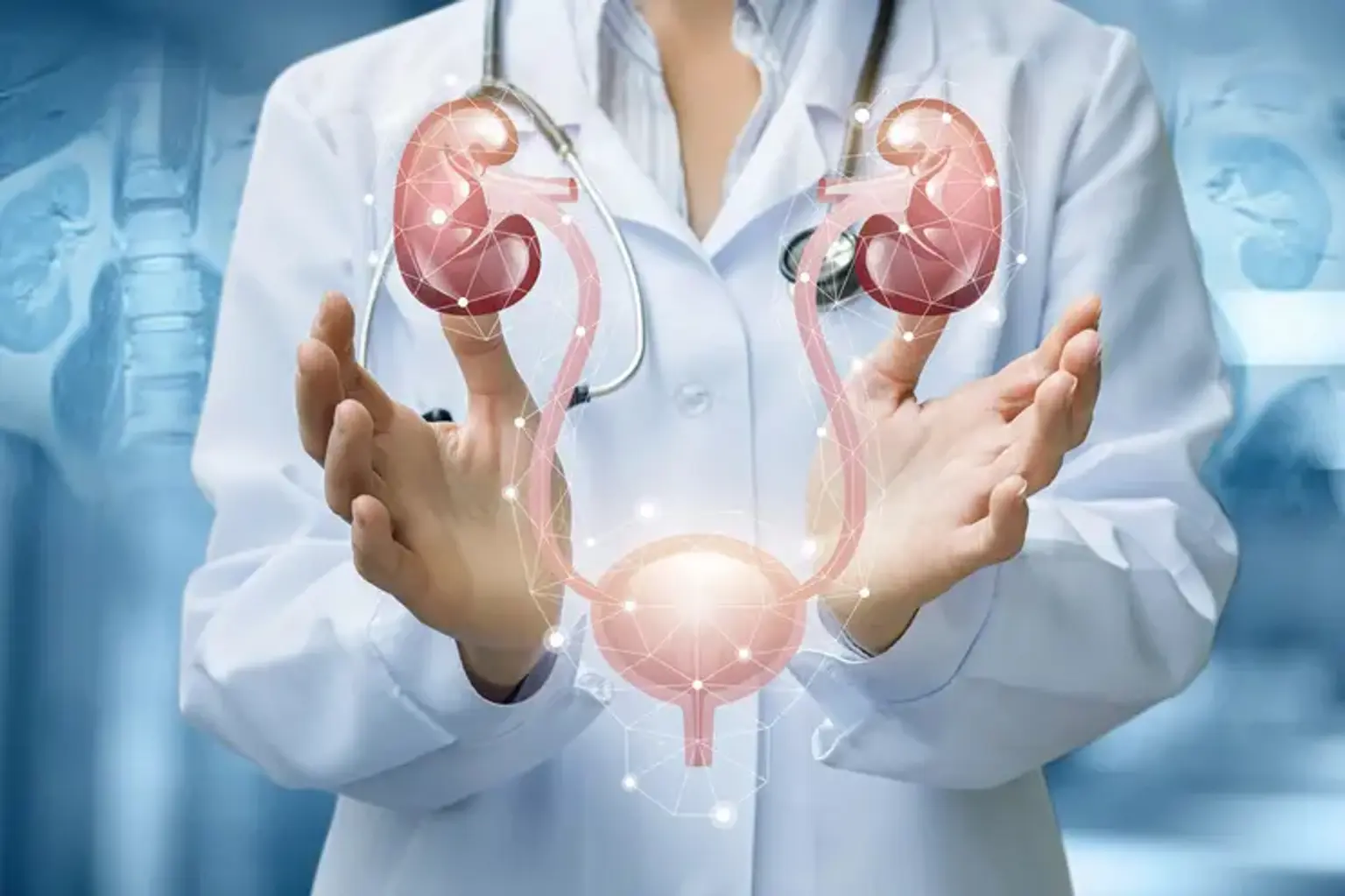
Normally, the urinary system consists of two kidneys, two ureters, a urinary bladder, and a urethra:
- The kidneys filter your blood and excrete waste and water via urine.
- Urine passes from the kidney to the bladder via tubes known as ureters.
- Urine is held in the urinary bladder before passing through the urethra and out of the body when you urinate.
When the bladder is removed, urine must escape the body in a different route, through a urinary diversion. In all forms of urinary diversions, a section of the intestine is surgically transformed into either a route tube for urine to depart the body or a reservoir for urine storage (like a normal bladder). Regardless of the surgical procedure used, urine and feces are kept totally separate. (They are two distinct systems, the urinary and digestive, respectively).
There are three main types of urinary diversion surgeries:
- Ileal conduit urinary diversion.
- Indiana pouch reservoir.
- Neobladder-to-urethra diversion.
For all of these procedures, a portion of the small and/or large bowel is disconnected from the fecal stream and used for reconstruction.
What is Artificial bladder?
The name given to the organ that results from the compulsory removal of the urinary bladder for cancer or any other cause, by using the patient's own small intestine and converting it into the bladder, is artificial bladder. We might refer to it as bowel bladder or orthotopic bladder. The ureter tubes are linked to the neobladder separately during this procedure. The urethral tube is also linked to the traditional external urinary tract.
Neobladder reconstruction is a surgical treatment that involves the development of a new bladder. A surgeon can develop a new channel for urine to depart the body if a bladder is no longer functioning correctly or is removed to treat another ailment (urinary diversion). Urinary diversion can be treated through neobladder reconstruction. During the process, a surgeon creates a new bladder from a piece of intestine. The redesigned bladder enables people to urinate on their own and decide when they urinate.
Who needs Artificial bladder?
An artificial bladder is required to replace the bladder following removal, particularly in bladder cancers with muscle involvement or that do not respond to high-grade common BCG therapy, and in infections that cause TB (bladder tuberculosis) or severe bladder loss. The capacity of the urine bladder reduces quickly after head traumas or spinal cord injuries, and the patient's comfort is fully compromised.
When a bladder is physically removed because it is diseased or no longer functions correctly, neobladder reconstruction is a possibility. People have their bladders removed for a variety of reasons, including:
- Bladder cancer
- A bladder that no longer works properly, which can be caused by radiation therapy, neurological conditions, chronic inflammatory disease or other disease
- Urinary incontinence that hasn't responded to other treatment
- Conditions present at birth that cannot be repaired
- Trauma to the bladder
What are the types of urinary diversion?
When bladder cancer is initially discovered, it is treated by TUR-T surgery. Following the pathology report, the course to take is determined based on the tumor size, extent, and degree. If the patient has muscle involvement or high-grade widespread cancer that is refractory to BCG therapy, a radical cystectomy (removal of the urinary bladder) and novel bladder procedures to replace the bladder are discussed. There are three primary approaches.
Ileal conduit urinary diversion:
A section of the gut transfers urine into an external collecting bag via a stoma. By this surgery, the ureters (the tubes that transport urine from the kidneys to the bladder) drain freely into a section of the ileum (the last segment of the small intestine). The ureters' draining end of the ileum is then taken out through a hole in the abdominal wall. This opening is known as a stoma, and it is covered with a bag that collects urine as it drains from the ileal conduit.
The advantages of the ileal conduit urinary diversion surgery are:
- The operation is relatively easy.
- It necessitates less surgical time (compared with other surgical methods).
- There is no need for catheterization on a regular basis (use of a tube to drain the urine)
The disadvantages of the ileal conduit urinary diversion are:
- There is a change in body image.
- It collects urine using an external bag, which may leak or have a bad smell.
Indiana pouch reservoir:
A pouch comprised of intestines retains urine until it is emptied by a catheter put through the stoma.
A reservoir or pouch is created from a part of the large intestine (the ascending colon on the right side of the belly) and a section of the ileum in this type of operation (the last segment of the small intestine). The ureters are rearranged such that they drain into the pouch. Urine flows easily from the kidneys into the pouch in a downward direction. This placement keeps urine from backing up into the kidneys, protecting them from infection. The small intestine is then extracted through a tiny incision in the abdominal wall (a stoma). Because the stoma is so tiny, it may be covered with an adhesive bandage.
In contrast to the ileal conduit, no external bag is required. To retain the urine inside the pouch, a one-way valve is surgically established. A short, thin catheter must be inserted through the stoma and into the pouch several times each day (typically every four hours around the clock) to empty the urine. At all other times, an adhesive bandage is applied over the stoma (when not actively emptying the pouch).
Most insurance coverage will cover enough catheters for you to use a fresh one each time. If your insurance does not cover it, or if you run out, catheters may be cleansed and reused. Catheters do not need to be sterilized. They may be brought on travels or to social gatherings and stored in a plastic bag.
The advantages of the Indiana pouch reservoir surgery are:
- Urine is stored inside the body, in the reservoir, until it is time to empty it.
- No external bag is necessary.
- There is no odor.
- The risk of urine leaking is minimal.
- The small stoma can be covered with an adhesive bandage.
The disadvantages of the Indiana pouch are:
- The surgery takes longer compared with the ileal conduit.
- Catheterization (the passage of tube into the stoma to empty the pouch) is required every four hours around the clock.
Neobladder-to-urethra diversion:
The gut is converted into a reservoir and connects to the urethra. This technique is most similar to the storing function of the urine bladder. A little portion of the small intestine is formed into a reservoir or pouch and attached to the urethra. The ureters are rearranged such that they drain into the pouch. Pee can travel from the kidneys to the ureters, to the pouch, and into the urethra in the same way as regular urine does. You must contract (tighten) your abdominal muscles to empty the pouch.
To be a candidate for this procedure, you must have a low risk of urethral cancer recurrence (return). Occasionally, people are unable to fully empty by squeezing their abdominal muscles. In certain circumstances, they must insert a catheter into the urethra up to six times each day to empty the pouch. If this is not something you are willing or able to accomplish, you should generally avoid this form of diversion.
The advantages of the neobladder-to-urethra diversion are:
- The process of urination most closely matches normal urination.
- No stoma is needed.
The disadvantages of the neobladder-to-urethra diversion are:
- Surgery time is slightly longer than the ileal conduit urinary diversion procedure.
- Urinary incontinence (leakage of urine) is typical following surgery while recovering control of urination, although it can linger up to six months. In addition, around 20% of patients at night and 5% to 10% of patients during the day are incontinent (leak urine) and must wear a pad.
- Despite the operation, some patients may be unable to empty their bladder properly and will require intermittent catheterization (putting tubing through the urethra into the pouch every four hours) for an extended length of time following surgery, if not forever.
How is pre-operative preparation for artificial bladder?
Patients are admitted to the hospital one day before the procedure because they require preoperative preparation. They only consume liquid meals and have an enema three times every day. There are current standard laboratory results such as urea creatinine, sodium potassium, and blood sugar on the morning of the surgery. Because the procedure will take a lengthy period, compression stockings are worn and used.
Your doctor will conduct testing to ensure you don't have a urinary tract infection. You may also undergo an imaging test of your urinary system, such as a CT scan, to ensure that the ureters — the tubes that transfer urine from the kidneys to the bladder — are in great condition.
Food and medications:
Your surgeon may instruct you to follow a clear liquid diet for 1 to 2 days prior to surgery. You should avoid eating and drinking after midnight the night before your surgery. Inform your doctor about all drugs, vitamins, and dietary supplements you are using. In certain situations, you may need to discontinue these drugs prior to surgery.
Learning to self-catheterize:
A possible consequence of neobladder repair is the inability to completely empty the bladder (urinary retention). If this occurs, you must be willing to perform self-catheterization. Self-catheterization involves inserting a small tube (catheter) into the opening of your bladder (urethra) to empty pee and reduce bladder pressure. You will be taught how to self-catheterize by a nurse or other health care practitioner.
How Artificial bladder is done?
The general steps in neobladder reconstruction include:
- Removing the damaged or nonfunctioning bladder
- Removing a piece of the large intestine, small intestine, or both
- Creating a spherical bladder out of intestinal tissue
- Placing the replacement bladder in the same place as the old bladder
- Integrating the replacement bladder into the ureters
- Connecting the replacement bladder to the tube that regulates the body's urine output (urethra)
- Intestinal Reconstruction
- Placing a temporary catheter in the urethra to empty the bladder while the patient recovers
Your surgeon may execute the surgery using a single abdominal incision or through a laparoscopic method. Laparoscopic surgery requires multiple extremely small holes in the lower belly for imaging and surgical equipment to pass through. This procedure can also be conducted with robotic instruments.
Postoperative care for Artificial bladder
The average hospital stay following neobladder repair is 3 to 5 days. A nurse or other health care worker will give you written instructions and discuss the following with you before you leave the hospital:
- Wound care
- Catheter management
- A regular schedule for draining your bladder
- Exercises to strengthen the pelvic floor
- Follow-up appointments for monitoring recovery and how well the new bladder is working
It may take some time for the neobladder to function properly. You may have difficulty controlling your bladder immediately after surgery and may suffer involuntary urine loss (urinary incontinence). This may continue until the neobladder extends to its normal size and the muscles that support it strengthen.
During the first 6 to 12 months following surgery, daytime bladder control (continence) normally improves. Nighttime continence may improve more in the second year. Persistent incontinence issues are more likely at night. Following a neobladder reconstruction, patients must be monitored for the rest of their lives. Inquire with your doctor about how frequently you should return for follow-up visits.
What are the possible complications of Artificial bladder surgery?
As with every operation, problems may occur based on the patient's age, other disorders, and overall health, as well as difficulties particular to this procedure. Many sutures are used in this procedure to convert the colon into a bladder. Furthermore, sutures are used to link the upper and lower urinary tracts to the neobladder.
Urine leaks may develop from these sutures in the early stages. The bulk of them, however, generally improve over time. Renal inflammation and kidney failure, known as pyelonephritis, can occur. Furthermore, issues such as reduced iron absorption owing to the removal of portions of the intestines may occur.
Living with Artificial bladder
Although there is no direct urine sensation as there is in the regular bladder, the artificial bladder filling causes bloating in the belly. Discharge can be achieved by raising intra-abdominal pressure and exerting pressure to the abdominal region as needed.
After peeing, no urine should be left within. Due to intestinal release material, mucus-like materials may appear in the urine. The goal of creating a bladder from the colon is to have no pee left inside after urinating, to urinate comfortably, to have no incontinence throughout the day or night, and to avoid infection, particularly kidney inflammation.
It takes one to two months to feel healthy and restore strength after urinary reconstruction and diversion. If you need help or have questions, don't hesitate to contact your doctor or other members of your healthcare team. Their objective for you is to return to your normal routine as quickly as feasible.
Urinary diversions frequently allow people to resume their old lives, jobs, and hobbies:
- Work: On average, most people can return to work after one or two months. If you have any worries about your employment or any potential dangers, see your doctor.
- Activities: Exercising and involvement in sports and other activities are advised following the post-operative period. Consult your doctor or a member of your healthcare team.
- Diet: There are no dietary constraints. Inquire with your doctor or a member of your healthcare team if you have any unique dietary needs.
- Travel: There are no limits on travel. Because you may not be able to obtain all required goods at your location, you should travel well prepared.
What about prosthetic bladder?
Following cystectomy, bladder function is replaced by conduits and pouches made of autogenous intestinal segments. These strategies are effective, but not ideal. Many experimental and clinical researchers have sought to develop a completely alloplastic "artificial urinary bladder."
Prosthetic alloplastic materials can be used to supplement (for example, "patch") or totally replace the original bladder. Bladder "patches" are relatively ineffective. Alloplastic porous patches successfully bind with detrusor muscle, increasing bladder capacity for short-term function. Within a year, such patches are extruded intra-luminally and coated with stone-forming minerals. Numerous experimental studies corroborate to this general finding.
Ideal characteristics of a prosthetic bladder:
- Forms biological connections with the urethra, ureters, and gut.
- Provides sufficient volume (200-500 ml)
- Enables full, voluntary urine evacuation, ideally through the urethra
- Can withstand intraluminal bacterial invasion
- Is resistant to extraluminal bacterial infection
- Drains the upper urinary tract consistently
- Is mechanically reliable
- Requires simple, inexpensive maintenance
- Allows for easy management of intraluminal bacterial colonization
- Is more acceptable visually and functionally than alternatives
- Is associated with few side effects and complications
Engineering problems associated with a prosthetic bladder:
- Parts of the urinary bladder are mobile
- Alloplasts bond poorly to mobile tissues
- Urine contains minerals that can crystallize and encrust foreign things.
- Foreign bodies harbor and perpetuate bacteriuria
- Encrusted foreign bodies grow into stones and can obstruct urinary passages
- Urine is an excellent growth medium for bacteria
- Bacterial colonization of bladder urine is common
- Bacteria-colonized urine incites inflammatory changes in adjacent tissues
- Urease-producing bacteria cause struvite encrustation and struvite stones
- Bacteria block fibroblastic ingrowth into alloplastic materials
- Alloplasts can cause local or systemic side effects months or years after implantation
A "distensible-volume model" or a "fixed-volume model" can be used to completely replace the urine bladder. The distensible-volume reservoir seeks to imitate the human model, whereas the fixed-volume reservoir substitutes a basic, hard wall reservoir for the human model.
Conclusion
Bladder substitution may be performed in individuals who have had a complete cystectomy for bladder cancer or a pelvic exenteration for cervical or rectal cancer. The number of new bladder cancer patients in the United States is estimated to be 37,000 in 2017. About 20% to 30% will ultimately lose their bladders and require a replacement.
Bladder reconstruction may be necessary in a number of clinical disorders, including bladder cancer, irradiation cystitis, interstitial cystitis, TB, and a variety of congenital malformations. At the moment, bladder repair is performed using an autogenous bowel segment. For many decades, the use of a whole prosthetic bladder as an intracorporeal urine reservoir has been an elusive objective. Many exploratory and clinical efforts have been conducted in an effort to build a near-normal bladder prosthesis using alloplastic materials. The optimal prosthetic bladder has yet to be constructed. However, accumulated experimental research indicates that many, if not all, of the desirable functional features of a whole prosthetic bladder, are feasible.
Urological surgeons have been researching methods to replace the function of the urinary bladder when it needs to be removed or has been made useless or harmful by illness for over a century. Simple anastomosis of the ureters to the rectosigmoid colon I or the development of simple intestinal conduits linking ureters to the skin were first proposed as solutions. Urologists have lately tried bladder repair with intestinal segments, the development of continent diversions, and increasingly sophisticated kinds of uretero-sigmoid anastomosis. Work on more exotic options, such as entirely artificial bladders, continues.
An artificial bladder should allow for appropriate urine storage, voluntary full evacuation of urine, and the preservation of renal function. Furthermore, it must be biocompatible, resistant to urinary encrustation, and resistant to bacterial infection. To meet these various needs, several methods have been offered over the years. However, the majority of these ideas and prototypes did not get beyond the level of a preliminary report of experimental results.
An adjustment phase is usual following major surgery, as it is with any big life change. It's normal to feel a bit down or disheartened. Discuss your feelings with your friends and relatives. Other members of a support group may be able to assist you to deal with your feelings if you join one. (Ask a member of your healthcare team about local support groups.)