Astrocytoma

Astrocytoma

Overview

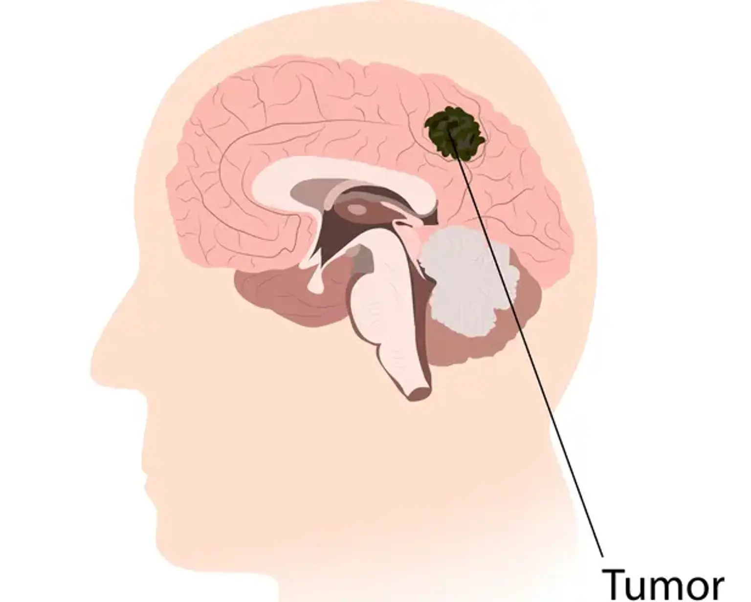
Astrocytoma develops from astrocytes, which are star-shaped glial cells in the cerebrum. It is the most frequent kind of glioma, mostly affecting the brain and occasionally the spinal cord. Glial tumors account for 60 percent of all brain tumors. They are a leading cause of death and morbidity in both young and old people. Astrocytomas are a kind of brain tumor that is rather prevalent. To avoid the significant morbidity and mortality associated with this disorder, they must be detected and treated as soon as possible.


What is an Astrocytoma?

Astrocytoma

Astrocytomas are the most frequent kind of primary brain tumor and are a subset of gliomas, a wider category of brain tumors. Astrocytomas develop from astrocytes, which are brain cells. These cells maintain and protect the brain's nerves (neurons) and aid in the transmission of signals between them. Because these cells are located throughout the brain, astrocytomas can form in a variety of locations.


Astrocytoma Causes

Astrocytoma Causes

The majority of primary brain tumors have no known origin, with the sole documented risk factor being ionizing radiation exposure. Evidence for a relationship with additional variables such as electromagnetic field exposure (cell phones), brain injury, or occupational risk factors is lacking.

Astrocytomas are, for the most part, sporadic tumors, which means they develop by chance or are unknown why they occur. There are just two scenarios that have been proved to induce the tumor:

1. Hereditary syndromes (i.e. caused by inherited DNA mutations):

  • Li-Fraumeni syndrome is caused by a mutation in the tumor suppressor gene p53 and is characterized by the advent of many malignancies at a young age, including breast cancer, bone cancers, leukemias, and astrocytomas.
  • Turcot syndrome: characterized by early start of colon cancer and astrocytomas due to mutations in numerous tumor suppressor genes, including APC and MMR.
  • Neurofibromatosis 1: caused by a mutation in the tumor suppressor NF1, this condition is characterized by the early emergence of astrocytomas, peripheral nerve tumors, skin freckling, and light-brown areas on the skin.
  • Tuberous sclerosis: is a rare genetic disorder associated with mental retardation and early onset of subependymal giant cell astrocytoma (SEGA).


2. Environmental:

  • Ionizing radiations: Ionizing radiation exposure has been linked to a delayed development of astrocytomas. Individuals who were exposed to therapeutic radiation to the head and neck region as children are more vulnerable (i.e. for treatment of leukemias or other brain tumors). The time between radiation exposure and the beginning of astrocytoma might be as long as 20-30 years.
  • Warfare chemicals: There is some speculation that exposure to Agent Orange during the Vietnam War may have contributed to the delayed start of astrocytomas among veterans.
  • Cellular phones: Despite some speculation, particularly about excessive use, there is no evidence to establish a causal risk for astrocytomas.


Types of astrocytoma

Types of astrocytoma

Astrocytomas are classified as grade 1 (most benign) to grade 4 (most malignant) by the World Health Organization (WHO). This grade, which is determined by examining tumor cells under a microscope, is based on the following characteristics:

  1. How abnormal the cells look like (atypia);
  2. How much they grow (mitosis);
  3. Presence of newly made blood vessels within the tumor (vascular proliferation). 

This is supplemented with an examination of the tumor's genetic characteristics, i.e. a DNA examination of the tumor cells. With the exception of grade 1 tumors, which are most frequent in children, most astrocytomas affect people over the age of 40. Furthermore, the older the patient, the more likely the astrocytoma would be of a greater grade.


Grade 1

  1. Pilocytic Astrocytoma is a well-defined tumor that develops slowly. The cerebellum, or the portion of the brain located in the rear of the head, just above the neck, is the most commonly affected. Because it does not infiltrate the surrounding brain, it is considered cured when totally removed and does not require chemotherapy or radiation.
  2. Pleomorphic Xantoastrocytoma is most usually seen in the temporal lobes and is frequently linked with seizures. Its cells can have a variety of forms (pleomorphic), however they normally do not proliferate. Surgery is almost always curative.
  3. Subependymal Giant Cell Astrocytoma (SEGA) is most frequent in children, generally in conjunction with the well-known condition tuberous sclerosis. It often develops inside the ventricles, which are fluid-filled areas deep within the brain, and can frequently obstruct the natural outflow of this fluid, resulting in hydrocephalus. Resection surgery is frequently curative.


Grade 2

Because Diffuse Astrocytoma is an invasive tumor, there is no apparent separation from the surrounding brain, and surgery alone may not be adequate to cure it (this depends on several other factors described below). The appearance of the tissue is only marginally different from that of a normal brain, although cells seem aberrant and slightly increased in number under the microscope.


Grade 3

Anaplastic Astrocytoma is a more malignant progression of a previously lower grade astrocytoma that has gained more aggressive characteristics, such as a faster rate of growth and more invasion into the brain. In compared to grade 2 tumors, it has a higher degree of cellular abnormalities and signs of cell proliferation (mitoses). For these tumors, surgery is never considered curative and must be followed by radiation and, nearly invariably, chemotherapy.


Grade 4

Glioblastoma (GBM) is the most dangerous, aggressive, and prevalent kind of astrocytoma (60%). Histologically, it is distinguished by extremely abnormal-looking cells, proliferation, regions of dead tissue, and the creation of new capillaries. GBM can emerge as a malignant development of a previously existing lower grade astrocytoma (about 10% of cases) or as a grade 4 tumor (90% of cases). The former situation is more typical in younger individuals, whereas the latter occurs after the age of 60. Regardless of how it appears, this tumor is a very aggressive malignancy with significant brain invasion and damage and a rapid progression.


Astrocytoma Symptoms

Astrocytoma Symptoms

Astrocytomas' clinical appearance is determined far more by their location inside the brain than by their biologic properties. There are certain areas of the brain that may accept extremely big tumors before they become symptomatic (for example, the regions in the forehead), while others can produce issues early on, such as limb weakness or difficulty with speech or vision.

In general, low-grade astrocytomas are larger before they become symptomatic than more aggressive, higher grade astrocytomas. This is due to the fact that lower-grade tumors tend to relocate the brain rather than kill it, and they are also linked with less brain edema than malignant tumors.

Common symptoms of astrocytomas are the following:

  • Persistent headaches
  • Headaches that are worse in the morning or cause awakening from sleep ( a sign of increased intracranial pressure)
  • Double or blurred vision
  • Speech problems
  • Decreased cognitive abilities
  • Grasp or limb weakness
  • New seizures


Diagnostic Imaging

Diagnostic Imaging

The sole test required to diagnose a brain tumor is neuroimaging. The best imaging for this is MRI. When possible, gadolinium contrast-enhanced MR imaging should be employed. If an MRI is not possible due to a contraindication, such as joint implants or pacemakers in place, computed tomography, or CT, may be used. Lower-grade gliomas do not contrast enhance, hence MRI fluid-attenuated inversion recovery sequences are used. If a tumor is discovered, it must be biopsied by a neurosurgeon.

This simply entails removing a tiny quantity of tumor tissue, which is subsequently examined and graded by a neuropathologist. The CT appearance of lowgrade astrocytomas is likewise not always conclusive. They are homogenous and poorly defined, appearing as poorly defined and non-contrast enhancing lesions. There may be some contrast enhancement in anaplastic astrocytomas. Because there is a possibility of metastatic illness, whole-body imaging should be considered to check for an alternative primary.

Tumor vascularity is critical, hence novel approaches for detecting it are being developed. ASL (arterial spin labeling) and DCE (dynamic contrast enhancement) MRI are two examples. Functional MRI is a new imaging modality on the horizon. It is beneficial to demarcate specific parts of the brain based on functioning prior to surgery. PET scan, MRS (magnetic resonance spectroscopy), and perfusion are some of the other modalities.

These may reveal information regarding the tumor's metabolic activity, blood flow characteristics, and structure. This can be used to evaluate if the lesion is progressing or necrosed following chemotherapy and radiation.

MRS (MRI spectroscopy): It is an MRI-based imaging technology that offers information on the chemical makeup of the tumor. It operates on the principle that some chemicals are plentiful in the normal brain while others are prevalent in tumors (for example, choline).


Management

brain tumor

The therapy for astrocytomas is determined on the tumor's grade, size, and location. Astrocytomas are often treated using neurosurgery to remove as much of the tumor as feasible. This is usually followed by radiation and, in certain cases, chemotherapy.


Surgery

Surgery is the initial step in the treatment of astrocytomas because it offers two significant advantages: To begin, it obtains tumor tissue in order to make a diagnosis. Second, it allows for the removal of as much tumor as is safely possible in order to decrease mass effect, minimize edema, and enhance responsiveness to adjuvant therapy when necessary. The choice to perform a basic biopsy or a thorough resection is influenced by a number of factors, including the patient's clinical and medical circumstances, as well as the tumor's projected amount of resectability.

Important tools to maximize efficiency and safety of surgery are:

  • Neuronavigation is essentially a GPS system for the brain, allowing the surgeon to see his/her location within the patient's brain in real time on the MRI. This considerably improves accuracy while lowering the chance of damaging the normal brain.
  • Awake surgery: This procedure is very beneficial for resecting tumors in speech regions, as well as when they are near to the main motor cortex on both sides. The patient is sedated but not intubated, allowing him or her to talk and execute orders as needed. The surgeon may continually assess the patient's functions while removing the tumor in this manner.
  • Motor mapping during general anesthesia: Even when the patient is sleeping, the parts of the brain that govern movement can be activated using an electrode. A stimulator is used to deliver currents to the brain cortex directly, and muscle responses are recorded. Positive reactions are viewed as brain regions that should not be removed.
  • Fluorescent dyes: Tumors, particularly those of higher grade, have the ability to avidly absorb certain dyes that are administered intravenously to the patient soon before surgery. As a result, the tumor tissue is colored by the particular dye, whilst the normal brain is not. This allows for a considerably more accurate description of what should and should not be excised. When viewed with suitable lenses, the most reliable dyes are 5-ALA, which colors the tumor violet, and Fluoresceine, which colors the tumor yellow.


Established adjuvant therapies

  • Steroids: Dexamethasone is the medicine of choice for relieving symptoms caused by brain swelling, which is frequently linked with tumors. It is a highly effective medication that works swiftly and consistently. Unfortunately, it has little impact on tumors and is linked with serious adverse effects when used for more than 2-3 weeks: weight gain, high blood sugars, hypertension, higher risk of infection, irritability.
  • Chemotherapy with TMZ: Temozolomide (TMZ) is an oral medication that acts by gently altering the DNA of tumor cells. This action on the DNA causes it to break, resulting in cell death unless DNA repair processes counteract the damage. TMZ is now a well-established first-line therapy for all astrocytomas of grade 3 or 4, and it is occasionally used for grade 2 tumors (when they are not completely resected in surgery or if their genetic analysis is not favorable). It is typically taken daily for 5 days, followed by a three-week break before beginning another cycle. 
  • Radiotherapy: For the past 50 years, radiation has been the cornerstone of astrocytoma treatment, and it is extremely effective, at least for the first few months after treatment. Radiation, like chemotherapy, operates by disrupting the DNA of tumor cells, causing them to die. Standard guidelines call for tiny doses of radiation in the tumor's vicinity five days a week for six weeks. Local hair loss (usually temporary) and fatigue are common side effects. Long-term negative effects include brain necrosis in the treated region and cognitive problems.
  • Bevacizumab (Avastin): Bevacizumab is a medication that prevents tumors from recruiting blood vessels, allowing them to feed themselves and develop. The Food and Drug Administration (FDA) authorized Avastin in 2013 for the treatment of recurrent glioblastomas. It is quite efficient in lowering tumor swelling and frequently helps improve symptoms. In this regard, it serves as an effective alternative to steroids. Unfortunately, unlike radiation and TMZ, it does not greatly improve survival.
  • Tumor treating electrical fields The FDA approved the use of a unique device worn on the scalp as a helmet in 2011, which creates low current electric fields that have been shown to slow tumor development. They can be used to treat recurrent glioblastomas as well as freshly diagnosed glioblastomas since 2015. This gadget must be worn for at least 18 hours every day in order to be effective. The majority of the side effects are skin irritation.
  • Antiseizure Drugs: The most often used medicine for this purpose is levetiracetam (Keppra). It is normally reserved for people who have already had at least one seizure.


Experimental Therapies

Astrocytomas, particularly glioblastomas, are the focus of intense research, and several clinical trials are conducted each year to find new strategies to improve survival. The following are the most active areas of investigation:

  • Targeted therapies: Nowadays, the majority of treatment institutions do extensive genetic analysis of any tumor tissue retrieved after surgery in order to gather patient-specific molecular signatures that can aid in determining the appropriate medicine for that tumor.
  • Immunotherapy: A compromised immune system is essential for the formation and progression of astrocytomas. As a result, a huge effort is presently underway to develop techniques to increase the immune system's response to the tumor. This is now being researched with the use of anticancer vaccinations, genetically engineered immune cells supplied to the patient via intravenous infusion, or medications that increase immune system activation.
  • Virus therapy: The use of viruses, once supplied to patients (typically directly injected into the tumor by the neurosurgeon), can selectively infect and destroy tumor cells while causing no damage to the surrounding normal brain, is a promising and increasing method. Herpes Simplex was historically the first virus to be thoroughly studied (the cold sore virus). Recently, some hopeful outcomes with the use of Poliovirus have been seen, albeit the effect was only shown in a limited number of patients.


Prognosis

diagnosis of astrocytoma

The major factors determining length of survival after a diagnosis of astrocytoma are the following:

  1. Tumor grade/histology
    • Grade 1 tumors are largely cured (96% survival rate at 5 years), usually by surgery only.
    • Grade 2 tumors: Overall median survival is 8 years. Presence of IDH1 mutation is associated with longer survival.
    • Grade 3 tumors: Median survival is 3-5 years
    • Grade 4 tumors: Median survival is 15 months.
  2. Extent of surgical resection: Despite the fact that complete microscopic excision of grade 2-4 tumors is unachievable due to their extensive infiltration into the normal brain, macroscopically comprehensive resection is linked with much higher survival. In the case of GBM, there is an increase in survival once at least 80% of the tumor is removed, and there is a stepwise improvement in prognosis as the amount of resection reaches 95-100% of the contrast-enhancing component.
  3. Use of adjuvant radiotherapy and chemotherapy.
  4. Age: young age is associated with longer survival.
  5. Functional status: minimal symptoms/normal neurological function are associated with longer survival.


Complications

Complications of Astrocytomas

Surgical problems are always a possibility with neurosurgical operations. Difficulties of chemotherapy and radiation are also observed, as with cancer treatment, although effective medications have been developed to address these complications.


Conclusion 

Astrocytomas

Astrocytomas, and especially their most severe form, glioblastoma, are the most aggressive primary brain tumors with the worst prognosis. Standard therapy only marginally increases patient survival. As a result, new treatments are required.