Basalioma
Last updated date: 30-May-2023
Originally Written in English
Basalioma
What is Basalioma?
Basalioma is the most common skin tumor; it is also known as basal cell carcinoma (BCC) and accounts for 80% of all skin malignancies. It is more common on photo-exposed (i.e., light-exposed) skin and in adults, however there are younger variations. This tumor often manifests as an infiltrating, non-metastatic growth.
Basal cells are one of three major types of cells in the skin's top layer; basal cells shed when new ones develop. BCC most commonly forms when DNA damage caused by UV radiation from the sun or indoor tanning causes alterations in basal cells in the skin's outermost layer (epidermis), leading in uncontrolled development.
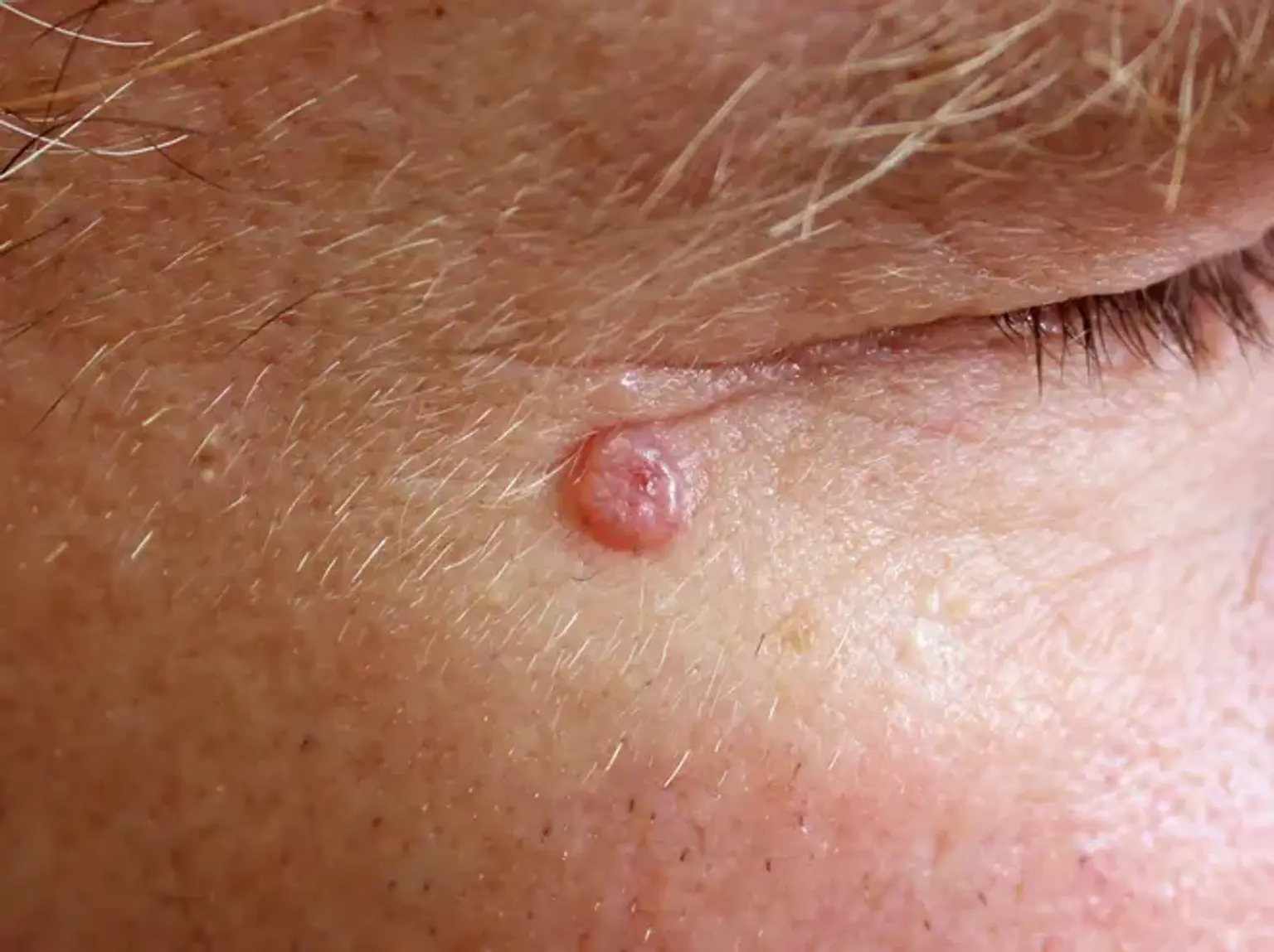
Basal cell carcinoma is a type of skin cancer. Basal cell carcinoma develops in the basal cells, which are skin cells that create new skin cells when old ones die. Basal cell carcinoma is most commonly seen as a somewhat translucent lump on the skin, although it can also take different forms.
Basal cell carcinoma is most common on sun-exposed parts of skin, such as your head and neck. Long-term exposure to ultraviolet (UV) radiation from sunlight is considered to cause the majority of basal cell carcinomas. Using sunscreen and avoiding the sun may help guard against basal cell carcinoma.
Epidemiology of Basal cell carcinoma
Basal cell carcinoma (BCC) is the most prevalent kind of skin cancer in humans, with a growing global incidence rate. Men are more likely than women to get BCC. BCC is more common in areas with increased UV exposure, such as those at higher or lower latitudes. A history of squamous cell carcinoma (SCC) or BCC is the most prevalent predictor of BCC development. Those with a BCC history are at least 10 times more likely to acquire a second BCC than patients without a history of non-melanoma skin cancer.
Estimated incidence rates have climbed between 20% and 80% in the previous 30 years. BCC incidence rates rise with age, with the median age of diagnosis being 68 years. BCC-related death is infrequent and mostly happens in immunocompromised persons. Metastatic BCC (1% of all cases) is more likely to be caused by malignancies with aggressive histopathologic characteristics. When a BCC spreads, it frequently includes regional lymph nodes, bone, the lungs, and the skin. The mean age of death is greater than in squamous cell carcinoma, and the age-adjusted mortality rate is estimated to be 0.12 per 100 000. The risk of death increases with age, male gender (more than double the incidence of occurrence compared to women), and White racial phenotype.
What causes Basal cell carcinoma (Basalioma)?
Exposure to UV radiation, particularly UVB wavelengths, is the most important etiological component in the development of basal cell carcinoma, but UVA wavelengths can also play a role. A thorough evaluation of the literature, including meta-analysis and sensitivity analysis, reveals that outdoor workers have a much greater risk, with an inverse connection between occupational UV exposure and BCC risk with latitude. Among White race individuals, the Fitzpatrick skin type is a strong predictor of the relative risk of BCC.
The cumulative UV dosage and skin type are not the only predictors of BCC development; exposure length and intensity, particularly in early infancy and adolescence, also play a role. Recreational sun exposure and the usage of indoor tanning salons are factors that contribute to the development of BCCs. UV light treatment may potentially result in the development of BCC. Intermittent severe sun exposure, as evidenced by past sunburns; a positive family history of BCC; a pale complexion, particularly red hair; facile sunburning (skin types I or II); and childhood blistering sunburns are all risk factors for the formation of BCC.
Because 20% of BCC develop on non-sun-exposed skin, ultraviolet light exposure is not the main risk factor. Other causes of BCCs include ionizing radiation exposure, arsenic exposure, immunosuppression, and genetic susceptibility. Xeroderma pigmentosum, basal cell nevus syndrome (also known as Gorlin syndrome), Bazex-Dupre-Christol syndrome, and Rombo syndrome are several genetic diseases linked to an elevated incidence of BCCs. There is no link to food, however smoking appears to be a risk factor in women.
Risk factors for Basal cell carcinoma:
A variety of risk factors might increase the probability of acquiring BCC. Some of these risk factors are as follows:
- Having a family history of BCC
- Having light skin
- Having skin that freckles or burns easily
- Having inherited syndromes that cause skin cancer, like disorders of the skin, nervous system, or endocrine glands
- Having fair skin, red or blonde hair, or light-colored eyes
- Being a man
Other, nongenetic risk factors exist. These are some examples:
- Age, with increased age correlating with increased risk
- Chronic sun exposure
- Severe sunburn, especially during childhood
- Living in a higher altitude or sunny location
- Exposure to radiation therapy
- Exposure to arsenic
- Taking immunosuppressing drugs, especially after a transplant surgery
What are the types of Basal cell carcinoma?
BCCs are usually not painful. The only symptom is skin growth or alteration. There are several forms of BCC. Each has a unique appearance:
- Pigmented BCC: This kind manifests as a brown, blue, or black lesion with a transparent and elevated border.
- Superficial BCC: This form appears as a reddish area on the skin, which is generally flat and scaly. It continues to expand and frequently has a raised edge.
- Nonulcerative BCC: This kind shows as a white, flesh-colored, or pink lump on the skin. It is frequently transparent, with visible blood vessels beneath. This is the most prevalent kind of BCC. It is most frequent on the neck, ears, and face. It has the potential to burst, hemorrhage, and scab over.
- Morpheaform BCC: This is the least prevalent kind of BCC. It resembles a scar-like lesion with a white and waxy look with no distinct boundary. This sort of carcinoma can signify a more aggressive form of BCC that is more likely to be disfiguring.
- Basosquamous BCC: This carcinoma has characteristics of both BCC and squamous cell carcinoma, another kind of skin cancer. It is exceedingly rare, yet it is more prone to spread than other forms of skin cancer.
What are the symptoms of Basal cell carcinoma?
Almost all BCCs form in regions of the body that are often exposed to sunlight. Tumors on the face, ears, shoulders, neck, scalp, and arms are all possible. Tumors can form in places that are seldom exposed to sunlight. Symptoms of basal cell carcinoma include:
- Lumps, bumps, pimples, scabs or scaly lesions on your skin.
- The lump may be slightly see-through (translucent) and close to your normal skin color or white to pink, brown to black or black to blue.
- The lump may appear shinier than the skin around it with tiny visible blood vessels.
- The lump may grow slowly over time.
- The lump may be itchy or painful.
- The lump may form an ulcer, which can ooze clear fluid or bleed with contact.
Basal cell carcinoma diagnosis
History and physical exam:
Your healthcare professional may quickly suspect basal cell carcinoma based on the look of the skin lesion. Your provider will do a physical exam and ask you questions about your symptoms to confirm the diagnosis:
- When did the bump or lesion on your skin appear?
- Did the size of the lesion change?
- Does the lesion appear different today than it did when you initially observed it?
- Does the lesion hurt or itch?
- Have you ever had skin cancer before?
Tests:
Following a physical examination, your physician may recommend testing to confirm a diagnosis, which may include:
- Skin biopsy: A portion of the damaged skin region (lesion) will be removed and examined under a microscope by your physician.
- Imaging tests: It is exceedingly unlikely that basal cell carcinoma will spread throughout your body. If your doctor believes that your cancer has spread to other parts of your body, he or she may order an MRI or a CT scan to look for cancer in lymph nodes or internal organs.
After doing a physical exam and evaluating the findings of your tests, your physician will decide the stage of your illness. Qualifications for the stage include:
- Determining the extent of the lesion (tumor) and if it has spread further into your tissues.
- Examine your lymph nodes for symptoms of malignancy.
- Examining other places of your body to check if the disease has spread (metastasized).
- Measuring the size, shape, and location of the lesion, as well as the rate at which it grows.
What is the treatment of Basal cell carcinoma?
The therapy for a BCC is determined by its kind, size, and location, the number of patients to be treated, patient variables, and the doctor's choice or skill. The majority of BCCs are surgically treated. Long-term surveillance is advised to look for new lesions and recurrence; the latter may be avoided if histology shows wide clean margins.
Excision biopsy:
Excision entails removing the lesion and stitching up the skin.
- The best therapy for nodular, infiltrative, and morphoeic BCCs.
- A 3 to 5 mm margin of normal skin should surround the tumor.
- Large lesions may need a flap or skin graft to fix the defect.
- Pathologist will report deep and lateral margins.
- For lesions that have not been entirely removed, further surgery is indicated.
Mohs micrographically controlled excision:
Mohs micrographic surgery is meticulously marking removed tissue and studying it under a microscope layer by layer to guarantee complete excision:
Very high cure rates achieved by trained Mohs surgeons.
- Used in high-risk facial regions such as the eyes, lips, and nose.
- Suitable for subtypes that are ill-defined, morphoeic, infiltrative, or recurring.
- A flap or skin transplant is used to treat large defects.
Superficial skin surgery:
Shaving, curettage, and electrocautery are all part of superficial skin surgery. It is a quick procedure that uses local anesthetic and does not require stitches.
- It is appropriate for tiny, well-defined nodular or superficial BCCs.
- Lesions are usually located on trunk or limbs.
- Wound is left open to heal by secondary intention.
- Moist wound dressings lead to healing within a few weeks.
- Eventual scar quality variable.
Cryotherapy:
Cryotherapy is the freezing of a superficial skin lesion, commonly using liquid nitrogen.
- Suitable for small superficial BCCs on covered areas of trunk and limbs.
- Best avoided for BCCs on head and neck, and distal to knees.
- Double freeze-thaw technique.
- Results in a blister that crusts over and heals within several weeks.
- Leaves permanent white mark.
Photodynamic therapy:
Photodynamic treatment (PDT) is a procedure that involves treating BCC with a photosensitizing agent and then exposing it to light several hours later.
- Aminolevulinic acid lotion and methyl aminolevulinate cream are examples of topical photosensitizers.
- Suitable for low-risk small, superficial BCCs.
- Best avoided if tumor in site at high risk of recurrence.
- Results in inflammatory reaction, maximal 3–4 days after procedure.
- Treatment repeated 7 days after initial treatment.
- Excellent cosmetic results.
Imiquimod cream:
Imiquimod is an immune response modifier.
- Best used for superficial BCCs less than 2 cm diameter.
- Applied three to five times each week, for 6–16 weeks.
- Results in a variable inflammatory reaction, maximal at three weeks.
- Minimal scarring is usual.
Fluorouracil cream:
5-Fluorouracil cream is a topical cytotoxic agent (destroys cancer cells).
- Used to treat small superficial basal cell carcinomas.
- Requires prolonged course e.g., twice daily for 6–12 weeks.
- Causes inflammatory reaction.
- Has high recurrence rates.
Radiotherapy:
Treatment with radiotherapy or X-rays can be used to treat primary BCCs or as an adjuvant treatment if margins are insufficient.
- Mainly used if surgery is not suitable.
- Best avoided in young patients and in genetic conditions predisposing to skin cancer.
- Best cosmetic results achieved using multiple fractions.
- Typically, patient attends once-weekly for several weeks.
- Causes inflammatory reaction followed by scar.
- Risk of radiodermatitis, late recurrence, and new tumors.
treatment for advanced or metastatic basal cell carcinoma:
Multidisciplinary consultation is required for locally advanced primary, recurring, or metastatic BCC. A mix of therapies is frequently employed.
- Surgery
- Radiotherapy
- Targeted therapy.
Vismodegib and sonidegib, which suppress the hedgehog signaling system, are examples of targeted treatment. These medications include significant risks and negative effects.
What are the complications of Basal cell carcinoma?
Recurrence:
It is not rare for BCC to recur after first therapy. Recurrent BCC often exhibits the following characteristics:
- Incomplete excision or narrow margins at primary excision
- Morphoeic, micronodular, and infiltrative subtypes
- Location on head and neck.
Advanced BCC:
Advanced BCCs are large, often neglected tumors.
- They may be several centimeters in diameter
- They may be deeply infiltrating into tissues below the skin
- They are difficult or impossible to treat surgically
Metastatic BCC:
- Very rare
- The primary tumor is frequently big, neglected, or recurring, and is found on the head and neck, with an aggressive subtype.
- May have had multiple prior treatments
- May arise in site exposed to ionizing radiation
- Can be fatal
How to prevent Basal cell carcinoma?
The most essential technique to avoid BCC is to avoid becoming sunburned. This is especially true during youth and early adulthood. Individuals with fair skin and those with a personal or familial history of BCC should protect their skin from sun exposure on a daily, year-round, and lifetime basis.
- Stay indoors or under the shade in the middle of the day
- Wear covering clothing
- Apply high protection factor SPF50+ broad-spectrum sunscreens generously to exposed skin if outdoors
- Avoid indoor tanning (sun beds, solaria)
- BCCs may be reduced in quantity and severity by taking 500 mg of nicotinamide (vitamin B3) twice day.
Prognosis of Basal cell carcinoma
The majority of BCCs may be cured with therapy. Cure is most probable if therapy begins when the lesion is tiny. Approximately half of persons with BCC acquire a second one within three years of the first. They are also more likely to develop other skin malignancies, including melanoma. Regular self-examinations of the skin, as well as long-term yearly skin checks by an experienced health expert, are advised.
What are the other types of skin cancer?
Squamous cell carcinoma:
Squamous cell carcinomas account for approximately two out of every ten skin malignancies (also called squamous cell cancers). These malignancies begin in the flat cells of the epidermis's top (outer) layer. These malignancies most typically arise on sun-exposed body parts such as the face, ears, neck, lips, and backs of the hands. They can also appear in scars or persistent skin lesions.
They can begin in actinic keratoses (described below). They occur less often in the skin of the vaginal region. Squamous cell cancers can typically be removed entirely (or treated in other ways), but they are more prone than basal cell tumors to spread to other regions of the body and develop into deeper layers of skin.
Melanoma:
Melanocytes, the pigment-producing cells present in the epidermis, are the source of these malignancies. Melanomas are significantly less prevalent than basal and squamous cell cancers, but if left untreated, they are more likely to advance and spread.
Less common types:
Other forms of skin cancer are significantly less prevalent and require different treatment. These kinds together account for fewer than 1% of all skin malignancies. They are as follows:
- Merkel cell carcinoma
- Kaposi sarcoma
- Cutaneous (skin) lymphoma
- Skin adnexal tumors (tumors that start in hair follicles or skin glands)
- Various types of sarcomas
Other precancerous conditions of the skin:
Actinic keratosis (solar keratosis):
Actinic keratosis (AK), also known as solar keratosis, is a skin disease induced by excessive sun exposure. AKs are little (less than a quarter-inch wide), rough or scaly patches that can be pink-red or flesh-colored. They usually begin on the cheeks, ears, backs of the hands, and arms of middle-aged or older adults with pale skin, but they can appear on other sun-exposed locations as well. People who have them often have more than one.
AKs develop slowly and normally do not produce symptoms (although some might be itchy or sore). They occasionally leave on their own, but they may return.
A tiny number of AKs develop into squamous cell skin malignancies. Most AKs do not progress to malignancy, but because it can be difficult to distinguish them from real skin malignancies at times, doctors frequently advise treating them. If they are not treated, you and your doctor should check them on a regular basis for changes that might indicate skin cancer.
Squamous cell carcinoma in situ (Bowen disease):
The earliest form of squamous cell skin cancer is squamous cell carcinoma in situ, often known as Bowen disease. The term "in situ" refers to cancer cells that are still solely in the epidermis (the top layer of skin) and have not spread to deeper layers.
Bowen disease manifests itself as reddish spots. Bowen disease patches are bigger, redder, scalier, and occasionally crusty when compared to AKs. Bowen illness, like AK, normally does not cause symptoms, however it may be itchy or uncomfortable.
These patches, like most other skin cancers (including AKs), commonly occur in sun-exposed locations. Bowen disease can also affect the skin in the anal and vaginal regions. This is frequently associated with sexually transmitted infection with human papillomaviruses (HPVs), which can also cause genital warts.
Bowen disease, which can sometimes lead to aggressive squamous cell skin cancer, is routinely treated. People who have these are also at a higher risk of developing other skin malignancies, therefore regular check-ups with a doctor are essential.
Keratoacanthoma:
Keratoacanthomas are sun-exposed skin tumors that are dome-shaped. They may develop rapidly at first, but their growth normally slows down. Many keratoacanthomas diminish or even disappear without therapy over time. Some, however, continue to develop and may even spread to other regions of the body. They can be difficult to distinguish from squamous cell skin cancer, and their progress is frequently unpredictable, thus many skin cancer specialists advocate treating them (typically with surgery).
Conclusion
Basal cell carcinoma is the most prevalent kind of cutaneous cancer, affecting about one in every five Americans. Although basal cell carcinoma is seldom lethal, it can be severely damaging and disfigure local tissues if therapy is poor or delayed.
Basal cell carcinoma is a kind of skin cancer that develops on exposed areas of your skin. It's normal to be concerned when your doctor diagnoses you with it, but bear in mind that it's the least dangerous sort of skin cancer. You can be treated if you catch it early.
This cancer is unlikely to migrate from your skin to the rest of your body, although it may expand nearby into bone or other tissue beneath your skin. Several therapies are available to prevent this from occurring and to eradicate the malignancy. Tumors begin as little, sparkly lumps on your nose or other regions of your face. However, they can appear on any area of your body, including your trunk, legs, and arms. If you have fair skin, you are more prone to get this type of skin cancer.