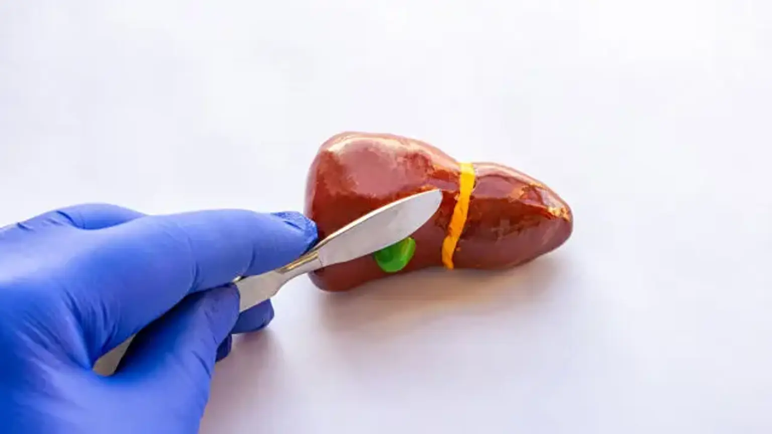
Bile duct stones
Overview
The presence of one or more gallstones in the common bile duct is referred to as bile duct stones (Choledocholithiasis). This often happens when a gallstone moves from the gallbladder into the common bile duct. Cholangitis, jaundice, and direct detection of stones with ultrasonography are the greatest indicators of the existence of common bile duct stones (CBDS).
What is Choledocholithiasis?
Choledocholithiasis, commonly known as common bile duct stones, is a biliary system blockage caused by gallstones in the common bile duct. The common bile duct is a tube that connects the common hepatic duct with the cystic duct to transport bile from the liver to the small intestine.
Epidemiology
Choledocholithiasis has been discovered in 4.6 percent to 18.8 percent of cholecystectomy patients. The prevalence of choledocholithiasis in cholelithiasis patients rises with age. Cholelithiasis is more frequent in women, pregnant women, the elderly, and individuals with high blood lipid levels.
Cholesterol stones are most commonly detected in obese people with little physical activity or in patients who have recently reduced weight on purpose. Patients with cirrhosis, those receiving complete parental nourishment, and those who have had an ileal resection all develop black pigment stones. The brown pigment main common bile duct stones are caused by nucleating agents such as bacteria.
Bile duct stones causes
Choledocholithiasis arises as a result of either stone development in the common bile duct or gallstone transit into the CBD from the gallbladder. Some of the variables that contribute to the creation of these stones are bile stasis, bactibilia, chemical imbalances, increased bilirubin excretion, pH imbalances, and the formation of sludge.
Stones can also develop in the intrahepatic biliary tree, which is known as primary hepatolithiasis and can progress to choledocholithiasis. Stones that are too big to pass through the ampulla of Vater linger in the distal common bile duct, producing obstructive jaundice and perhaps pancreatitis, hepatitis, or cholangitis. The composition of gallstones distinguishes them.
Cholesterol stones are mostly constituted of cholesterol, whereas black pigment stones are primarily composed of pigment, and brown pigment stones are primarily built of a mixture of pigment and bile lipids. Cholesterol stones account for around 75% of secondary common bile duct stones in the United States, with black pigment stones accounting for the balance. Brown pigment stones are the most prevalent kind of primary common bile duct stone. Gallstones obstructing the CBD cause symptoms and consequences such as pain, jaundice, and infection.
Gallstones in bile duct pathophysiology
Gallstones can develop as a result of bile produced in the liver and retained in the gallbladder. Gallstones in certain cases will travel from the gallbladder into the cystic duct and subsequently into the common bile duct. The majority of choledocholithiasis cases are caused by gallstones passing from the gallbladder into the CBD. Primary choledocholithiasis, or the production of stones within the common bile duct, occurs less often.
Primary choledocholithiasis develops in the presence of bile stasis, resulting in intraductal stone development. The bile duct grows in size with age. Primary bile duct stones are more likely to occur in older people who have dilated bile ducts and biliary diverticula. Complicated Mirizzi syndrome and hepatolithiasis are less prevalent causes of choledocholithiasis. Stones in the common bile duct restrict bile flow, resulting in obstructive jaundice and perhaps hepatitis.
Bactibilia and ascending cholangitis can also result from stagnant bile. Because a bacterial biofilm often covers common bile duct stones, choledocholithiasis patients are more likely to develop cholelithiasis and sepsis than those with other types of bile duct blockage. Because the pancreatic duct connects to the common bile duct near the duodenum, the pancreas may become inflamed as a result of pancreatic enzyme blockage. Gallstone pancreatitis is the medical name for this condition.
Bile duct stones symptoms
A comprehensive history and physical examination must be performed by the treating practitioner. This involves inquiring about the patient's stomach discomfort's onset, timing, and severity, as well as any past incidences of comparable pain. The discomfort is colicky, in the right upper quadrant of the belly, and of moderate intensity. The discomfort is sporadic and recurring. Patients frequently report episodes of epigastric, right upper quadrant, or epigastric pain.
A complete system examination will disclose that the patient may have observed a yellowing of his eyes or skin, as well as pruritus and even nausea or vomiting. When the stones impede the CBD, conjugated bilirubin enters the circulation, causing jaundice. Such individuals have a history of clay-colored stools and urine that becomes tea-colored. Jaundice might appear during episodes. Cholangitis patients may also have a fever, chills, and potentially impaired mental condition (Charcot triad or Reynolds pentad).
Gallstones are responsible for almost half of all pancreatitis cases. When CBD is obstructed at the level of the ampulla of Vater, pancreatitis develops. Pancreatic discomfort is felt in the epigastric and midabdominal regions, is constant (unlike colicky pain in choledocholithiasis), and spreads to the back. There is also nausea and vomiting. Some people have occasional discomfort as a result of a transitory obstruction in the common bile duct. When there is a transient obstruction in the bile duct, it is caused by floating stones or debris.
The provider should examine the patient thoroughly, paying special attention to the patient's overall appearance, skin, vital signs, and abdomen. There is tenderness in the right upper quadrant of the abdomen. Systemic symptoms, such as fever, hypotension, and flushed skin, indicate infection or sepsis.
The Courvoisier sign is the presence of palpable gallbladder on the exam and is found when gallbladder dilatation occurs as a result of a common bile duct occlusion. Any heat, diaphoresis, jaundice, scleral icterus, tachycardia, hypotension, tachypnea, or right upper quadrant stomach soreness should be noted.
Bile duct stones Diagnosis
A white blood cell count, hemoglobin/hematocrit, platelet count, total bilirubin, direct bilirubin, alkaline phosphatase, aspartate aminotransferase, and alanine aminotransferase should be ordered by the practitioner. Total bilirubin levels greater than 3 mg/dL to 4 mg/dL are closely related with choledocholithiasis in cholelithiasis patients. Gamma-glutamyl transpeptidase levels are also high.
Serum alanine aminotransferase (ALT) and aspartate aminotransferase (AST) concentrations are high in cholestatic biliary obstruction, with increases in alkaline phosphatase, serum bilirubin, and gamma-glutamyl transpeptidase (GGT) outweighing serum AST and ALT elevations. Because liver tests are high owing to a number of different etiologies, the positive predictive value of elevated liver tests is low. As a result, normal levels aid in the exclusion of choledocholithiasis.
Symptom relief, along with declining liver function tests, implies that the gallstone was passed naturally. A lipase test should also be performed to rule out gallstone pancreatitis. An INR with prothrombin time can also be ordered to evaluate intrinsic liver function.
The first test that should be conducted for any patient suspected of having biliary illness, including choledocholithiasis, is a transabdominal ultrasound. An abdominal ultrasound will often reveal a dilated common bile duct (greater than 6 mm) and stones inside the common bile ducts. The presence of gas in the duodenum usually makes it difficult to detect CBD stones, however ultrasonography may reliably diagnose CBD dilatation with up to 90% accuracy.
CBD stones can be detected via abdominal ultrasonography, which has a sensitivity of 15-40%. In the face of a negative ultrasound, a magnetic resonance cholangiopancreatography (MRCP) might be requested if there is still a high suspicion based on history, physical, and laboratory results.
MRCP is also a noninvasive test with a sensitivity of 92% and a specificity of 100%. Endoscopic ultrasonography can also be used to diagnose suspected choledocholithiasis; however, it is more intrusive than transabdominal ultrasound or MRCP. This requires inserting an ultrasonic probe into the duodenum while being guided via an endoscope. The sensitivity and specificity of CBD stone detection for MRCP,
Although diagnostic endoscopic retrograde cholangiopancreatography (ERCP) is more sensitive, it is no longer done frequently due to a 10% risk of post-procedure pancreatitis.
An intraoperative cholangiogram can be used to evaluate for choledocholithiasis in patients having laparoscopic or open cholecystectomy. It is done by placing a catheter into the cystic duct, then injecting contrast material to define the biliary tree. Films are taken to look for filling flaws and the passage of contrast into the duodenum. Choledocholithiasis can also be detected with intraoperative or laparoscopic ultrasonography. This approach, however, is operator-dependent and is not widely used by general surgeons.
Bile duct stone Management
Endoscopic removal of the blocking stones is used to treat choledocholithiasis. Under general anesthesia, an ERCP can be conducted with the patient in either the prone, left lateral, or supine position, albeit the prone position is the most commonly employed. After that, the endoscopist will insert a duodenoscope into the second segment of the duodenum and insert a catheter and guidewire into the common bile duct. A sphincterotome is then used to cauterize the papilla and expand the ampulla of Vater.
This motion frequently results in the stones being loosened. A variety of snares and baskets can be employed to grab and remove the stones as needed. A balloon catheter can also be used to sweep the common bile duct for stones. The endoscopist can also insert a stent into the common bile duct, which serves two functions. First, any residual stones will be softened, perhaps making them simpler to remove with a second ERCP. Second, the stent will allow for bile drainage, which will avoid obstructive jaundice.
Surgical removal is recommended if the stones are big, stuck, or there are several stones within the biliary tree. To remove any stones that cannot be removed using endoscopic procedures, a laparoscopic or open common bile duct exploration is required. During the same hospitalization, an elective cholecystectomy is also advised to avoid future occurrences of choledocholithiasis.
Cholecystectomy in choledocholithiasis patients is still debatable, however most specialists advocate it. Arguments against cholecystectomy can be offered in people who cannot endure surgery well (for example, due to age or medical difficulties), as long as the organ is asymptomatic.
Cholecystectomy is not recommended for first-time CBD stones. Open choledochotomy, transcystic exploration (a procedure used to eliminate stones from the CBD during laparoscopic cholecystectomy), percutaneous extraction, and extracorporeal shock wave lithotripsy are further surgical possibilities.
Treatment options for choledocholithiasis discovered during cholelithiasis or cholecystitis surgery include intraoperative common bile duct exploration, intraoperative ERCP, and postoperative ERCP. If consent was acquired before to the surgery, the intraoperative procedure can be conducted. Otherwise, ERCP is advised at a later date but during the same hospitalization.
There are no drugs available to treat choledocholithiasis. However, if the pancreatic duct was manipulated during an ERCP, a one-time dosage of 50 mg to 100 mg rectal indomethacin can be given to avoid post-procedure pancreatitis. Antibiotics are seldom required for choledocholithiasis unless the patient simultaneously has cholecystitis or cholangitis.
Prognosis
The prognosis of choledocholithiasis is determined by the existence and severity of complications. Approximately 45 percent of choledocholithiasis patients are asymptomatic. Only 55% of patients who decline surgery or are deemed ineligible for surgery endure varied degrees of problems. Even after completing therapeutic treatments, less than 20% of individuals have recurrence of symptoms. Under normal conditions, the prognosis is positive if therapy is commenced at the appropriate time.
Can bile duct stones pass on their own?
Small stones can travel into the duodenum on their own in rare situations. The majority of patients, however, need stone removal through ERCP or surgery.
ERCP employs an endoscopic guide to make a tiny hole in the biliary path, allowing the stones to move more readily. Although ERCP is quite effective, problems such as pancreatitis, bleeding, and duct rupture might occur. To avoid stone recurrence, stone removal is usually followed by cholecystectomy.
Bile duct stones food to avoid
The gallbladder diet seeks to alleviate the stress that food can have on the gallbladder, either by simplifying digestion or by supporting the gallbladder. A nutritious diet includes plenty of fresh fruits and vegetables, fruit juice, low-fat dairy products, whole grains, nuts, spices, and legumes.
A diet heavy in processed meat, soft drinks, refined grains, red meat, high-fat dairy products, sugar, tea, solid fat, baked potato, snacks, egg, salt, pickled food, and sauerkraut is unhealthy. People who ate a well-balanced diet were less likely to develop gallbladder disease.
Complications
Choledocholithiasis and its treatment can lead to several complications, such as:
- Post-ERCP Pancreatitis
- Sepsis
- Wound infection
- Cholangitis
- Retained and impacted stones
- Cholangitis
- Gallstone pancreatitis
- Respiratory insufficiency
- Biliary duct injury
- Renal failure
- Liver failure and cirrhosis
- Hepatic vascular injury
Cholangitis
In acute cholangitis, bile duct blockage permits germs from the duodenum to climb. The majority of cases (85%) are caused by common bile duct stones, however bile duct blockage can also be caused by tumors or other disorders.
Common infecting organisms include
- gram-negative bacteria (eg, Escherichia coli, Klebsiella species, Enterobacter species);
- less common are gram-positive bacteria (eg, Enterococcus species) and
- mixed anaerobes (eg, Bacteroides species, Clostridia species).
Abdominal discomfort, jaundice, and fever or chills are all symptoms (Charcot triad). The abdomen is uncomfortable, and the liver is frequently sensitive and swollen (possibly containing abscesses). Confusion and hypotension, stomach discomfort, jaundice, and fever or chills (Reynolds' pentad) all indicate a 50% fatality probability and significant morbidity.
Preventing bile duct stones
There is no way to completely avoid gallstones, which can develop in even the healthiest, youthful, or most active persons. People can, however, take precautions to lessen their risk. These precautions are especially critical for those with a family history of gallstones or other risk factors, such as obesity.
Preventive measures include:
- Eating a lower fat diet: People might concentrate on avoiding items that are high in fat but poor in nutritional value, such as potato chips and fried dishes. They can substitute more healthful fats, such as avocado and fish oil, for these items.
- Adding more fiber: This may be accomplished by consuming more whole grains, fruits, and vegetables.
- Attaining a moderate body weight: If required, a mix of nutrition and exercise, with an emphasis on becoming more physically active, can help people lose weight.
- Avoiding crash diets and extremely low calorie diets: These diets may actually increase the likelihood of developing gallstones.
- Talking with a doctor about medications: Some medications, such as birth control pills, have been linked to an increased risk of gallstones.
- Seeking excellent prenatal care during pregnancy: People should also notify their doctor if they have any odd or new stomach discomfort while pregnant.
Conclusion
The common bile duct stone (CBD stone) is a reasonably common condition, with an incidence of 10-20% in individuals with gallstones. This is also linked to severe consequences such as obstructive jaundice, acute suppurative cholangitis, and acute pancreatitis. The most crucial aspect of CBD stone management is early diagnosis and timely treatment. Endoscopic ultrasonography and magnetic resonance cholangiopancreatography show great sensitivity, specificity, and accuracy for the identification of CBD stones, according to a recent meta-analysis.
Endoscopic ultrasonography, in particular, has been shown to be more sensitive than others. A therapeutic regimen for individuals with symptomatic gallstones is given according on whether they are at low, moderate, or high risk of CBD stones.
In high-risk patients with gallstones and CBD stones, if competence, operating time, and tools are available, single-stage laparoscopic CBD exploration and cholecystectomy is preferable to ERCP with laparoscopic cholecystectomy in terms of technical success and shorter hospital stay. In several hospitals, patients with CBD stones and gallstones are treated with ERCP combined laparoscopic cholecystectomy.