Biliary system disease

Last updated date: 03-Mar-2023

Originally Written in English

Biliary system disease

Overview

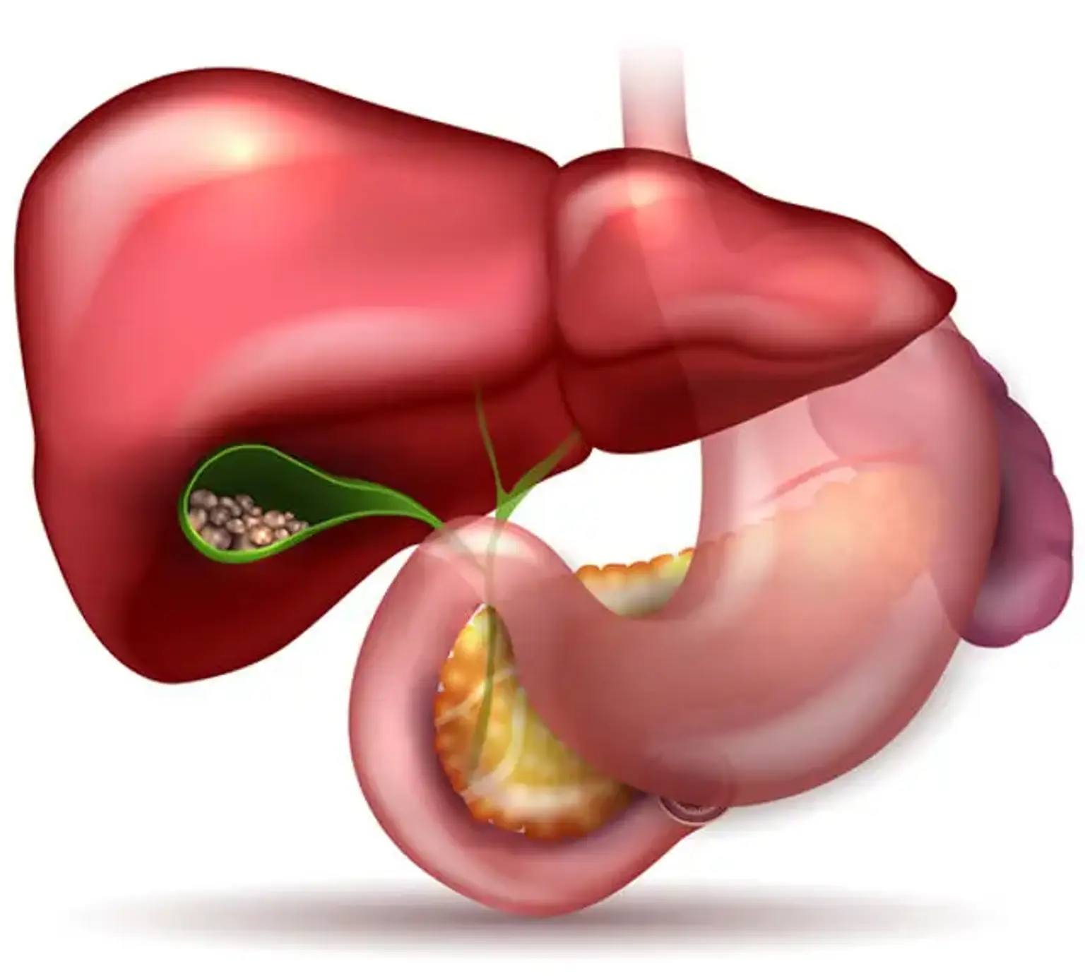
The biliary system is affected by a wide range of disorders, many of which show with similar clinical signs and symptoms. Gallstones, acute calculus cholecystitis, acute acalculus cholecystitis, Mirizzi syndrome, chronic cholecystitis, cholangitis (recurrent pyogenic, primary sclerosing, primary biliary, autoimmune), biliary tract cancers, biliary tract cysts, and other disorders are examples of these conditions.


Epidemiology

Gallstone disease is one of the most prevalent and expensive digestive disorders. According to the third National Health and Nutrition Examination Survey, 6.3 million men and 14.2 million women aged 20 to 74 years in the United States have gallbladder disease.

A million new instances of gallstones are diagnosed each year. In the United States, there are 20 million instances. There are around 2-7 incidences of primary sclerosing cholangitis (PSC) per 100,000 people. PSC affects about 5% of people with chronic ulcerative colitis. Gallbladder cancer occurs at a rate of 2.5 occurrences per 100,000 people. 


Pathophysiology

liver produces bile

The liver produces bile, which is then routed into the digestive tract through the biliary ductal system for fat emulsification and absorption. Abnormalities in bile composition, architecture, or function cause biliary illness. The chemical composition of bile is determined by the liver, and this can be changed subsequently by the gallbladder and the biliary epithelium. Cholesterol, which is normally insoluble in water, enters solution by the formation of vesicles containing phospholipids (mostly lecithin) or mixed micelles with bile salts and phospholipids.

Gallstone etiology cannot be explained only by cholesterol supersaturation. The transformation of cholesterol from a soluble state to a solid crystalline form is the first stage in gallstone formation. Biologic compounds in gallbladder bile have an impact on the process, either positively or negatively.

Mucus, for example, may encourage nucleation, whereas bile-specific glycoproteins may prevent nucleation. Mucin hypersecretion by the gallbladder mucosa results in the formation of a viscoelastic gel that promotes nucleation. Arachidonyl lecithin, which is absorbed and released into the bile, enhances prostanoid production by gallbladder mucosa and promotes mucus hypersecretion, whereas prostaglandin inhibitors decrease mucus secretion.

Finally, gallbladder hypomotility and bile stasis appear to enhance gallstone development and growth, which may be relevant in diabetes, pregnancy, oral contraceptive usage in women, and extended fasting in critically sick patients receiving complete parenteral nutrition.


Biliary system disease causes 

Gallstones

Gallstones

Gallstones are clinically silent in around 80% of patients. Approximately 20% of people acquire symptoms over a period of 15-20 years, or approximately 1% every year, and virtually all become symptomatic before problems emerge. The most common clinical manifestation is biliary-type discomfort caused by a blockage of the bile duct lumen. Other symptoms (for example, sensitivity to fatty foods, indigestion) have too low a predictive value to be therapeutically useful. Gallbladder cancer is uncommon in the setting of cholelithiasis, occurring at a rate of around 0.1 percent per year. Gallstones are classified into two categories.

  • Cholesterol stones (85%)

These are divided into two subtypes:

  1. Pure (90%-100% cholesterol) or 
  2. Mixed (50%-90% cholesterol).

Pure stones are usually single, white, and greater than 2.5 cm in diameter. Mixed stones are often smaller, many in number, and come in a variety of forms and hues. They are often organized in concentric layers of laminated layers of alternating thicker whitish cholesterol and thinner dark pigment in a concentric pattern around a pigmented core. These stones are more common in Westerners, and they are generally detected in the gallbladder.

Obesity, a high-calorie diet, clofibrate therapy, gastrointestinal disorders involving major bile acid malabsorption, cystic fibrosis with pancreatic insufficiency, female sex, and the use of oral contraceptives and other estrogenic medications are all risk factors for the development of cholesterol gallstones. Coffee and ascorbic acid have been linked to a lower likelihood of symptomatic cholesterol gallstones.


  • Pigment stones (15%)

Pigment stones occur in 2 subtypes

  1. Brown and
  2. Black.

Calcium bilirubinate and calcium-soaps are the main components of brown stones. Bacteria contribute to their creation by secreting beta glucuronidase and phospholipase. Bacterial glycocalyx binds to bile pigment and precipitates out of solution. These stones, which are more frequent in Asia, originate within the bile ducts. They are usually seen in conjunction with periampullary duodenal diverticula.

When excess bilirubin enters the bile and polymerizes into calcium bilirubinate, black stones develop in the gallbladder. Patients with persistent hemolysis, alcoholic cirrhosis, and advanced age are more likely to develop these stones.


Acute calculus cholecystitis

Acute calculus cholecystitis is an inflammation of the gallbladder caused by a clogged cystic or bile duct. It commonly manifests itself after 5 hours of biliary-type discomfort. Chemical irritation causes the initial inflammation, and bacterial infection is most likely a subsequent occurrence.

A shift in pain perception, often a migration to the right upper quadrant, indicates transmural gallbladder inflammation with parietal peritoneum involvement. Nausea and vomiting are typical accompanying symptoms, and the majority of patients are afebrile early in the disease's progression.


Mirizzi syndrome

Mirizzi syndrome is a kind of common hepatic duct blockage caused by an impacted stone in the cystic duct. It is estimated that it occurs in 0.7% to 1.4% of all cholecystectomies. It is frequently overlooked prior to surgery, which can result in substantial morbidity and biliary damage, especially with laparoscopic surgery.


Acute acalculous cholecystitis

The existence of an inflamed gallbladder in the absence of a clogged cystic or common bile duct is referred to as acute acalculous cholecystitis. It is more common in severely sick patients (e.g., severe burns, multiple traumas, protracted postoperative care, prolonged intensive care) and accounts for 5% of cholecystectomies.

Because stomach discomfort, fever, and leukocytosis are widespread in these individuals, and the signs and symptoms are not specific for acalculous cholecystitis, the clinician must have a high index of suspicion to make the diagnosis. The etiology is assumed to be ischemic, with a gangrenous gallbladder as a consequence. This disease is associated with an elevated risk of morbidity and death.

A rare variety called as acute emphysematous cholecystitis is often characterized by clostridial organism infection and obstruction of the cystic artery linked with atherosclerotic vascular disease and, in many cases, diabetes.


Chronic cholecystitis

Chronic cholecystitis is a prevalent condition that is often accompanied with gallstones. The clinical symptoms are vague, although cholescintigraphy may first point to a diagnosis. The pathophysiology is unknown, however it might be due to aberrant bile composition, which causes chemical harm to the gallbladder mucosa. The diagnosis is confirmed by histologic evidence of a mononuclear infiltration, fibrosis, and epithelial metaplasia. A subgroup of individuals develops dystrophic calcifications inside the fibrosis, resulting in a porcelain gallbladder, which is associated with gallbladder malignancy.


Cholangitis

Cholangitis

Cholangitis is a biliary infection that can aggravate both benign and malignant biliary blockage. The clinical presentation varies greatly depending on the type of the sickness, the patient's age, and the patient's condition. The Charcot trinity (fever, right upper quadrant discomfort, and jaundice) occurs in only 20% to 70% of patients. Reynolds (1959) defined a pentad in which severe illness is accompanied by hypotension and alterations in mental function.

Typically detected organisms are of intestinal origin, most notably Escherichia coli, Streptococcus faecalis, Clostridium species, and Enterobacter species. They most likely reach the biliary system by portal bacteremia. The presence or absence of pus in the biliary system has no effect on the severity of the clinical symptoms; nevertheless, suppurative cholangitis is associated with a greater death rate.


Primary sclerosing cholangitis

PSC is a chronic cholestatic biliary illness characterized by nonsuppurative biliary ductal inflammation and fibrosis. The etiology is unknown, however it has been linked to autoimmune inflammatory disorders such as chronic ulcerative colitis and (less often) Crohn's disease, as well as uncommon ailments such as Riedel thyroiditis and retroperitoneal fibrosis.

The majority of individuals appear with weariness, pruritus, and, on rare occasions, jaundice. The natural history is diverse, but it entails progressive bile duct damage, which leads to cirrhosis and liver failure. Unless the biliary system has been instrumented, the clinical characteristics of cholangitis (fever, right upper quadrant discomfort, and jaundice) are unusual.


Primary biliary cholangitis

Primary biliary cholangitis

Primary biliary cholangitis (PBC), formerly known as primary biliary cirrhosis, is a cholestatic biliary illness characterized by tiredness, itching, or asymptomatic increase of alkaline phosphatase. The term change reflects the reality that cirrhosis only develops in its late stages, and hence does not appropriately identify people with early-stage illness. Jaundice develops as a result of the increasing deterioration of bile ductules, which leads to liver cirrhosis and hepatic failure. 


Autoimmune cholangitis

Autoimmune chollangitis is a rare and unique disease condition. While it has similar characteristics with PBC, AMA tests are negative, gamma globulin and IgM levels are lower, and tests for fluorescent antinuclear antibody (FANA) and anti–smooth muscle antibody (ASMA) are more frequently positive.


Neoplasms of the biliary tract

Carcinoma of the biliary system manifests with clinical symptoms of:

  • Weight loss (77%), 
  • Nausea (60%)
  • Anorexia (56%)
  • Abdominal pain (56%),
  • Fatigue (63%), 
  • Pruritus (51%), 
  • Fever (21%),
  • Malaise (19%),
  • Diarrhea (19%),
  • Constipation (16%), and 
  • Abdominal fullness (16%).

Before obstructive jaundice emerges, symptomatic patients generally have advanced illness with dissemination to hilar lymph nodes. It is linked to a bad prognosis.


This rare cancer affects 2.5 people out of every 100,000. It accounts for 54% of biliary tract malignancies, and more than 6500 people die from it in the United States each year. Cancer that develops in the gallbladder's infundibulum (neck) can cause gallbladder hydrops that is clinically indistinguishable from an obstructing stone.


  • Cholangiocarcinoma

Cholangiocarcinoma

Cholangiocarcinoma is a kind of bile duct adenocarcinoma. It can develop without any risk factors, however it is more usually associated with chronic cholestatic liver disease, such as PSC, choledochal cysts, oriental cholangiohepatitis, and asbestos exposure at work. Cholangiocarcinoma is responsible for 25% of all biliary tract malignancies. Patients often appear with jaundice, anorexia, weight loss, and pruritus, as well as a nonspecific upper or right upper quadrant abdominal discomfort.


Biliary tract cysts

Cystic dilatation of the biliary tree is a rare condition. Approximately half of the patients had some combination of jaundice, abdominal discomfort, and an abdominal mass. These cysts are frequently linked with an abnormal junction of the pancreatic and biliary ductal systems. This shows that pancreatic juice penetrates the bile duct, causing a proteolytic and inflammatory damage to the duct wall and resulting in the creation of biliary cysts.


Biliary disease symptoms

Biliary disease symptoms

Biliary illness manifests in a variety of ways, ranging from no symptoms to a constellation of signs and symptoms of different severity and combination. An accurate diagnosis, then, begins with paying attentive attention to the patient. Accurate diagnosis is supported by clinical expertise and frequently incorporates imaging investigations.

When abdominal discomfort is the primary symptom, try to figure out when it started and what happened after that. Clarify the patient's perception of pain; visceral pain is regarded as a hazy, dull, gnawing, burning, or aching feeling, whereas parietal pain is sharper in quality and more localized. Pain perception may be enhanced by psychological factors (e.g., fear, stress), but pain perception may be lowered by diminished awareness.

  • Biliary-type pain

Upper abdominal discomfort is a common symptom of biliary illness. The pain is piercing, painful, or tight, and is usually intense and localized in the epigastrium. The experience is typically difficult to define; it may appear unexpectedly, persist for 15 minutes to several hours, and then go abruptly. Although the name biliary colic is widely used, it is a misnomer because the pain pattern is consistent.

The discomfort is produced by a biliary lumen blockage, with distension, and is clinically similar to when the obstruction occurs at the cystic duct or another level of the common bile duct. Referred pain in the posterior scapula or right shoulder area may be experienced when unpleasant visceral sensations grow more acute, and may be accompanied by nausea and vomiting.

  • Jaundice

The hepatobiliary tract is primarily in charge of bilirubin metabolism and transport. When the serum bilirubin level climbs beyond 3 mg/dL, a yellow coloring of the skin appears, which is referred to as jaundice. Abnormalities that cause jaundice might develop at any stage of the process.

  • Jaundice and abdominal pain

The combination of jaundice and abdominal pain suggests a subacute obstruction of the biliary ductal system. In elderly patients, however, biliary tract obstruction may be painless. Rarely, acute viral hepatitis can be confused with biliary-type pain.

  • Painless jaundice

The development of jaundice in the absence of stomach discomfort suggests a malignant bile duct blockage. Jaundice appears gradually in this case and may be coupled with anorexia, weight loss, and acholic, soft or loose stools. Nonbiliary reasons should be examined, such as increased bilirubin production (as a result of hemolysis, blood transfusions, or inadequate erythropoieses) and reduced bilirubin clearance owing to genetic abnormalities. 

  • Pruritus

Itching is a painful sensation in the skin that is accompanied by a strong urge to scratch. While there are numerous reasons, itching is linked to cholestasis and may become the patient's most irritating symptom. Itching may begin in the hands and feet, but it frequently spreads to the rest of the body and is worst at night. Itching does not discriminate between hepatic and biliary cholestasis.

  • Fatigue

In illnesses affecting the intrahepatic bile ducts, such as primary biliary cholangitis, primary sclerosing cholangitis, and vanishing bile duct syndrome, the gradual onset of tiredness, followed by pruritus and eventually jaundice, is noticed to variable degrees.

  • Weight loss

A history of weight loss is linked to more significant biliary tract illnesses. Weight loss may be caused by insufficient nutritional intake (e.g., anorexia) or fat malabsorption (eg, a paucity of bile in cholestatic diseases or prolonged biliary obstruction).


Physical Examination

The patient suffering from acute biliary-type pain is frequently restless, worried, and disappointed as a result of futile attempts to find a comfortable posture. Acute severe pain is generally accompanied by grimacing of the face. Patients who are writhing and diaphoretic are frequently severely and critically sick; nevertheless, some patients with peritonitis may lie quiet, with a troubled look, and avoid being touched or jostled.

Skin color in persons with light skin might indicate not just jaundice but also the cause; a yellow discoloration is related with indirect hyperbilirubinemia, an orange hue with hepatocellular jaundice, and a dark green tint with protracted biliary blockage. Evidence of easy bruisability may suggest cirrhosis-related coagulopathy. Excoriation of the skin (from scratching, often sparing the mid back), melanin pigmentation, and xanthomas of the eyelids and extensor surfaces are all symptoms of cholestasis.

The abdomen should be examined initially to identify whether it is scaphoid, flat, bloated, or asymmetric. Auscultation may reveal nonexistent bowel sounds, which indicate ileus, hyperactive bowel noises (borborygmi), or high-pitched tinkling, which indicates intestinal blockage. Peritonitis is indicated by the elicitation of discomfort and involuntary guarding with light probing or shaking of the abdomen. A bulk or fullness in the right upper quadrant may be palpable.

An enlarged gallbladder in a jaundiced patient indicates a malignant blockage of the bile duct. In the absence of jaundice, a palpable lump in the right upper quadrant might be a gallbladder tumor or chronic cystic duct blockage.


Diagnosis

Diagnosis of biliary system disease

Patients with uncomplicated cholelithiasis or simple biliary colic frequently have normal laboratory test findings; laboratory investigations are not usually required until complications are expected. When indicated, blood testing may include the following:

  • Complete blood count (CBC) with differential
  • Liver function panel
  • Amylase
  • Lipase

Imaging modalities that may be useful include the following:

  • Abdominal radiography (upright and supine) – Used primarily to exclude other causes of abdominal pain 
  • Ultrasonography – The procedure of choice in suspected gallbladder or biliary disease
  • Endoscopic ultrasonography (EUS) – An accurate and relatively noninvasive means of identifying stones in the distal CBD
  • Laparoscopic ultrasonography – Promising as a potential method for bile duct imaging during laparoscopic cholecystectomy
  • Computed tomography (CT) – For identifying gallbladder stones, it is more costly and less sensitive than ultrasonography, although it is excellent for displaying stones in the distal CBD.
  • Magnetic resonance imaging (MRI) combined with magnetic resonance cholangiopancreatography (MRCP) - Typically reserved for cases of probable choledocholithiasis.
  • Endoscopic retrograde cholangiopancreatography (ERCP)
  • Percutaneous transhepatic cholangiography (PTC)


Management

Gallstones

Gallstones

With the introduction of laparoscopic cholecystectomy, the medical therapy of gallstone disease has lately decreased. Cholecystectomy remains the favored treatment due to its lower cost, definitive nature, safety, and greater patient convenience.

In selected individuals with symptomatic gallstone disease, medical treatment may be a viable alternative to cholecystectomy. For the nonsurgical care of individuals with gallstone disease, three approaches are available, which can be used alone or in combination:

  1. Oral bile salt therapy, primarily ursodeoxycholic acid,
  2. Contact dissolution, and 
  3. Extracorporeal shock-wave lithotripsy. 

Several years of therapy may be required to completely dissolve a stone, and there is a 50% possibility of stone recurrence during a 5-year period. Some evidence supports the use of nonsteroidal anti-inflammatory medicines (NSAIDs) in the treatment of recurrent biliary pain; prostaglandin inhibition may stop the inflammatory process and minimize the risk of developing acute cholecystitis.

There is general agreement that asymptomatic gallstones detected by chance should not be treated. When a patient develops symptoms or difficulties from gallstones (for example, acute cholecystitis), therapy to remove the gallstones is advised since the chance of further symptoms or complications is high. According to the National Cooperative Gallstone Study, the probability of recurring symptoms was roughly 70% during the two years after the initial presentation.


Acute cholecystitis

Patients with a clinical diagnosis of acute cholecystitis should be hospitalized to the hospital and given nothing by mouth (NPO) and IV fluid hydration; correction of any related electrolyte abnormalities is a critical first step. Most occurrences of inflammation are caused by a mechanical blockage of the cystic duct and are mediated by a chemical irritation of the gallbladder mucosa.

Broad-spectrum antibiotics are recommended when the sickness has a bad clinical presentation, fever, and leukocytosis. Empiric antibiotic therapy should target the organisms that are most frequently recovered. While second-generation cephalosporins, such as cefuroxime, are often used, they do not treat Enterococcus. As an empiric therapy, a combination of ampicillin and gentamicin might be utilized.

Although NSAIDs are effective in reducing inflammation, narcotic analgesics are frequently required to relieve pain. Once their situation has stabilized, these patients should have a cholecystectomy unless they have serious concomitant illness.


Cholangitis

Cholangitis is treated similarly to acute cholecystitis, with the exception that endoscopic intervention is required. Antibiotics and the creation of biliary drainage are the pillars of treatment. Initially, broad-spectrum antibiotics are recommended to treat gram-negative bacteria and Enterococcus.

In contrast to acute cholecystitis, metronidazole is frequently used to cover anaerobes in individuals with a more toxic look, even though the contribution of anaerobes to infection is limited in patients who have not undergone previous biliary system instrumentation or surgery.

Endoscopic retrograde cholangiopancreatography (ERCP)–guided sphincterotomy and stone extraction or biliary stent insertion are used to create biliary drainage depending on the etiology of the cholangitis and the patient's comorbid state. In a sepsis patient, the least aggressive technique to create biliary drainage is frequently the best, with intentions for final therapy (cholecystectomy) when the patient's condition has stabilized.

When ERCP is unavailable, ineffective, or contraindicated, percutaneous drainage should be explored. In individuals with an intact gallbladder, a percutaneous cholecystostomy tube may be a possibility.


Primary sclerosing cholangitis (PSC)

The two main aims of treatment in PSC are disease slowing and reversal, as well as management of advancing illness and associated consequences. In individuals with PSC, immunosuppressive and anti-inflammatory medications such as ursodeoxycholic acid (UDCA), corticosteroids, cyclosporine, methotrexate, azathioprine/6-mercaptopurine, tacrolimus, and D-penicillamine have been explored.

Endoscopic treatment to dilate and/or stent dominating strictures is feasible in sites with extensive therapeutic biliary endoscopy competence. Relief of jaundice and pruritus is a key possible therapeutic benefit, albeit a benefit on disease progression has not been convincingly shown.

For patients with severe liver damage caused by PSC, liver transplantation is currently the therapy of choice.


Complications

Fatigue, pruritus, steatorrhea, fat-soluble vitamin deficiencies (A, D, E, and K), metabolic bone disease, hypercholesterolemia, xanthomas, hypothyroidism, and anemia are all typical consequences of chronic cholestatic liver illnesses such as PSC and PBC. PBC has been linked to Sjögren's syndrome, Raynaud phenomenon, and sicca symptoms.

Around 20% of PSC patients develop a prominent stricture in the intrahepatic or extrahepatic biliary tree. Medical treatment for biliary strictures has been futile. Nonsurgical techniques for biliary obstruction relief, such as endoscopic or radiologically guided balloon dilatation of strictures or insertion of prosthetic stents across strictures, should be tried first.

Choledocholithiasis and cholelithiasis caused by cholesterol and/or pigment stones may occur in up to one-third of PSC patients. Bacterial cholangitis can develop in PSC patients.

Cholangiocarcinoma occurs in around 20% of PSC patients, primarily late in the course of long-standing ulcerative colitis and the cirrhotic stage of biliary illness. Approximately half of PSC patients are identified with cholangiocarcinoma within two years after their original diagnosis, with a dismal prognosis due to advanced illness at the time of diagnosis. As indicated by the discovery of cholangiocarcinoma in 10% of patients having liver transplantation for PSC, this complication is difficult to detect.

Hepatocellular carcinoma is more common in persons with PBC who have had stage IV illness for a long time. PSC and ulcerative colitis patients are at an elevated risk of colon cancer and the advancement of neoplastic transformation.


Conclusion 

Gallbladder and bile duct diseases are prevalent, with gallstones and related consequences being the most common. Biliary strictures, which are frequently malignant, are produced by adenocarcinomas of the pancreas, bile ducts, ampulla of Vater, and gallbladder. Sclerosing cholangitis and a range of congenital diseases are uncommon.

Gallstones, which induce biliary colic and cholecystitis by obstructing the cystic duct, and bile duct obstruction (cholestasis), which causes jaundice, dark urine, and pale feces, itching, and occasionally persistent right hypochondrial discomfort, are examples of biliary system disorders. Fevers and rigors are symptoms of a bacterial infection of the biliary system (cholangitis), which commonly occurs in conjunction with partial blockage.

Bilirubinuria is evident, and the absence of urobilinogen in the urine indicates full biliary blockage. Imaging is essential in the diagnosis of biliary illness, beginning with ultrasonography and progressing to CT and MRI in more severe patients. However, these investigations may not provide enough anatomical detail for diagnosis or treatment planning, necessitating additional imaging with cholangiographic techniques such as magnetic resonance cholangiopancreatography, endoscopic retrograde cholangiopancreatography (ERCP), or percutaneous transhepatic cholangiography (PTC).