Biventricular pacemaker (Heart failure pacing)
Overview
Heart failure is one of the leading causes of morbidity and death across the world, and it is associated with a lower life expectancy, a worse quality of life, and a greater cost burden on the healthcare system.
Heart failure can be caused by a variety of factors, but the most common is left ventricular systolic dysfunction. Over the last three decades, breakthroughs in the medical therapy of patients with heart failure with lower ejection fraction have improved patient survival, but heart failure morbidity and death have remained high.
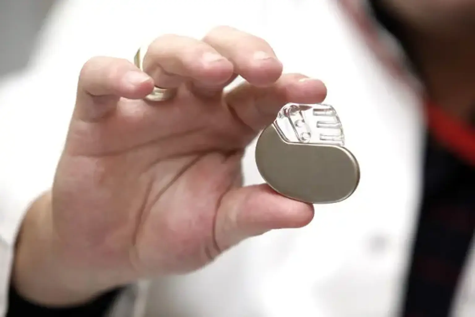
A cardiac pacemaker (or artificial pacemaker, to distinguish it from the natural pacemaker of the heart) is a medical device that make electrical impulses delivered by electrodes to cause the heart muscle chambers to contract and thus pump blood; by doing so, this device replaces and/or regulates the function of the heart's electrical conduction system.
The fundamental function of a pacemaker is to keep the heart rate stable, either because the heart's natural pacemaker is too slow or because there is a block in the electrical conduction system. Modern pacemakers are externally programmable, allowing a cardiologist, specifically a cardiac electrophysiologist, to choose the best pacing modes for each patient. Modern devices are demand pacemakers, which stimulate the heart depending on the circulatory system's dynamic demand.
A defibrillator is a form of pacemaker that combines pacemaker and defibrillator capabilities in a single implanted device, which should be named a defibrillator for clarity. Others, known as biventricular pacemakers, use numerous electrodes to stimulate different locations inside the lower heart chambers in order to enhance synchronization of the ventricles, or bottom chambers of the heart.
Anatomy & physiology of coronary sinus
One of the most difficult technical components of cardiac resynchronization treatment (CRT) device implantation is the optimum placement of a left ventricular lead in a tributary of the coronary sinus. The coronary sinus is the major vein of the larger venous system that flows in the atrioventricular groove's posterior portion.
When the great cardiac vein connects with the major posterior lateral vein, the coronary sinus is produced. The inferior left ventricular vein and the intermediate cardiac vein, which drain the posterior part of the left ventricle, are two more significant tributaries that enter the coronary sinus. The middle cardiac vein is the coronary sinus's greatest tributary, receiving communications from the anterior veins as well as the septal and inferior walls of both ventricles.
Pathophysiology of Dyssynchrony
Ventricular dyssynchrony is a disorder in which the ventricles (the heart's lowest two chambers) do not beat in synchronization. This disorder can cause blood to become trapped in the heart and not properly circulate throughout the body. For patients suffering from ventricular dyssynchrony, there are several diagnostic tests and therapies available.
Reduced left ventricular systolic function is linked to neurohumoral activation, which results in ventricular remodeling and dilatation. The majority of cardiomyopathy patients have conduction delays, such as a left bundle branch block (LBBB), resulting in electromechanical dyssynchrony. The ensuing contractile dyssynchrony causes regional variation in myocardial work, with early stimulated regions having lower load and late activated territory having higher load.
The transfer of blood from early to late activation sites and back to early activation sites leads in a net decrease in expelled stroke volume. This volume shift is substantially more pronounced when the variations in muscle activation between early and late contracting zones are largest.
Electromechanical dyssynchrony hence worsens morbidity and mortality in heart failure, where the underlying function is already weakened. CRT's simultaneous bi-ventricular pre-excitation restores coordinated contraction and hence improves net systolic performance by increasing chamber ejection and effort.
Indications of usage CRT
Biventricular pacing device (CRT) is indicated in the following patients:
1. It is a class I recommendation for patients with;
- Left ventricular ejection fraction of less than 36%.
- Sinus rhythm with LBBB morphology.
- QRS duration of >149ms.
- New York Heart Association (NYHA) II, III, or ambulatory IV symptoms on optimal medical therapy.
- Acceptable non-cardiac health.
2. It is a class IIa recommendation in patients with:
- Left ventricular ejection fraction of less than 36%.
- Sinus rhythm with LBBB morphology.
- QRS duration of 120 to 149ms.
- New York Heart Association (NYHA) II, III, or ambulatory IV symptoms on optimal medical therapy.
- Acceptable non-cardiac health.
3. It is a class IIa recommendation in patients with:
- Left ventricular ejection fraction of less than 36%
- Sinus rhythm with non-LBBB pattern.
- QRS duration of >149ms.
- New York Heart Association (NYHA) III, or ambulatory IV symptoms on optimal medical therapy.
- Acceptable non-cardiac health.
4. It is a class IIa recommendation in patients with:
- Left ventricular ejection fraction of less than or equal to 35%, on optimal medical therapy
- Atrial fibrillation and requires ventricular pacing.
- Rate control will end up on 100% ventricular pacing
- Acceptable non-cardiac health.
5. It is a class IIb recommendation in patients with:
- Left ventricular ejection fraction of less than 36%
- Sinus rhythm with non-LBBB pattern
- QRS duration of 120 to 149ms
- New York Heart Association (NYHA) III, or ambulatory IV symptoms on optimal medical therapy.
- Acceptable non-cardiac health.
6. It is a class IIb recommendation in patients with:
- Left ventricular ejection fraction of less than 36%, on optimal medical therapy.
- Undergoing a pacing device implantation for other indications
- Anticipated to have >40% ventricular pacing.
- Acceptable non-cardiac health.
Contraindication of CRT
In suitably chosen individuals, there are no proven absolute contraindications to CRT.
Relative contraindications may include:
- Dementia
- Advanced malignancy requiring palliative care
- Chronic disease with a life expectancy of less than one year
- Acute decompensated heart failure
- Active infection or sepsis
- Coagulopathy
Equipment used in CRT
Following equipment is required for implanting a biventricular pacing device.
- Cardiac catheterization laboratory with fluoroscopy and hemodynamic monitors
- Ultrasound machine (for access)
- Needles and sheaths
- Pacing leads and generator
- Device programmer
- Sutures.
Preoperative preparation of the chosen patient
Preparation is similar to other cardiac catheterization procedures.
Patients should be thoroughly examined for the operation and given detailed information about the risks, benefits, and consequences.
Before checking the coagulation profile, blood tests are performed, especially if the patient is taking anticoagulants.
The patient is required to fast for at least six hours before to the treatment. Intravenous cannulas are kept in place, and antibiotics with anti-staphylococcal coverage are given as a preventative measure against implant infection.
When the patient arrives at the catheterization laboratory, the access site is cleaned with an antiseptic solution and draped.
Before the operation, intravenous sedation is administered, and local anesthetic drugs are administered at the site of venous puncture and pocket formation.
Technique of how Biventricular pacemaker inserted.
Venous Access:
The CRT device insertion technology has advanced fast since its inception in the early 1990s. Initially, a restricted thoracotomy method was employed for epicardial lead implantation, but developments in left ventricular (LV) transvenous lead delivery systems have resulted in the completely transvenous implanted system. Despite advancements, technical obstacles of implanting three leads (right atrial, right ventricular, and LV epicardial) exist when the site of approach is considered.
Subclavian, axillary, and cephalic veins are common venous approaches. The cephalic vein, followed by the subclavian vein, is the preferred venous entry for lead implantation. Subclavian vein access or intrathoracic access can be used to get access to the subclavian vein.
Some surgeons prefer combined cephalic and subclavian vein access for easier catheter management and coronary sinus cannulation. During sequential lead implantation, this approach reduces lead contact and the possibility of lead dislodgement.
However, when the leads run through the ligamentous tissues between the medial end of the clavicle and the first rib, there is a substantial danger of pneumothorax and densely fibrotic tracts. Furthermore, fibrosis may make percutaneous lead extraction difficult.
CRT Leads Implantation:
Three long hydrophilic guidewires are put in the accessible vein in the event of CRT-D (CRT devices paired with defibrillator) (cephalic or subclavian). A guide catheter is then inserted through the sheath into the coronary sinus, followed by a hydrophilic wire or deflectable catheter for the insertion of the left ventricular lead.
A balloon-tip catheter put into the guide catheter can be used to perform coronary sinus (CS) venography. Fluoroscopy is used to guide the placement of a left ventricular lead in an appropriate tributary of the CS. Finally, the right atrial (RA) lead is implanted. Once the three leads are in place. The sheath is cut open, and the RA and RV leads are sutured to the pectoral muscle.
Successful resynchronization is achieved by placing the LV lead in an appropriate tributary of the CS, ideally in the proximal third to the middle third of the LV, with a negative effect if the LV lead is placed in the apical area.
CRT Programming:
To maximize the benefits of resynchronization, an individually tailored AV interval is required. Optimized AV interval programming synchronizes atrial and ventricular contraction, increases atrial contribution to diastolic filling of the left ventricle, and reduces presystolic mitral regurgitation. VV interval modification can improve interventricular synchronization and LV contraction even more, while the efficacy of VV optimization on CRT result is still being debated. Although clinical result evidence does not support non-invasive AV and VV interval optimization strategies using electro- and echocardiography, these methods can be evaluated.
Special cautions should be considered in chosen patient
Insertion:
A pacemaker can be installed while the patient is awake, using local anesthetic to numb the skin with or without sedation, or while the patient is unconscious, using a general anesthetic. Antibiotics are often used to lower the risk of infection. Pacemakers are often placed in the front of the chest, around the left or right shoulder. Before washing the skin with a disinfectant such as chlorhexidine, the skin is prepped by cutting or removing any hair above the implant site. An incision is made below the collarbone, and a space or pocket beneath the skin is formed to house the pacemaker generator.
This pocket is often formed slightly above the pectoralis major muscle (prepectoral), however in certain circumstances, the device may be placed beneath the muscle (submuscular). The lead or leads are inserted into the heart via a big vein that is guided by X-ray imaging (fluoroscopy). Depending on the kind of pacemaker required, the leads' tips may be placed in the right ventricle, right atrium, or coronary sinus.
Typically, surgery takes between 30 and 90 minutes to perform. The surgical incision should be kept clean and dry after implantation until it heals. Excessive shoulder movement should be avoided during the first few weeks to decrease the risk of dislodging the pacemaker leads.
A pacemaker generator's batteries normally last 5 to 10 years. When the batteries reach the end of their useful life, the generator is changed in a process that is typically less invasive than implanting a new implant. Making an incision to remove the existing device, disconnecting the leads from the old device and attaching them to a new generator, reinserting the new device, and sealing the skin are all steps in the replacement process.
Periodic pacemaker check-up:
After the pacemaker is implanted, it is tested on a regular basis to verify that it is operational and operating properly. The device can be examined as frequently as necessary, depending on the frequency established by the following physician. Routine pacemaker checkups are usually performed in-office every six months, however this might vary based on the patient/device state and remote monitoring availability.
Newer pacemaker devices may also be remotely probed, with the patient providing pacemaker data through an at-home transmitter linked to their regional cellular network.
At the time of in-office follow-up, the device will be interrogated to perform diagnostic testing. These tests include:
- Sensing: the ability of the device to evaluate intrinsic cardiac activity.
- Impedance: A test for determining the lead's integrity. Large and/or rapid increases in impedance can indicate a lead fracture, whereas large and/or sudden drops in impedance can indicate a lead insulation breach.
- Threshold amplitude: The smallest amount of energy (usually measured in hundredths of a volt) necessary to pace the atrium or ventricle linked to the lead.
- Threshold duration: The amount of time required for the device to pace the atrium or ventricle linked to the lead at the predetermined amplitude.
- Percentage of pacing: The proportion of time that the pacemaker has been actively pacing since the previous device interrogation defines how dependent the patient is on the device.
- Estimated battery life at current rate: Because current pacemakers are "on-demand," meaning they only pace when needed, the device's lifetime is determined by how frequently it is used. Other elements influencing device lifetime include programmed output and algorithms (features) that consume more current from the battery.
- Any events that were stored since the last follow-up: Specifically, arrhythmias such as atrial fibrillation. These are often saved in accordance with particular criteria established by the physician and unique to the patient. Some devices can display intracardiac electrograms from the event's beginning as well as the event itself. This is extremely useful in determining the root cause of an occurrence and making any required programming adjustments.
Magnetic fields, MRIs, and other lifestyle issues:
After a pacemaker is implanted, a patient's lifestyle is typically not significantly altered. Full-contact sports and activities using powerful magnetic fields are examples of risky pursuits.
Some daily activities may need to be changed for the pacemaker patient. A car seatbelt's shoulder strap, for example, may be painful if it falls over the pacemaker insertion site. Women will be unable to wear bras for a period of time following the procedure and may be required to wear bras with broad shoulder straps in the future.
If the patient wishes to participate in any sport or physical activity, specific pacemaker protection can be worn to avoid physical injuries or damage to the pacemaker leads.
Any activity using powerful electro-magnetic fields should be avoided. This may involve tasks such as arc welding with particular types of equipment or maintaining large equipment that generates powerful magnetic fields (such as an MRI machine).
Turning off the pacemaker:
A panel of The Heart Rhythm Society, a professional group located in Washington, DC, determined that it was legal and ethical to fulfill requests to deactivate implanted cardiac devices made by patients or those with legal power to make choices for patients. Lawyers believe the legal position is comparable to removing a feeding tube, although there is presently no legal precedence in the United States of America with pacemakers.
In the United States, a patient is believed to have the right to refuse or quit treatment, even a pacemaker that keeps him or her alive. Physicians have the right to refuse to turn it off, but the HRS panel advises them to send the patient to a physician who will. Some patients think that hopeless, debilitating diseases, such as those caused by severe strokes or late-stage dementia, might cause so much pain that they would prefer not to have supporting measures, such as cardiac devices, implanted.
Possible complications associated with CRT
Major complications associated with CRT device implantation include:
- Access site hemorrhage and pocket hematoma: In clinical studies, the incidence of access site hemorrhage and pocket hematoma has been observed to be up to 2.5 %. However, because the studies only reported hematomas that required care, the real prevalence of pocket hematomas in ordinary clinical practice may be greater. Pocket hematoma and early re-intervention for pocket hemorrhage are linked to an increase in device infection.
- Lead dislodgement: In CRT trials, the rate of lead dislodgement ranged from 2.9 to 10%. The incidence of left ventricular lead dislodgement is greater than that of right atrial and right ventricular leads.
- Infection: It is one of the most difficult problems associated with CRT and other cardiac implanted devices. The incidence of device-related infection for CRT implantation has been reported to be as high as 3.3 percent. Male gender, past device-related infection, and re-implantation have all been linked to an increased risk of device-related infection.
- Pneumothorax: It is an uncommon complication that has been documented in up to 0.66 percent of cardiac pacing device implantations. A greater pneumothorax incidence has been recorded with subclavian access, chronic obstructive lung disease, and advanced age (>80 years) upon implantation. To avoid this issue, the cephalic vein cut-down procedure should be utilized whenever possible.
- Coronary sinus perforation/dissection: Coronary venous dissection is an uncommon but well-known consequence of left ventricular lead insertion after cardiac resynchronization therapy device installation. It has been observed to increase post-procedural hospital stay by up to 0.28 %.
Conclusion
Heart failure is one of the leading causes of morbidity and death globally, and it is associated with a lower life expectancy, a worse quality of life, and a greater cost burden on the healthcare system. Heart failure can be caused by a variety of factors, but the most common cause is left ventricular systolic dysfunction.
The majority of cardiomyopathy patients have conduction delays, such as a left bundle branch block (LBBB), which causes electromechanical dyssynchrony. The resulting contractile dyssynchrony causes significant regional variation in myocardial work, with early activated regions having lower load and territories of late activation having higher lead.
Such patients benefit from the use of a cardiac resynchronization treatment (CRT) device. Aside from the survival effect, CRT improves functional class, quality of life ratings, physiological parameters such as peak VO, and hospitalizations in patients with severe heart failure.
CRT implantation is a complicated process that necessitates operating skills and competence, as well as careful patient selection and preparation. As a result, a system is required to ensure that all components of patient care are completed. In pre-procedure care, the evaluation must be performed to confirm that the patient satisfies the prescribed criteria for the operation, in addition to addressing the risks and advantages of the surgery with the patient and family. Proper sterile procedure is critical to achieving better results in terms of device-related infection.