Bloodless percutaneous vertebroplasty

    Last updated date: 22-Aug-2023

    Originally Written in English

    Bloodless percutaneous vertebroplasty

    Bloodless percutaneous vertebroplasty

    Overview

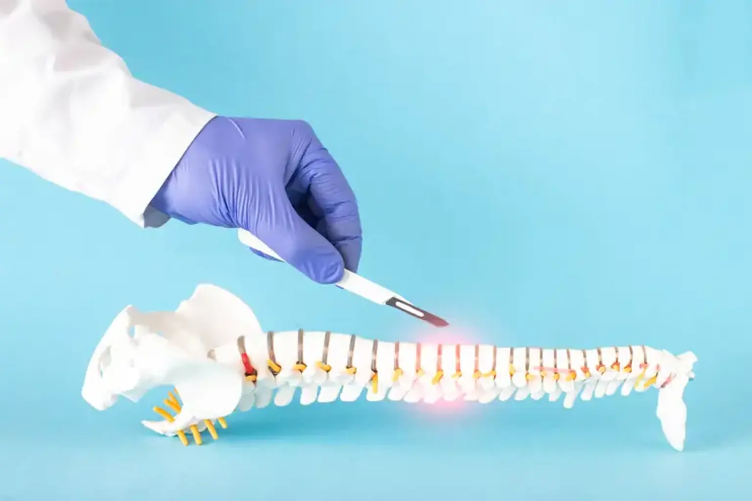
    Image-guided spinal surgeries have been performed for many years by interventional radiologists. Percutaneous vertebroplasty is a recent method that involves injecting a medical grade cement through a needle into a painfully damaged vertebral body. This stabilizes the fracture, enabling most patients to stop or reduce analgesics and resume regular activities.


    What is Percutaneous Vertebroplasty?

    Percutaneous Vertebroplasty

     Percutaneous cementoplasty (also known as vertebral packing or vertebroplasty) is a treatment used to treat patients with pathologic vertebral bodies in order to prevent vertebral body collapse and discomfort. Percutaneous cementoplasty is a potential pain management therapy in individuals with bone loss. Deramond et al, the method's inventors, conducted the first percutaneous cementoplasty in 1984.

    Percutaneous cementoplasty has gained in popularity since then, and several technological advancements have been accomplished. Percutaneous cementoplasty has gained acceptance, and the number of documented instances is growing.

    This technique may have a major influence on the morbidity and cost of symptomatic osteoporotic spinal compression fractures in the United States. Patients who do not respond to conservative treatment options such as bed rest, analgesics, and back bracing should be considered for vertebroplasty. Patients with active infection, untreated coagulopathy, and specific forms of fracture morphology are not candidates for this surgery.

    Because many patients have many chronic fractures, there should be a significant relationship between physical examination signs and symptoms and cross-sectional imaging findings. This treatment has a success rate of 73 to 90% in treating osteoporotic fractures. Vertebroplasty can effectively treat aggressive vertebral hemangiomas and may be palliative in individuals with malignant pathologic fractures. Significant problems from the operation occur in less than 1% of cases.

    Percutaneous vertebroplasty may be performed to relieve pain in patients suffering from severe painful osteoporosis with loss of height and/or compression fractures of the vertebral body, as well as in patients suffering from symptomatic vertebral hemangioma and painful vertebral body tumors (metastasis and myeloma).


    Relevant anatomy

    Bloodless percutaneous vertebroplasty anatomy

    A typical thoracic spine has 12 vertebrae and is immovable. A body, pedicles, laminae, spinous processes, and facet joints are all crucial anatomic landmarks to examine. Importantly, the lateral processes of the thoracic vertebrae establish the articulation with the paired 12 ribs on either side. The chest cavity is formed by the 12 vertebrae, 24 ribs, and sternum, which allows negative-pressure breathing and protects the chest wall.

    The lumbar spine follows the thoracic spine and is more movable than the thoracic. The lumbar spine is made up of five major vertebrae and other significant features, such as the body, pedicles, lamina, spinous processes, facet joints, and lateral processes. All articulations of the lumbar spine contribute to flexion-extension, bending, and rotation, allowing for truncal motion.

    This architecture must be considered in three dimensions and be clearly discernible on X-rays in order to permit appropriate needle insertion under fluoroscopic guidance. Comprehending radiographic anatomy is a critical step in understanding these techniques.


    Indications for Percutaneous Vertebroplasty

    Indications for Percutaneous Vertebroplasty

    Percutaneous vertebroplasty is indicated for painful, non-healing osteoporotic or neoplastic vertebral compression fractures that are unresponsive to conventional treatment. When these disorders cause a loss of normal vertebral body height, balloon kyphoplasty or vertebral augmentation with implants can be used to restore normal spinal alignment while simultaneously relieving pain.

    Medical therapy alternatives are available for people who are not candidates for vertebroplasty or who prefer not to seek surgical surgery. Bed rest, pain management with nonsteroidal anti-inflammatory drugs, calcitonin, opioids, and external bracing are all part of the medical therapy for compression fractures. Patients whose compression fractures do not heal after completing the aforementioned conservative protocol over a 3-6 week period should be evaluated for vertebroplasty.

    Patients who do not have fractures but are suffering from pain caused by lytic metastatic tumor or a rare symptomatic hemangioma are also candidates for vertebroplasty. Vertebroplasty can also be done as a preventative measure to stabilize a compromised vertebra before surgery.

    In the case of spinal malignancies, vertebroplasty serves a palliative purpose. In general, radiation is utilized to relieve spine pain in 70% of patients of previously untreated severe vertebral metastases. This impact, however, is delayed and might take up to 2-6 weeks to offer relief. Vertebroplasty can achieve the same pain alleviation aim with an almost instantaneous analgesic impact.

    • Vertebral hemangioma

    Vertebroplasty, as first described by Galibert in 1984, has effectively treated severe localized spinal pain caused by radiologically unaggressive vertebral (body) hemangioma.


    Osteoporotic vertebral fracture

    Osteoporotic vertebral fracture

    Vertebral fracture is one of the most prevalent consequences of osteoporosis. It can occur spontaneously or, more commonly, as a result of slight trauma. Compression fractures affect around 500,000 people in the United States each year, with women having a 16% lifetime risk and males having a 5% lifetime risk.

    These vertebral fractures are frequently extremely painful and can result in pain-limited immobility, which leads to demineralization and the perpetuation of a vicious cycle of ongoing demineralization and increasing fracture risk. Although the early symptoms usually go away in 4-6 weeks, some people experience severe, chronic, incapacitating pain despite medical treatment. Untreated fractures result in decreased height, decreased normal thoracic kyphosis, and persistent back discomfort.

    The VERTOS trial, which directly compared vertebroplasty to medical care, is an essential element in the issue of vertebroplasty. The VERTOS research compared osteoporotic compression fracture therapy of 18 patients with vertebroplasty to 16 patients with appropriate medical care and concluded that the vertebroplasty group had better pain alleviation. 

    Two large trials compared vertebroplasty to sham treatments and found no significant improvement with vertebroplasty. This conflicting information has spurred heated discussion over the procedure's proper purpose.


    Pre procedure patient evaluation

    Bloodless percutaneous vertebroplasty evaluation

    An accurate history and physical exam are the most critical aspects in planning this treatment. It has been demonstrated that palpating soreness does not correlate with a better vertebroplasty result. A thorough history of a patient with an acute spinal compression fracture might provide valuable information.

    • Deep pain with sudden onset
    • Midline location
    • Exacerbation by motion and standing


    • Imaging

    Plain film X-ray, CT scan, and MRI of the spine are all studies to consider when evaluating individuals for vertebroplasty. These three techniques will aid in determining which vertebra to inject.

    An MRI will be used to assess canal narrowing or osteomyelitis. Plain films can be used for procedure planning and can be used to evaluate lytic or blastic bone lesions. Informed consent and a full discussion of the risks, benefits, and alternatives are critical components of every surgery.


    • Setup and positioning

    The procedure time might vary, however it usually lasts between 40 and 120 minutes.

    • Conscious sedation and local anesthesia are used in the majority of cases. Use of anesthesia has also been described.
    • Prophylactic antibiotics such as cefazolin should be considered.
    • Place the patient in the prone position.
    • Align posterior ribs to obtain a good lateral view.
    • On the anteroposterior (AP) view, find the obliquity that projects the pedicle over the upper outer third of the vertebral body. Mark this point.


    • Radiation exposure

    When doing these treatments, it is critical to consider radiation exposure. In a study of 11 vertebroplasty instances, patient effective doses were assessed to be 34mGy, with average fluoroscopy periods of 28 minutes (in comparison, a typical CT abdomen and pelvis effective dose is 8–14 mGy). Operator exposures were highest in the hands and chest, despite the fact that using mobile shielding reduced occupational dosage by 76%. An operator might do 150 operations per year before exceeding yearly dose restrictions, according to estimates.


    Technique

    Bloodless percutaneous vertebroplasty process

    • Needle placement

    Lidocaine with epinephrine is a typical local anesthetic used in conjunction with a spinal needle to anesthetize the periosteum. AP and lateral fluoroscopic images can be used to confirm the correct trajectory. Different gauge needles might be used depending on the size of the pedicle. For lumbar and lower thoracic pedicles, an 11-gauge needle is preferred, however a 13-gauge needle will sufficient for midthoracic pedicles.

    A big clamp will maintain tension along the back, allowing for simpler needle insertion. To gently tap the needle, use a sterilized hammer. Insert the needle into the spinal body, utilizing the AP and lateral views to confirm.

    Transpedicular approaches:

    • Advance the needle into the anterior third of the central vertebral body for a unipedicular approach.
    • Advance needles into the midportion of the hemivertebrae for a bipedicular approach.
    • A parapedicular method may also be explored.


    • Polymethylmethacrylate mixing and injection procedure

    Depending on the producer, the cement may be made in a variety of ways. Both polymethylmethacrylate (PMMA) and barium sulfate (opacifying agent) should be prepared according to the manufacturer's instructions. Slowly inject this combination into the vertebral body, stopping before PMMA seeps posteriorly into the epidural region or considerably fills a vein.

    The amount of cement utilized to fill the vertebral body is an essential concern. Because pain alleviation is unrelated to cement volume, aiming to completely fill the vertebral body is unnecessary.


    Titanium-implantable vertebral augmentation devices versus vertebroplasty or kyphoplasty

    kyphoplasty

    Even after balloon kyphoplasty, osteoporotic compression fractures continue to collapse, resulting in height loss and the development of angulation and deformity. This appears to raise the likelihood of adjacent level fractures. The SpineJack system is made up of bilateral expandable titanium implants and bone cement.

    When compared to balloon kyphoplasty, this technique provides more symmetric and balanced lateral and anterior support while using less bone cement. Clinicians may now obtain improved pain management, restore vertebral body height, restore spinal alignment, and lower the likelihood of adjacent level fractures by using this technique.

    The ability to execute kyphoplasty safely in patients with mild-moderate retropulsion of the posterior endplate without neurological impairment is a significant benefit of SpineJack kyphoplasty over traditional balloon kyphoplasty or vertebroplasty.

    In some cases, the SpineJack device has been utilized to raise broken endplates, therefore reducing the retropulsed bone fragment. SpineJack is better in cases where patients have a retropulsed bone fragment for the reasons stated above. Balloon kyphoplasty is contraindicated in the presence of retropulsed bone pieces.

    A research comparing SpineJack kyphoplasty to balloon kyphoplasty found that SpineJack patients experienced superior pain relief at 1 month and 6 months after treatment.


    Postprocedural care

    Percutaneous vertebroplasty

    Patients are usually kept in bed for 1-2 hours and given analgesics for postoperative pain as well as muscle relaxants for spasms. Patients with osteoporosis should be medically controlled and properly monitored following the operation. Without systemic osteoporosis medication, 20% of fracture patients experience a second fracture.

    DEXA scans, lifestyle changes (smoking cessation, alcohol moderation, exercise), nutritional supplements (calcium, vitamin D), and medical care are all part of the treatment for osteoporosis (antiresorptive).


    Percutaneous vertebroplasty complications

    Percutaneous vertebroplasty complications

    Percutaneous vertebroplasty, among other procedures, has become a standard treatment for osteoporotic and malignant vertebral fractures. When doing vertebroplasty, it is critical to pay close attention to the anatomic structures. Mild problems include post-procedure discomfort and cement leaking into the intervertebral disc space and paravertebral soft tissues.

    Infection and cement leak into the epidural area are examples of moderate consequences. Leaks into the paravertebral veins might result in pulmonary embolism, cerebral embolism, or heart perforation. There are three types of vertebroplasty complications: mild, moderate, and severe.

    • Mild complications 

    Mild problems include: a short increase in discomfort following the treatment; transitory arterial hypotension; cement leaking into the intervertebral disc space, which has little or no clinical significance, or into the paravertebral soft tissues; and an increased risk of future fractures.

    It is uncommon to see a temporary increase in discomfort after vertebroplasty. It is the result of an inflammatory response caused by the heat created by the polymerization of methyl methacrylate. 1 Although discomfort seldom lasts more than a few hours, analgesics may readily handle it (steroidal or nonsteroidal).

    Transient arterial hypotension is an uncommon complication2 that can be caused by polymethyl methacrylate injection during percutaneous vertebroplasty; nevertheless, it responds well to supportive interventions and is self-limiting.

    Although cement leakage into the disc during vertebroplasty has no definite clinical consequences, it theoretically increases the risk of subsequent fractures of adjacent vertebral bodies; therefore, in a centrally located fracture, it is advised to place the needle laterally and far from the center of the vertebra, and to adjust the cement consistency so that the cement is more viscous, making it less prone to leak.

     Furthermore, adapting to lesser quantities of cement may reduce the likelihood of leaking (while still providing enough stability and pain relief); additionally, stopping injection promptly when disc leakage develops may be beneficial.

    Cement leakage into the paravertebral soft tissues occurs in 6% to 52% of cases and is typically asymptomatic, with just a few instances of peripheral neuropathy. After the surgery, a significant proportion of individuals with osteoporosis have new fractures; two-thirds of these new fractures occur in a vertebra close to those previously treated.

    • Moderate complications 

    Infection, cement leak into the epidural/foraminal area, and a misdirected needle in the tract are examples. Infection is a possibility with every percutaneous operation. It is a complication that can be avoided or reduced. Infection can cause discitis, osteomyelitis, or even an epidural infection, which can be fatal if surgery is not performed to remove the cement that acts as a nidus for infection.

    Cement extravasation into the epidural or foraminal area is an uncommon consequence. Most instances are clinically quiet unless a post-procedure CT is performed. The prevalence might reach 40%. Only 0.4% of individuals will get paraplegia as a result of cement-related spinal cord compression. Needle traversing of the lamina rather than the pedicle is possible, particularly in the thoracic vertebra where the pedicle is smaller, and can result in disastrous consequences.

    Serious complications Severe problems are typically caused by cement leakage into the paravertebral veins, which can result in pulmonary embolism, heart perforation, cerebral embolism, and even death. A combination of highly vascular lesions and the liquid nature of the cement might cause a paravertebral venous leak. Radicular discomfort may be caused by this leak.

    An uncommon consequence of vertebroplasty is pulmonary cement embolism. It is asymptomatic in up to 4.6% of cases and is directly connected to the frequency of paravertebral venous leak, but not to the number of vertebral bodies treated. If the patient is symptomatic, traditional signs such as chest pain, hypotension, and dyspnea will be noticed. The onset might be quick or delayed, and it can eventually lead to death. It is treated with oxygen inhalation, short-term anticoagulant medication, intravenous steroid therapy, or, in extreme situations, intravenous steroid therapy combined with catheter-aided interventional methods.


    Conclusion 

    In a large majority of patients with osteoporotic fractures, percutaneous vertebroplasty offered considerable pain alleviation. The treatment stabilized the spine in cancer patients but did not give continuous pain alleviation. Minor and occasional complications occurred. Percutaneous vertebroplasty is a potential treatment for osteoporotic fractures and some vertebral column neoplasms.