Introduction
Bone fractures are common injuries that can significantly impact daily life. A fracture occurs when a bone breaks or cracks, typically due to trauma like falls, accidents, or sports injuries. Proper treatment is essential to ensure proper healing and reduce the risk of complications. Without the right care, a bone fracture could lead to long-term pain, deformity, or impaired function. The treatment approach for fractures varies based on the fracture type and severity, but it generally involves immobilization, pain management, and rehabilitation.
Seeking medical attention immediately after a fracture is crucial to prevent further injury and begin the healing process as soon as possible. The faster the treatment, the better the chances of complete recovery.
Initial Steps in Fracture Management
Recognizing the symptoms of a bone fracture is essential for early treatment. Common signs include intense pain, swelling, bruising, and sometimes a visible deformity. Here’s what to do immediately after a fracture:
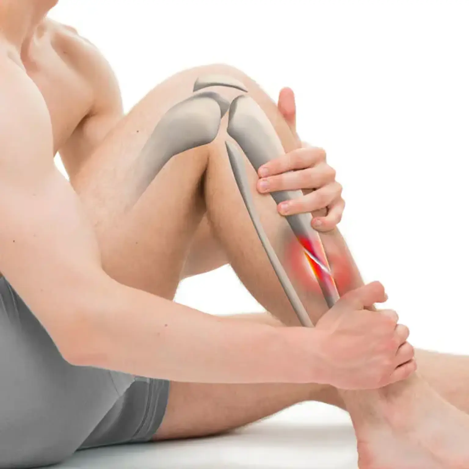
Immobilize the Bone: Keep the affected area still to prevent further damage. Use splints, clothing, or any available materials to support the fracture site until medical help arrives.
Apply Ice: Ice helps reduce swelling and pain. Wrap ice in a cloth and apply it to the injured area, but avoid direct contact with the skin.
Elevate the Injured Area: If possible, raise the affected limb above heart level to reduce swelling.
Seek Immediate Medical Care: It’s crucial to get medical attention quickly for a proper diagnosis and treatment plan. Delayed treatment can lead to complications like infections or improper bone healing.
Following these steps minimizes the risk of further damage and sets the stage for proper treatment.
Diagnosis of Bone Fractures
Accurate diagnosis is key to selecting the best treatment plan for a bone fracture. During a medical consultation, a healthcare provider will perform a physical examination and assess the symptoms, such as swelling, bruising, or deformity. However, imaging techniques are often needed to confirm the fracture type and extent.
X-rays: The most common diagnostic tool, X-rays allow doctors to view the bone structure and determine the location and severity of the fracture.
CT Scan: In complex cases, a CT scan may be used to provide a more detailed image, especially if the fracture is not clearly visible on an X-ray.
MRI: For fractures involving soft tissues, ligaments, or cartilage, an MRI might be necessary.
By accurately diagnosing the fracture, doctors can create an effective treatment plan, whether that involves non-surgical approaches like casting or splinting or requires surgery for more severe cases.
Types of Bone Fractures
Bone fractures come in various forms, each requiring a different treatment strategy. Understanding the different types of fractures can help guide appropriate care:
Closed Fracture: The bone breaks but does not puncture the skin. This is the most common type and usually treated with immobilization through a cast or splint.
Open Fracture: The bone breaks and punctures the skin, exposing the bone to external elements. This type of fracture carries a higher risk of infection and typically requires surgical intervention.
Comminuted Fracture: The bone shatters into several pieces, often requiring surgical repair and longer recovery times.
Greenstick Fracture: Common in children, this is an incomplete fracture where the bone bends and cracks but doesn’t break all the way through.
Stress Fracture: Small cracks in the bone due to repetitive force or overuse, typically seen in athletes.
Each fracture type requires tailored treatment to ensure optimal healing. Fracture severity also determines whether non-surgical or surgical treatments are necessary.
Surgical Treatment for Bone Fractures
In some cases, surgery is necessary, particularly when fractures are severe, displaced, or involve multiple bone fragments. Surgical treatment aims to restore the bone’s original alignment and ensure proper healing. Common surgical options include:
Internal Fixation: Involves using metal pins, plates, or screws to hold the bone together. This method is common for fractures that are displaced or unstable.
External Fixation: A set of pins or screws is inserted into the bone, and an external frame is used to hold everything in place. This method is often used for complex or open fractures.
Bone Grafting: Sometimes used when bone loss occurs, where healthy bone tissue is taken from another area of the body or a donor site to promote healing.
Surgical intervention often involves a longer recovery period, including physical therapy to regain strength and mobility.
Fracture Immobilization Techniques
Immobilization is key to bone healing, as it prevents the broken bones from moving and causing further injury. There are several immobilization techniques used based on the type and location of the fracture:
Casts: Hard or soft casts are used to keep bones in place and protect them from further injury. They are most commonly used for fractures of the limbs.
Splints: A splint is a temporary form of immobilization that may be used in the emergency room before a cast is applied. It supports the injured area without fully enclosing it.
Braces: In some cases, a brace may be used, especially for fractures around joints. It provides support while still allowing some movement.
The length of time a fracture needs to be immobilized depends on the severity and location of the break. Generally, immobilization lasts for a few weeks, but more severe fractures may require longer treatment.
Bone Fracture Healing Process
Bone healing is a gradual process that occurs in three main stages:
Inflammation (1-2 weeks): After a fracture, blood vessels around the broken bone are damaged, causing swelling and inflammation. This phase sets the foundation for healing, as a blood clot forms at the fracture site.
Repair (2-6 weeks): During this phase, the body starts creating a "callus" made of collagen that bridges the gap between the broken bone ends. New bone tissue begins to form, and the bone is gradually stabilized.
Remodeling (months to years): Over time, the callus is replaced with mature bone tissue. The bone continues to strengthen, and its shape returns to normal. This phase can take several months to years, depending on the fracture's complexity and the patient’s age and health.
Factors such as age, nutrition, and overall health can affect the speed and quality of bone healing. Proper nutrition, including adequate calcium and vitamin D intake, is essential to support the healing process.
Post-Fracture Care: Pain Management and Infection Prevention
Pain management is an important aspect of fracture recovery. It’s common to experience discomfort during the healing process, but effective pain management can help you stay comfortable and support the healing process. Some strategies include:
Over-the-counter medications: Pain relievers like ibuprofen or acetaminophen can help reduce pain and inflammation.
Prescription painkillers: For more severe pain, a doctor may prescribe stronger medications, but these are typically used for a short period to avoid dependency.
Ice and Elevation: Applying ice and elevating the injured area can also reduce pain and swelling.
For open fractures, infection prevention is critical. Proper wound care, such as cleaning the wound and applying prescribed antibiotics, can prevent infections. In some cases, especially with surgical treatments, antibiotics may be used to reduce the risk of infection.
Physical Therapy After Bone Fractures
Once a fracture starts to heal, physical therapy becomes an essential part of the recovery process. Therapy helps restore strength, flexibility, and mobility, and it can prevent stiffness and weakness in the affected area. Common physical therapy exercises include:
Strengthening exercises: These help rebuild muscle strength around the fractured bone, preventing atrophy.
Stretching exercises: These are used to improve flexibility and range of motion once the fracture is stable.
Weight-bearing exercises: Depending on the type of fracture, some patients may be advised to gradually start putting weight on the affected bone to stimulate healing.
The length of physical therapy varies based on the severity of the fracture and the patient’s age. Regular sessions and consistent exercises are crucial to full recovery.
Non-Surgical Fracture Treatment Options
Many bone fractures can heal without the need for surgery. Non-surgical treatments are typically used for fractures that are stable, not displaced, and not causing serious complications. The primary goal is to immobilize the broken bone and promote healing. Common non-surgical options include:
Casting and Splinting: These are the most common ways to immobilize fractures. A cast or splint keeps the bone in the correct position, preventing movement that could hinder healing.
Braces: For some fractures, especially in joints, a brace can provide support while allowing some limited movement.
Rest and Elevation: In addition to immobilization, resting the affected area and elevating it above heart level can help reduce swelling and pain.
Non-surgical treatment typically requires regular follow-up visits to ensure the bone is healing properly and the fracture remains aligned.
Lifestyle Adjustments During Fracture Recovery
During fracture recovery, making certain lifestyle adjustments can help speed up healing and maintain comfort. These include:
Rest and mobility aids: Adequate rest is important, but mobility aids like crutches, walkers, or canes may be necessary to avoid putting weight on the injured area.
Diet and nutrition: Proper nutrition supports bone healing. A diet rich in calcium, vitamin D, and protein can accelerate the repair process. Foods like dairy, leafy greens, and fish can help.
Mental health support: Bone fractures can cause frustration due to limited movement, so maintaining a positive outlook is important. Staying connected with family and friends, engaging in hobbies, and practicing relaxation techniques can help boost mental well-being.
These adjustments can minimize discomfort and support a smoother recovery process.
Preventing Future Fractures
After recovering from a bone fracture, preventing future fractures is important, especially if you are at higher risk due to factors like age, bone density, or lifestyle. Here are a few strategies:
Strengthening exercises: Regular weight-bearing exercises, such as walking, jogging, or strength training, can help keep bones strong and less likely to fracture.
Fall prevention: If you’re prone to falls, consider making changes to your home to reduce the risk, like installing grab bars, improving lighting, and removing tripping hazards.
Diet and supplements: Consuming a balanced diet with adequate calcium and vitamin D, along with taking supplements if necessary, can help maintain bone health.
Regular check-ups: For individuals with conditions like osteoporosis, regular bone density scans and consultations with healthcare providers can help monitor bone health and prevent fractures.
By taking these preventive measures, you can reduce your risk of future fractures and maintain strong, healthy bones.
Special Considerations for Elderly Patients
Older adults are at a higher risk for fractures, particularly those caused by osteoporosis, falls, or weakened bones. Fractures in the elderly often require special care:
Slower healing: Aging bones take longer to heal, and recovery might be more complicated due to underlying health conditions, such as diabetes or cardiovascular disease.
Risk of complications: Older adults are more prone to complications like infections, blood clots, or prolonged immobility, which can affect overall recovery.
Supportive care: Elderly patients may require more assistance with daily activities and rehabilitation. Physical therapy is essential to regain strength and mobility, while caregivers may need to monitor progress closely.
Ensuring elderly patients have access to appropriate care, pain management, and rehabilitation is key to successful recovery and improving quality of life
How to Support a Loved One Recovering from a Fracture
If someone you know is recovering from a bone fracture, providing emotional and physical support can make a big difference in their recovery. Here’s how you can help:
Assist with mobility: Help your loved one move around, especially in the early stages of recovery when they may be using crutches or a walker.
Encourage rest: Healing takes time, so encourage them to rest and avoid overexertion, while offering assistance with daily activities like preparing meals or household chores.
Be empathetic: Fracture recovery can be frustrating and painful. A positive attitude and emotional support can boost their morale and help reduce anxiety.
Stay informed: Educate yourself on the fracture recovery process so you can provide practical assistance and understand the challenges they may face.
Being there for someone recovering from a fracture can help them navigate the healing journey with confidence and comfort.
The Role of Orthopedic Surgeons in Fracture Treatment
Orthopedic surgeons play a crucial role in treating bone fractures, especially when surgery is needed. These specialists are trained to handle complex fractures and use advanced techniques for bone repair. Their responsibilities include:
Diagnosis and treatment planning: Orthopedic surgeons use imaging and clinical evaluation to determine the best approach for each fracture, whether surgical or non-surgical.
Surgical intervention: When fractures require surgery, orthopedic surgeons perform procedures such as internal or external fixation to align and stabilize the bone.
Post-operative care: They monitor healing progress and make adjustments to the treatment plan if necessary, ensuring the bone heals properly.
Orthopedic surgeons work closely with other healthcare providers, including physical therapists, to guide patients through their recovery.
The Cost of Fracture Treatment and Insurance Considerations
Fracture treatment costs can vary widely depending on the type of fracture, treatment required, and location. For example, a simple fracture treated with a cast may be relatively inexpensive, while a complex fracture requiring surgery, rehabilitation, and physical therapy can be much more costly.
Insurance plays a significant role in covering fracture-related costs. Most health insurance plans cover diagnostic imaging, doctor visits, and surgeries related to fractures, but the extent of coverage can vary. It’s important to understand:
Out-of-pocket expenses: Depending on your insurance, you may still have co-pays or deductibles for services like emergency room visits, surgery, or follow-up care.
Rehabilitation coverage: Physical therapy often requires multiple sessions, so check if your insurance plan includes coverage for rehabilitation.
Cost-saving options: For those without insurance, there may be payment plans or financial assistance programs available through hospitals or clinics.
Planning ahead and understanding your insurance benefits can help manage the financial aspect of fracture treatment.
The Psychological Impact of Bone Fractures
Recovering from a bone fracture is not only a physical challenge but can also take a psychological toll. The sudden loss of mobility and the pain associated with the injury may lead to feelings of frustration, anxiety, and depression. Patients may struggle with:
Loss of independence: Difficulty performing everyday tasks can lead to feelings of helplessness or dependence on others.
Fear of re-injury: Especially for athletes or active individuals, there can be anxiety about returning to previous activities and the risk of further injury.
Body image concerns: In some cases, visible scars, deformities, or temporary changes in appearance can affect a person’s self-esteem.
Psychological support, such as counseling or support groups, can help individuals manage these emotional challenges and improve their mental well-being during recovery.
Understanding Fracture Complications
While most fractures heal well with proper treatment, complications can arise. It's important to be aware of potential risks and signs of complications during recovery:
Delayed Healing or Nonunion: Sometimes, bones take longer to heal, or they fail to heal completely. This can happen if the bone is not aligned properly or if there is insufficient blood supply.
Infection: For open fractures, infection is a significant concern. Symptoms like increased redness, warmth, or discharge from the wound should be addressed immediately.
Compartment Syndrome: A rare but serious complication, this occurs when pressure builds up in a muscle compartment, restricting blood flow. It requires immediate medical attention.
Post-traumatic Arthritis: If a fracture affects a joint, it can lead to long-term arthritis, causing pain and reduced range of motion.
Recognizing and treating these complications early can prevent more serious issues and ensure a smoother recovery process.
Preventing Bone Fractures in Children and Teens
Children and adolescents are particularly vulnerable to fractures due to active lifestyles and growing bones. While many fractures in younger individuals are due to falls or sports activities, there are steps that can be taken to minimize risk:
Proper equipment: Encourage the use of protective gear such as helmets, knee pads, and elbow pads during sports or recreational activities.
Bone health: Adequate nutrition, including calcium and vitamin D, is essential for bone growth and strength during the developmental years.
Safe environments: Ensure that living and play areas are safe, free from tripping hazards, and that children understand basic safety practices.
Supervision: Active children should be supervised during physical activities, especially those involving risk, such as biking, skateboarding, or climbing.
Fostering healthy habits and creating a safe environment can help reduce the risk of bone fractures in young individuals.
Conclusion
Bone fractures are a common yet complex medical condition that require careful treatment and attention. Whether managed through non-surgical methods or more advanced surgical interventions, proper care and rehabilitation are key to ensuring full recovery. Patients should be aware of potential complications and the importance of follow-up care, physical therapy, and lifestyle adjustments. Emotional support is also crucial, as the psychological impact of a fracture can be significant. By staying informed, adhering to treatment plans, and making preventive lifestyle changes, individuals can optimize their recovery and reduce the risk of future fractures. With advancements in medical technology, the future of bone fracture treatment looks promising, offering even more effective and personalized solutions.