Introduction
Brain imaging plays a crucial role in diagnosing and monitoring various neurological conditions. Among the most effective and widely used techniques are MRI (Magnetic Resonance Imaging) and MRA (Magnetic Resonance Angiography). These advanced imaging tools help doctors assess brain health, detect abnormalities, and plan effective treatments. Whether for routine health checks or diagnostic purposes, brain imaging offers invaluable insights into the brain’s structure and function.
As technology advances, the use of MRI and MRA in brain health is becoming increasingly popular, especially in countries like Korea, where cutting-edge healthcare services are widely available. These imaging techniques allow for a non-invasive look at the brain, enabling early detection of conditions like strokes, aneurysms, tumors, and even cognitive decline. Understanding the role of these imaging procedures is vital for anyone concerned about their brain health.
What is Brain Imaging?
Brain imaging refers to the use of specialized techniques to visualize the structure and function of the brain. It is a non-invasive procedure that helps doctors diagnose, monitor, and treat various neurological conditions. Brain imaging is essential for detecting issues that may not be evident through physical exams or symptoms alone, such as strokes, brain tumors, multiple sclerosis, and brain infections.
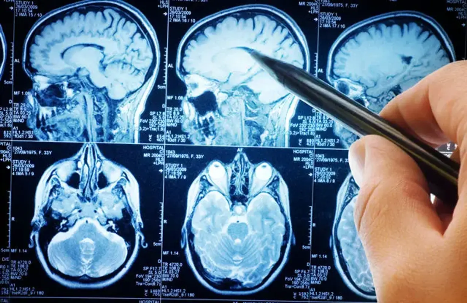
MRI and MRA are two primary forms of brain imaging that provide a detailed view of the brain’s anatomy, blood vessels, and functionality. MRI scans offer a detailed image of the brain’s soft tissues, while MRA focuses on the blood vessels within the brain, allowing doctors to detect any vascular abnormalities such as aneurysms or narrowed arteries.
Brain imaging can be used to assess a wide range of conditions, from neurological diseases like Alzheimer’s to vascular problems like stroke. By utilizing these imaging techniques, healthcare providers can offer more precise diagnoses and treatment plans, improving patient outcomes.
MRI (Magnetic Resonance Imaging) for Brain Health
MRI (Magnetic Resonance Imaging) is a non-invasive imaging technique that uses powerful magnets and radio waves to create detailed images of the brain's structure. During an MRI scan, the patient lies on a table, and the machine generates cross-sectional images of the brain. These images are then used to assess the brain's condition, identify abnormalities, and detect early signs of disease.
How MRI Works: MRI scans work by creating images using a magnetic field and radiofrequency waves. The magnetic field aligns the hydrogen atoms in the body, and when a radiofrequency pulse is applied, these atoms produce signals that the MRI machine captures. These signals are then processed to create detailed images of the brain’s internal structures.
MRI for Brain Conditions: MRI scans are particularly useful for detecting structural brain issues, including:
Brain tumors: MRI is highly sensitive in detecting even small brain tumors.
Multiple sclerosis: MRI helps to identify lesions in the brain caused by this autoimmune disease.
Brain inflammation or infection: MRI can highlight areas of swelling or infection in the brain.
Brain volume loss: MRI is used to detect changes in brain volume, which is especially important for conditions like Alzheimer's or other forms of cognitive decline.
MRI is non-invasive and offers a high level of detail without using ionizing radiation, making it a safe and effective method for brain health assessments.
MRA (Magnetic Resonance Angiography) in Brain Imaging
MRA (Magnetic Resonance Angiography) is a specialized type of MRI that focuses on the blood vessels of the brain. While standard MRI scans provide detailed images of the brain’s structure, MRA focuses on visualizing the brain's vascular system. This makes MRA particularly useful in diagnosing and monitoring vascular conditions, such as aneurysms, stenosis (narrowing of blood vessels), and vascular malformations.
How MRA Differs from MRI: While both MRI and MRA are used to assess the brain, the key difference lies in what they visualize. MRI focuses on soft tissue structures, while MRA is specifically designed to assess the blood vessels in the brain. MRA can highlight areas of reduced blood flow, blockages, or irregularities in blood vessels, which are often linked to stroke or aneurysms.
Applications of MRA in Brain Health: MRA scans are particularly beneficial for:
Detecting brain aneurysms: MRA can identify weak spots in blood vessels that may lead to aneurysms.
Evaluating vascular malformations: It helps identify abnormal blood vessel formations in the brain that may require surgical intervention.
Monitoring stroke risk: MRA can show areas of reduced blood flow, which can be indicative of an increased stroke risk.
Like MRI, MRA is non-invasive and does not require incisions or needles, providing a safe and effective method for monitoring brain vascular health.
Brain Health Imaging and Cognitive Conditions
Brain imaging, particularly MRI and MRA, plays a crucial role in diagnosing cognitive conditions. These scans help identify brain abnormalities that contribute to cognitive decline, such as Alzheimer’s disease, dementia, and vascular dementia.
MRI for Cognitive Health: MRI scans can detect changes in brain structure associated with cognitive decline. For instance, brain volume loss is a common sign in Alzheimer’s patients, and MRI can reveal these structural changes. Identifying these changes early helps doctors intervene before symptoms become severe.
Detecting Cognitive Decline: Cognitive decline often starts subtly, but brain imaging can highlight early signs, enabling timely treatment. MRI and MRA can assess brain health and identify areas where cognitive functions may be deteriorating, offering a proactive approach to brain health.
Neuroimaging and its Role in Brain Health
Neuroimaging refers to the use of various techniques, including MRI and MRA, to examine the brain's structure and function. It plays a pivotal role in diagnosing neurological diseases, tracking disease progression, and planning treatment.
Advanced Neuroimaging Techniques: Neuroimaging techniques, such as functional MRI (fMRI) and MRA, offer insights into both the structure and function of the brain. fMRI measures brain activity by detecting changes in blood flow, while MRA provides a detailed look at blood vessels in the brain.
Applications in Brain Health: Neuroimaging is essential for diagnosing diseases like stroke, aneurysms, brain tumors, and cognitive disorders. It also helps monitor conditions over time, providing doctors with vital information to adjust treatment plans as needed.
Brain Imaging in Korea
Korea is known for its advanced healthcare system, and brain imaging is no exception. The country uses the latest technology in MRI and MRA to diagnose and treat neurological conditions.
Brain MRI in Korea: Korean hospitals offer state-of-the-art MRI technology that provides high-resolution images, allowing for accurate diagnoses of brain conditions. Many clinics in Korea specialize in cognitive health, offering comprehensive brain health assessments using these imaging tools.
Popularity of Brain Imaging in Korea: The popularity of MRI and MRA scans in Korea is growing, thanks to the country's reputation for innovation in medical technology. Both local and international patients seek these advanced imaging services to detect brain conditions early and receive effective treatments.
Brain Health Test and Diagnosis Using MRI & MRA
Brain Health Test: A brain health test using MRI or MRA helps identify a variety of brain-related issues. This test typically includes imaging scans to assess the brain’s structure, volume, and blood flow, offering a comprehensive view of brain health.
Diagnostic Process: During a brain health test, patients undergo an MRI or MRA scan. MRI scans show structural changes in the brain, such as tumors or signs of cognitive decline, while MRA scans focus on blood vessels, detecting blockages or aneurysms.
Identifying Health Conditions: These brain health tests are crucial for diagnosing conditions like strokes, vascular malformations, brain tumors, and cognitive decline. Early detection through MRI and MRA can significantly improve treatment outcomes and quality of life.
Brain Volume Restoration through Imaging
Brain volume restoration refers to medical interventions aimed at regaining lost brain tissue, often due to aging or diseases like Alzheimer's. MRI and MRA play a significant role in detecting brain volume loss, which is crucial for initiating restorative treatments.
How MRI Detects Volume Loss: MRI scans can reveal brain shrinkage or loss of gray matter, which is often associated with cognitive decline. Early detection of volume loss is essential for doctors to recommend interventions that may help slow or reverse brain degeneration.
Restorative Treatments: Once MRI scans show signs of volume loss, treatments like regenerative medicine, brain stimulation therapies, or even procedures such as fat grafting are sometimes used to restore brain volume or improve brain function. These treatments, available in countries like Korea, aim to rejuvenate brain function and slow the progression of cognitive decline.
Brain Volume Restoration Treatment After MRI Scan
After detecting brain volume loss via MRI, patients may explore various treatment options aimed at restoring brain health and function. Brain volume restoration treatments focus on regenerating lost brain tissue or slowing its decline.
Restoration Procedures: In some cases, regenerative treatments like stem cell therapy or brain stimulation are used to encourage growth in areas of the brain that have shrunk. Additionally, fat grafting in cosmetic surgery is an emerging trend to restore both facial volume and potentially aid in improving cognitive health, as it stimulates tissue regeneration.
The Role of MRI in Post-Treatment Monitoring: MRI scans are often used post-treatment to track the effectiveness of brain restoration therapies, ensuring that the treatments are having the desired effect on brain volume and cognitive function.
Benefits of Brain Imaging for Early Diagnosis
One of the most significant advantages of MRI and MRA brain scans is the ability to diagnose brain conditions early, which can make a significant difference in the outcome of treatment.
Improved Accuracy and Early Detection: Brain imaging provides detailed, non-invasive insights into the brain’s structure, allowing doctors to detect abnormalities that may not be visible through physical exams. Early detection is crucial in preventing conditions like stroke, brain tumors, and cognitive decline from progressing to more severe stages.
Life-Saving Diagnostics: Early diagnosis through brain imaging can be life-saving, especially for conditions like aneurysms or brain tumors. By detecting these conditions early, doctors can implement treatments that prevent further damage and significantly improve patient outcomes.
Non-Invasive Brain Scans: MRI and MRA Benefits
One of the primary advantages of MRI and MRA scans is that they are non-invasive. This means that these procedures do not require surgery, needles, or incisions, making them safer and more comfortable for patients.
Safety and Comfort: MRI and MRA scans are generally safe, with minimal risks. They involve no exposure to radiation, unlike CT scans or X-rays. These imaging techniques provide a detailed view of the brain, offering valuable diagnostic insights without the need for invasive procedures.
Benefits of Non-Invasive Imaging: Non-invasive brain scans are particularly beneficial for routine monitoring of brain health, detecting issues early, and assessing brain volume and vascular conditions without discomfort. Patients can quickly return to their daily activities after the procedure, with no significant recovery time required.
Brain Imaging for Cognitive Wellness and Aging
As we age, brain health becomes increasingly important, and brain imaging plays a key role in monitoring cognitive wellness. MRI and MRA scans can identify early signs of cognitive decline, enabling preventive measures to be taken before serious conditions like Alzheimer’s or dementia develop.
Using MRI for Cognitive Wellness: MRI scans can detect brain changes that are common with aging, such as decreased brain volume. Regular brain health assessments help doctors identify at-risk individuals and offer early interventions, including lifestyle changes, medications, or cognitive therapies.
Impact of Early Diagnosis: Early detection of cognitive decline through brain imaging allows for more effective management. Treatment options such as brain exercises, medications, or even regenerative therapies can be employed to slow down or reverse cognitive decline, improving patients' quality of life as they age.
Understanding the Role of Brain Imaging in Facial Aging
Facial aging and brain health may seem unrelated, but recent research suggests a connection. As individuals age, both the brain and face experience changes in volume, which can impact overall health and appearance. Understanding this relationship is important for recognizing early signs of cognitive decline and facial volume loss.
Facial Volume Loss and Cognitive Decline: As facial volume diminishes with age, it can reflect changes in the brain. Brain volume loss, often detected through MRI scans, is linked to cognitive decline and diseases like Alzheimer’s. Interestingly, restoring facial volume through treatments like fat grafting may offer more than just cosmetic benefits. Some studies suggest that restoring volume to the face may have a positive impact on brain health by stimulating blood flow and potentially improving cognitive function.
Regenerative Treatments for Facial and Brain Health: In Korea, advanced procedures such as fat grafting and regenerative therapies aim to restore both facial and brain volume. These treatments may help rejuvenate facial appearance while offering potential cognitive benefits. The use of MRI scans to monitor brain volume before and after treatment ensures the effectiveness of these interventions.
Brain Health Diagnosis and Monitoring with MRI & MRA
MRI and MRA scans are integral to the diagnosis and monitoring of brain health. These imaging tools provide valuable insights into the brain’s structure, blood vessels, and functionality, offering a comprehensive view of an individual’s neurological health.
Diagnostic Process: Brain imaging, particularly MRI and MRA, is used to diagnose conditions like brain tumors, stroke, aneurysms, and cognitive decline. MRI scans offer a detailed look at brain tissue, while MRA scans focus on the vascular system, identifying issues that might affect brain health. Together, these scans offer a comprehensive understanding of an individual’s brain condition.
Long-Term Monitoring: Regular brain imaging helps doctors track changes over time. For patients at risk of cognitive decline or neurological disorders, frequent MRI and MRA scans can detect subtle changes early, allowing for timely treatment adjustments. This proactive monitoring can improve outcomes and reduce the risk of severe cognitive impairment.
Cognitive Decline Imaging: How MRI and MRA Help
Cognitive decline is a gradual process that can significantly impact an individual’s quality of life. Fortunately, MRI and MRA scans are powerful tools that help identify early signs of cognitive impairment, enabling early intervention and more effective treatment.
How MRI Detects Cognitive Decline: MRI scans can reveal changes in brain structure, such as shrinkage in areas responsible for memory, learning, and other cognitive functions. Brain volume loss often occurs in the hippocampus and other parts of the brain associated with cognitive health. Detecting these changes early through MRI imaging allows for interventions that may slow or prevent further decline.
The Role of MRA in Cognitive Health: MRA scans, by focusing on blood vessels, help detect vascular issues that can contribute to cognitive decline. Blocked or narrowed blood vessels in the brain can restrict blood flow, increasing the risk of stroke or cognitive impairment. By identifying these vascular issues early, MRA scans allow for treatments aimed at improving brain health and preventing further cognitive decline.
Advanced Brain Imaging Techniques in Korea
Korea is at the forefront of medical technology, including advanced brain imaging techniques. The country’s healthcare system offers some of the most sophisticated MRI and MRA services, making it a popular destination for patients seeking high-quality brain health assessments.
Technological Advancements in MRI & MRA: Korean clinics are equipped with the latest MRI machines that provide high-resolution images of the brain, enabling accurate diagnosis of even the smallest abnormalities. Additionally, functional MRI (fMRI) and diffusion tensor imaging (DTI) are increasingly used to assess brain activity and connectivity, offering more detailed insights into cognitive function.
Why Korea Leads in Brain Imaging: Korea’s expertise in brain imaging is driven by its cutting-edge technology, highly trained medical professionals, and emphasis on patient care. MRI and MRA scans in Korea are known for their precision, speed, and affordability, making them an excellent option for both local and international patients seeking top-notch diagnostic services.
Common Brain Imaging Procedures in Korea
Korea is renowned for its advanced medical technology, including cutting-edge brain imaging procedures. These procedures, particularly MRI and MRA scans, are commonly used to diagnose and monitor brain health.
Overview of Brain Imaging Procedures: In Korea, brain imaging services include high-quality MRI and MRA scans that offer detailed insights into brain structure and function. MRI is used to detect structural abnormalities like tumors, lesions, and brain volume loss, while MRA scans focus on blood vessels, helping identify issues like aneurysms or blockages that could lead to stroke.
Why Choose Korea for Brain Imaging?: The advanced MRI and MRA technology in Korea provides accurate, high-resolution images that help detect neurological conditions early. Additionally, the affordability and accessibility of these services make them an attractive option for international patients seeking reliable, expert care.
Common Concerns About MRI and MRA Scans
Although MRI and MRA scans are widely used and generally safe, patients often have questions about the procedures. Addressing common concerns can help alleviate anxiety and encourage individuals to undergo the tests if necessary.
Is Brain Imaging Safe?: MRI and MRA scans are non-invasive and do not involve the use of ionizing radiation, making them safer than other imaging techniques like X-rays or CT scans. However, individuals with metal implants or pacemakers may need to discuss alternative options with their healthcare provider.
What to Expect During the Scan: The procedure is typically painless, although the patient may experience some discomfort from the loud noise produced by the MRI machine. The scan usually takes between 30 to 60 minutes, during which patients must remain still to obtain clear images.
Are MRI and MRA Scans Expensive?: While costs can vary depending on the clinic and country, MRI and MRA scans are generally more affordable in countries like Korea, which has advanced medical infrastructure at competitive prices. Insurance may cover part or all of the cost, especially if the scans are medically necessary.
Conclusion
Brain imaging, particularly MRI and MRA, is a critical tool for diagnosing and managing neurological health. As technology continues to advance, the future of brain imaging looks promising, with even more precise and accessible diagnostic options on the horizon.
Advancements in Brain Imaging: The development of new imaging technologies, such as functional MRI (fMRI), diffusion tensor imaging (DTI), and higher-resolution MRIs, will allow for more detailed and accurate assessments of the brain. These advancements will further improve early detection, enabling doctors to diagnose conditions at even earlier stages.
The Role of Brain Imaging in Healthcare: Brain imaging will continue to play an essential role in healthcare, offering a non-invasive way to assess brain health, detect abnormalities, and track disease progression. Regular brain imaging can help individuals monitor cognitive health, especially as they age, providing peace of mind and enabling early interventions.
Embracing Cutting-Edge Technology: As countries like Korea continue to lead in brain imaging technology, these procedures will become more accessible and affordable for patients worldwide. Early detection through brain imaging has the potential to save lives and improve quality of life by providing timely treatments and interventions.