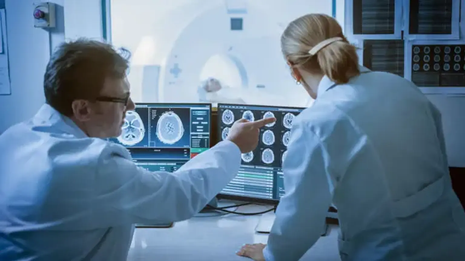
Brain MRI & MRA
Overview
An MRI and an MRA are both noninvasive and painless diagnostic methods for seeing tissues, bones, and organs within the body. An MRI (magnetic resonance imaging) is a type of imaging that produces detailed pictures of organs and tissues. The emphasis of an MRA (magnetic resonance angiography) is on the blood arteries rather than the surrounding tissue.
Stroke, temporal lobe epilepsy, infection, inflammation, tumor, multiple sclerosis (MS), dementia, post-trauma, metabolic disorders, congenital malformations, internal auditory canal pathology, vascular pathology, pituitary fossa pathology, nerve palsies, and metabolic disorders are all conditions for which brain MRI is recommended. Stenosis/occlusion associated to stroke, or carotid stenosis, Dissection (a rupture in the artery wall that can induce stroke), or AVM's when brain MRA is recommended (arteriovenous malformations).
Foreign bodies, mechanical heart valves, surgical implants, plates, screws, staples, and clips, and metal-containing prosthetics, pacemakers, cochlear implants, drug infusion ports, insulin pumps, deep-brain stimulators, and other electrical devices, metal tooth implants and fillings are all contraindications to using brain MRI and MRA.
