Introduction
Burn injuries can be devastating, leaving both physical and emotional scars. Severe burns often affect not only the skin but also the underlying tissues, requiring complex treatment. Burn reconstruction surgery plays a vital role in restoring a person’s appearance and function after significant burns. This surgical approach aims to improve the quality of life for burn survivors by addressing both aesthetic concerns and functional impairments.
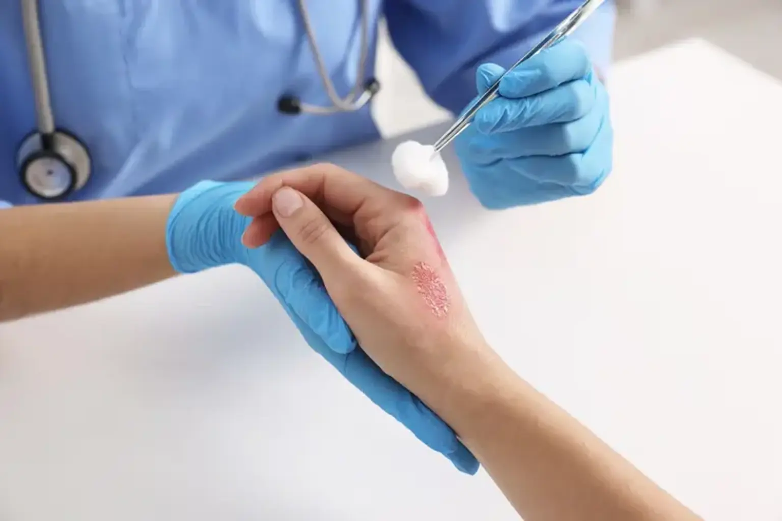
Burn reconstruction surgery is not just about cosmetic restoration; it's about helping patients regain confidence, mobility, and a sense of normalcy after traumatic burns. Whether the burns are from an accident, fire, or chemical exposure, reconstructive surgery can make a life-changing difference.
What is Burn Reconstruction Surgery?
Burn reconstruction surgery is a specialized form of plastic surgery designed to repair and reconstruct the skin and tissue after severe burn injuries. It focuses on restoring both the appearance and function of the affected areas, such as the face, hands, or joints. This surgery may involve techniques like skin grafts, tissue expansion, and flaps to rebuild the skin and restore movement.
Unlike initial burn treatments, which are aimed at stabilizing the injury and preventing infection, burn reconstruction is a long-term solution. The goal is to heal the body and address issues like scarring, loss of function, and cosmetic concerns caused by severe burns. This surgery helps reduce deformities, improve mobility, and enhance overall quality of life for burn survivors.
The Need for Burn Reconstruction
Severe burns, particularly third-degree burns, can leave lasting scars that affect a person’s mobility and appearance. These injuries may involve significant tissue damage that doesn’t heal properly on its own. In such cases, burn reconstruction surgery becomes essential. It helps in reconstructing areas where the skin has been damaged and where other treatments have failed to restore function or appearance.
The surgery is particularly necessary for burns that impact critical areas such as the face, hands, and joints, where skin and muscle damage can impair essential functions. Without reconstruction, scars could limit movement, cause deformities, or result in long-term disfigurement. For many burn survivors, reconstruction surgery offers a chance to regain independence and restore their self-esteem.
Types of Burns That Require Reconstruction Surgery
Burns are classified into three main types based on severity: first-degree, second-degree, and third-degree. While first and second-degree burns generally heal with little to no long-term effects, third-degree burns often require reconstructive surgery due to the extent of tissue damage.
First-degree burns: Affect only the outer layer of skin (epidermis) and usually heal within a week without scarring.
Second-degree burns: Affect the epidermis and part of the dermis. They cause blisters and may leave scars, but they can often heal with proper care.
Third-degree burns: Destroy both the epidermis and dermis, often extending to the deeper layers of the skin. These burns are the most severe and can result in permanent scarring and loss of skin function. Reconstruction surgery is often needed for these burns to restore function and appearance.
Reconstructive surgery is especially crucial for burns to sensitive areas such as the face, hands, and joints, where scarring could impede essential daily activities like eating, talking, or moving.
The Process of Burn Reconstruction Surgery
Burn reconstruction surgery begins with a thorough assessment of the patient's injuries. The surgeon evaluates the severity of the burns, the areas affected, and the overall health of the skin. Pre-surgical planning is essential to determine the best approach, including the use of skin grafts, tissue flaps, or other techniques to restore damaged skin.
The surgery typically takes place in stages, especially for severe burns. In some cases, skin grafts are used, where healthy skin is transplanted from another part of the body to cover the burn area. Other techniques include tissue expansion, where healthy skin is gradually stretched to create more tissue for grafting. Depending on the complexity, reconstructive surgery may involve multiple procedures over several months or even years to ensure optimal healing.
Techniques Used in Burn Reconstruction
There are several techniques used in burn reconstruction surgery, each chosen based on the type and location of the burn.
Skin Grafting: Skin grafts are the most common technique. A full-thickness or split-thickness graft may be taken from a donor site (often the thigh or abdomen) and applied to the burned area. Full-thickness grafts are used for deep burns, while split-thickness grafts are typically used for less severe burns.
Flap Surgery: In cases where more extensive tissue reconstruction is needed, flap surgery may be used. This involves moving healthy skin and tissue from one part of the body to another, along with the blood vessels that supply it.
Tissue Expansion: This technique involves using a tissue expander to stretch the skin over time, creating extra tissue that can later be used for grafting. This is often employed for burns on the scalp or other areas with limited skin availability.
These techniques are designed to improve both the function and appearance of the burned area, ensuring that the patient regains as much mobility and normal appearance as possible.
Risks and Complications of Burn Reconstruction Surgery
As with any major surgery, burn reconstruction carries some risks. While complications are relatively rare, it’s important for patients to understand what to expect.
Infection: The risk of infection is higher in burn victims due to compromised skin integrity. Surgeons take great care to sterilize the surgical area, and patients are typically prescribed antibiotics to minimize this risk.
Graft Rejection: In some cases, the body may not accept the transplanted skin, leading to graft rejection. This may require further procedures to replace or repair the graft.
Scarring: While burn reconstruction surgery is intended to minimize scarring, some level of scarring is inevitable. However, advanced techniques can help reduce visible scars, especially in critical areas like the face.
Decreased Mobility: In some cases, scar tissue can form in such a way that it limits movement, especially around joints. Physical therapy and regular follow-up appointments are critical to prevent this.
Understanding these risks helps patients prepare for the recovery process and make informed decisions about their treatment options.
Post-Surgical Care and Recovery
The recovery process after burn reconstruction surgery is gradual and requires ongoing care. After the surgery, the patient will need to follow specific wound care instructions to ensure proper healing. This includes keeping the area clean, avoiding excessive movement in the surgical site, and using ointments to prevent infections and promote skin healing.
Physical therapy is often recommended to help improve mobility, especially for burns that affect the hands, joints, or other areas with critical function. Stretching exercises and range-of-motion therapy are important to prevent contractures, where the skin tightens and restricts movement.
Pain management is another crucial aspect of recovery. Surgeons typically provide a combination of medications to manage pain and inflammation during the healing process. Swelling may also occur, especially in the first few weeks, but it typically subsides as healing progresses.
While the initial recovery may take several weeks, full recovery and the final results of the surgery can take several months to a year, depending on the severity of the burns and the complexity of the surgery. Regular follow-up visits with the surgeon are important to monitor progress and make adjustments to the recovery plan as necessary.
Expected Results of Burn Reconstruction Surgery
The outcomes of burn reconstruction surgery vary depending on the severity of the burns, the surgical techniques used, and the patient’s overall health. However, patients can typically expect significant improvements in both appearance and function.
Aesthetic Improvements: The primary goal of burn reconstruction is to reduce visible scarring and restore a more natural look. This can greatly enhance the patient's self-esteem and confidence. While scars may not disappear completely, advanced techniques such as skin grafts and flap surgery can minimize their visibility, especially on areas like the face, hands, and neck.
Functional Restoration: Rebuilding mobility and function is another key goal. Burns that affect joints or areas like the hands or feet may severely limit movement. Through reconstructive surgery, patients can regain a range of motion and functionality, which significantly enhances their ability to perform daily activities.
Recovery Timeline: While the skin and tissues may begin healing within weeks, the final results of burn reconstruction surgery can take months or even a year to fully emerge. Follow-up care is crucial during this period to monitor healing and optimize results.
Psychological and Emotional Impact of Burn Reconstruction Surgery
Burn injuries can be deeply traumatic, not only physically but emotionally as well. Many burn survivors struggle with body image issues, anxiety, and depression due to the scarring and disfigurement caused by severe burns. Burn reconstruction surgery plays a significant role in helping patients heal emotionally by restoring their appearance and confidence.
For many survivors, the ability to look in the mirror and see a less disfigured version of themselves can help reduce the emotional burden of burn injuries. Restoring function, such as regaining the ability to use their hands or facial muscles, can also have a profound impact on their psychological well-being.
While the physical results of surgery are important, the emotional recovery is equally critical. Counseling and support groups can complement the surgical process by providing patients with coping strategies and emotional support during their recovery.
Burn Reconstruction for Facial Burns
Facial burns are among the most emotionally and physically challenging injuries to treat. They affect both appearance and function, particularly around the eyes, mouth, and nose. Reconstructing facial burns requires a delicate and specialized approach due to the complexity of the facial muscles, skin, and nerves.
Advanced techniques such as microsurgery are often employed to reconstruct these areas. Microsurgical procedures allow for the precise transfer of tissue, helping to restore the skin’s function while minimizing scarring. The use of flaps and grafts can also improve facial appearance, helping to restore normal expression and movement.
In many cases, burn reconstruction surgery of the face not only addresses the functional aspects but also enhances emotional well-being, as the face is central to one’s self-image. For many burn survivors, the restoration of their facial appearance represents a major milestone in their recovery journey.
Burn Reconstruction Surgery for Hands and Extremities
Burns to the hands and extremities pose unique challenges, as they can severely limit mobility and functionality. The hands, for example, are crucial for everyday tasks such as eating, writing, and driving. When burns cause scarring or deformities in the hands or other extremities, burn reconstruction surgery can help restore both function and appearance.
Techniques such as skin grafting and flap surgery are used to replace damaged tissue. These procedures aim to preserve or restore the movement and dexterity of the hands and other affected areas. In some cases, reconstructive surgery may involve joint release or tendon repair to improve flexibility and function.
The recovery process is often longer for burns on hands and extremities because these areas are in constant use. However, the results of surgery can significantly improve a patient’s quality of life, allowing them to regain independence and return to normal activities.
Global Popularity and Access to Burn Reconstruction Surgery
Burn reconstruction surgery has become widely accessible worldwide, especially in developed countries with advanced healthcare systems. In regions with specialized burn centers, patients can access state-of-the-art treatments that offer significant recovery and rehabilitation.
However, the availability of these services can vary greatly depending on geographic location, healthcare infrastructure, and socioeconomic factors. In low-income areas or developing nations, access to burn reconstruction may be limited due to a lack of specialized surgeons, equipment, or medical facilities.
Global awareness of burn injury treatment has grown, leading to collaborations between international medical organizations and local hospitals to improve access to burn reconstruction, particularly in underserved regions.
Burn Reconstruction Surgery Costs
The cost of burn reconstruction surgery can be significant, depending on the complexity of the procedure, the number of surgeries required, and the type of hospital or clinic performing the surgery. Costs typically include surgeon’s fees, anesthesia, hospital stays, post-surgery care, and rehabilitation.
In high-income countries, the cost can range from several thousand to tens of thousands of dollars. However, many countries offer financial assistance, insurance coverage, or government subsidies to reduce the financial burden on patients.
In cases where reconstruction is deemed medically necessary, insurance may cover part of the procedure, but patients should verify coverage details with their insurance providers.
Advancements in Burn Reconstruction Surgery
Recent advancements in burn reconstruction surgery have significantly improved outcomes for burn survivors. Some notable innovations include:
3D Skin Printing: This technology is revolutionizing skin grafting by printing skin cells directly onto the burn site, potentially reducing rejection and promoting faster healing.
Tissue Engineering: Researchers are exploring lab-grown skin and synthetic grafts to improve tissue regeneration, reducing the need for donor site skin removal.
Stem Cell Therapy: Stem cells are being studied for their ability to regenerate skin cells, offering hope for quicker and more effective healing in the future.
These breakthroughs, along with improved surgical techniques and materials, are contributing to better functional and cosmetic results, reducing recovery time and enhancing patient satisfaction.
Choosing a Qualified Burn Reconstruction Surgeon
Choosing a qualified burn reconstruction surgeon is essential for the success of the procedure. Patients should seek surgeons who specialize in burn care and have experience with the specific techniques required for reconstructing burn injuries.
A good surgeon will thoroughly assess the patient's individual needs and develop a personalized treatment plan. It’s also important to ensure that the surgeon is board-certified and works in accredited facilities to guarantee safety and the highest standards of care.
Patients should feel comfortable discussing their goals, expectations, and any concerns with their surgeon. Seeking second opinions and reviewing before-and-after photos from past patients can help ensure the right choice of surgeon.
The Role of Physical Therapy in Burn Recovery
Physical therapy plays a critical role in the recovery process after burn reconstruction surgery. Burn injuries, especially those that affect joints, tendons, or muscles, can lead to stiffness and limited mobility. Physical therapy helps prevent complications like contractures, where skin tightens and restricts movement.
Therapists will work with patients to perform exercises that improve range of motion, flexibility, and strength. This may include stretching, strengthening, and functional exercises tailored to the patient’s needs. For burns that affect the hands or feet, specialized therapy can help patients regain fine motor skills and the ability to perform daily tasks.
Therapy is essential in ensuring that the physical function of the affected areas is fully restored and to reduce the risk of long-term disability.
Burn Reconstruction and Self-Esteem
The emotional and psychological impact of burn injuries can be profound, often leading to decreased self-esteem and body image concerns. Burn reconstruction surgery provides not only physical healing but also significant emotional relief.
Restoring a more natural appearance can help patients regain their confidence, allowing them to feel more comfortable in social and professional settings. For many burn survivors, seeing their reflection improve after surgery is a major milestone in their emotional recovery.
Support groups, counseling, and therapy can also play a role in boosting self-esteem, as emotional recovery often complements the physical recovery process.
Support Systems and Resources for Burn Survivors
Burn survivors often face significant challenges during their recovery journey, both physically and emotionally. Having a strong support system is vital to overcoming these hurdles. Family, friends, and healthcare providers can offer invaluable support by encouraging rehabilitation, providing emotional comfort, and assisting with daily tasks during recovery.
In addition to personal support, many organizations and charities provide resources for burn survivors. These include financial aid, support groups, and counseling services. National burn associations, such as the American Burn Association (ABA), offer information, resources, and connect survivors with community support.
Such networks can help survivors navigate the complexities of burn recovery and ensure they have the emotional and practical support they need to heal.
Frequently Asked Questions (FAQs) About Burn Reconstruction Surgery
1. How soon can I get burn reconstruction surgery after my injury? Reconstruction surgery typically takes place after the initial burn treatment has stabilized the injury, which can take weeks to months. Surgeons will assess when it’s safe to proceed based on healing progress.
2. Will I experience pain after the surgery? While some pain is expected after any surgery, modern pain management techniques help minimize discomfort. Most patients recover well with appropriate pain control during the healing period.
3. How long is the recovery period? Recovery time varies based on the severity of the burns and the surgery. Initial healing can take weeks, but full recovery, including functional restoration and scar maturation, can take months to a year.
4. Will my insurance cover the surgery? Insurance coverage varies by plan and location. Many insurance policies cover reconstructive surgery when it is medically necessary. Patients should check with their insurance provider to understand the details of their coverage.
5. Are there alternatives to surgery for burn reconstruction? Non-surgical options, such as pressure garments, silicone gel sheets, and steroid injections, can help reduce scarring and improve the appearance of burns. However, surgery is often the most effective method for functional and aesthetic restoration of severe burns.
Conclusion
Burn reconstruction surgery is a crucial procedure that not only helps restore the physical appearance of burn survivors but also significantly improves their functionality and quality of life. The advancements in surgical techniques, such as skin grafting, tissue flaps, and innovative therapies like 3D skin printing and stem cell research, are transforming the outcomes for patients, offering them better cosmetic results and faster recovery times.
While the surgery can be complex and may require multiple procedures over time, the emotional and physical benefits are profound. Burn reconstruction offers survivors the chance to regain mobility, confidence, and a sense of normalcy, with ongoing support systems and rehabilitation playing essential roles in recovery.
For individuals considering this surgery, choosing a qualified, experienced burn reconstruction surgeon is critical to achieving the best outcomes. With continued advancements in burn care and increased accessibility worldwide, burn reconstruction surgery is making a positive difference in the lives of countless individuals, offering them a path to healing, hope, and recovery.