Caesarean Section

Last updated date: 05-May-2023

Originally Written in English

Caesarean Section

Overview

Caesarean section, often known as C-section or caesarean birth, is a surgical technique in which one or more infants are born through an incision in the mother's belly, which is frequently used because vaginal delivery might endanger the baby or mother. Obstructed labor, twin pregnancy, high blood pressure in the mother, breech birth, and issues with the placenta or umbilical cord are all reasons for the procedure.

The curvature of the mother's pelvis or the history of a previous C-section may necessitate a caesarean birth. It is feasible to try vaginal delivery after a C-section. The World Health Organization advises against performing caesarean sections unless absolutely required. Some C-sections are performed without a medical cause, at the request of someone, most often the mother.
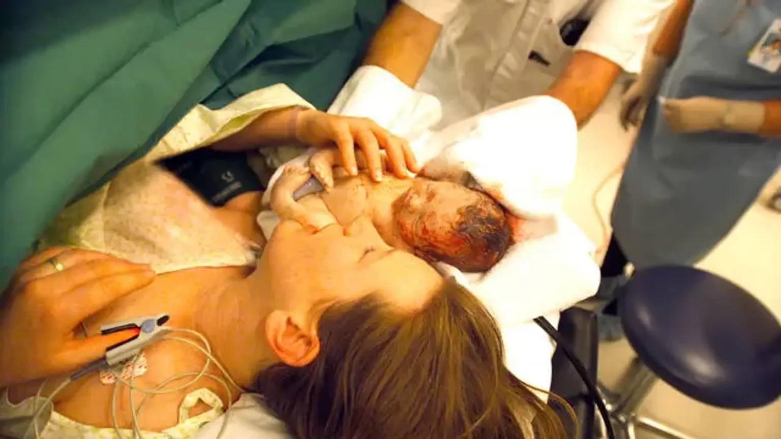
 

Caesarean section definition

Caesarean section definition

Cesarean section is a method of fetal birth that involves an open abdominal incision (laparotomy) and a uterine incision (hysterotomy). The first reported cesarean section took place in 1020 AD, and the process has progressed greatly since then. It is presently the most often performed procedure in the United States, with over 1 million women born via cesarean section each year. Cesarean delivery rates increased from 5% in 1970 to 31.9 percent in 2016.

Despite ongoing attempts to lower the number of cesarean sections, experts do not expect a major decline for at least a decade or two. While there are risks of both immediate and long-term consequences, cesarean birth may be the safest or perhaps the only route for some women to deliver a healthy infant.


Anatomy and Physiology

To perform a cesarean birth, the surgeon must cut through all of the layers that separate him or her from the fetus. The epidermis is incised first, then the subcutaneous tissues. The fascia that covers the rectus abdominis muscles is the next layer. The anterior abdominal fascia is normally divided into two layers.

The first is made up of aponeurosis from the external oblique rectus muscle, while the second is a fused layer made up of aponeuroses from the transverse abdominis and internal oblique muscles. The surgeon enters the abdominal cavity through the parietal peritoneum after separating the rectus muscles, which run from cephalad to caudal.

Unlike in a nongravid patient, the uterus is frequently seen at this location immediately following entrance into the abdomen in a gravid lady. If the patient has an adhesive condition from previous operations, the surgeon may find adhesions including tissues such as the omentum, colon, anterior abdominal wall, bladder, and anterior part of the uterus.

After locating the uterus, the surgeon can locate the vesicouterine peritoneum, also known as the vesicouterine serosa, which joins the bladder and the uterus. If the surgeon wishes to create a bladder flap, the vesicouterine peritoneum must be incised. The bladder may become harder to separate from the uterus in a patient who has had previous cesarean procedures.

The uterus is made up of three layers: the serosal outer layer (perimetrium), the muscle layer (myometrium), and the inner mucosal layer (endometrium). To create the uterine incision or hysterotomy, all three of these layers are incised. The uterine vessels run along the lateral aspects of the uterus on both sides, and care must be given to prevent harming these blood vessels when the uterine incision is created or expanded. The uterine arteries originate from the anterior division of the internal iliac artery.

During pregnancy, blood flow through these arteries is eight times quicker, with a unilateral flow of over 300 milliliters per minute at 36 weeks. The uterine arteries enter the uterus through the cardinal ligament after crossing the ureters anteriorly. The uterine arteries connect with the ovarian arteries, which emerge from the abdominal aorta, in the wide ligament.

Depending on the condition of the patient's amniotic membranes (whether her "water is broken" or intact), the surgeon may come across an amniotic sac during uterine incision. The amniotic sac is made up of two layers, the chorion and the amnion, which join together early in pregnancy. If present, the amniotic sac would be the final barrier between the surgeon and the fetus. The fetus is delivered at this moment, completing the primary purpose of the cesarean surgery.

The gravid uterus frequently obscures the remaining female reproductive anatomy. However, once the fetus is delivered, other structures may become apparent, especially if the surgeon exteriorizes the uterus for repair. The Fallopian tubes and ovaries may be appreciated by the surgeon, and tubal ligation may be performed if the patient has already indicated a desire and provided informed permission for this kind of contraception.

The wide ligament can also be seen. This structure is made up of two peritoneum leaves and connects the uterus to the pelvic sidewalls. If the medial leaf of the wide ligament is opened, the ureter can be found coursing. The cervix, which is positioned in the lowest region of the uterus, and the vagina are not normally visible.


Indications

Indications of Caesarean Section

A fetus cannot or should not be delivered vaginally for a variety of reasons. Some of these reasons are rigid, since a vaginal birth would be risky in certain clinical settings. For example, if the patient has a previous classical cesarean scar or uterine rupture, a cesarean delivery is frequently recommended. However, because of the possible risks of cesarean birth, significant research has been conducted in order to find techniques to minimize the cesarean rate.

There has been a focus on reducing the number of first-time cesareans, as many women who have one cesarean delivery will have the rest of their children delivered through cesarean. She may opt for another cesarean for a variety of reasons, or she may be ineligible for a repeat vaginal birth. For example, if the patient has an unfavorable cervix at term, cervical softening with drugs such as misoprostol is not advised due to an increased risk of uterine rupture.

Maternal Indications for Cesarean 

  • Prior cesarean delivery
  • Maternal request
  • Pelvic deformity or cephalopelvic disproportion
  • Previous perineal trauma
  • Prior pelvic or anal/rectal reconstructive surgery
  • Herpes simplex or HIV infection
  • Cardiac or pulmonary disease
  • Cerebral aneurysm or arteriovenous malformation
  • Pathology requiring concurrent intraabdominal surgery
  • Perimortem cesarean

Uterine/Anatomic Indications for Cesarean  

  • Abnormal placentation (such as placenta previa, placenta accreta)
  • Placental abruption
  • Prior classical hysterotomy
  • Prior full-thickness myomectomy
  • History of uterine incision dehiscence
  • Invasive cervical cancer
  • Prior trachelectomy
  • Genital tract obstructive mass
  • Permanent cerclage

Fetal Indications for Cesarean  

  • Nonreassuring fetal status (such as abnormal umbilical cord Doppler study) or abnormal fetal heart tracing
  • Umbilical cord prolapse
  • Failed operative vaginal delivery
  • Malpresentation
  • Macrosomia
  • Congenital anomaly
  • Thrombocytopenia
  • Prior neonatal birth trauma


How many c sections can you have?

Every patient is unique, as is every scenario. However, based on current medical research, most medical authorities advise that if several C-sections are planned, the expert guideline is to keep the total number of C-sections to three.


Contraindications

Contraindications for Caesarean Section

There has been a focus on reducing the number of first-time cesareans, as many women who have one cesarean delivery will have the rest of their children delivered through cesarean. She may opt for another cesarean for a variety of reasons, or she may be ineligible for a repeat vaginal birth. For example, if the patient has an unfavorable cervix at term, cervical softening with drugs such as misoprostol is not advised due to an increased risk of uterine rupture.

In certain clinical situations, a cesarean birth may not be the best option. Consider the following related contraindications. A pregnant patient, for example, may have significant coagulopathy, making surgery highly risky. Vaginal delivery may be desirable in this instance.

A patient with a long history of abdominal surgery, on the other hand, may be an unsuitable surgical candidate. In the event of fetal death, conducting a cesarean exposes the pregnant patient to the hazards of the procedure while providing no benefit to the fetus. The same principles apply if the fetus has serious defects that make life impossible.


Preparation

Patients and their partners should be educated about the likelihood of cesarean birth as part of their prenatal care. Before, during, and after the surgery, the patient should be informed of what to expect. If a cesarean section is planned, maybe owing to maternal or fetal difficulties, any maternal comorbidities (anemia, diabetes, hypertension, obesity) should be optimized preoperatively if feasible.

With cesarean birth, there is a risk of aspiration and subsequent pneumonitis. To avoid low stomach pH, antacids (sodium citrate) and a histamine H2 antagonist can be given before surgery. When it comes to fasting, it is customary to ask a patient to be "NPO after midnight." It is usual to ask a stable patient who has had an unplanned cesarean to fast for 6 hours.

Patients should be advised to consume clear liquids until 2 hours before surgery, and solid meals should be avoided for 6 hours beforehand. Additionally, nondiabetic patients may be given carbohydrate fluid supplementation up to 2 hours before surgery, which may enhance patient outcomes.

Patients should be instructed to drink clear liquids till 2 hours before surgery and to avoid substantial meals for 6 hours prior. Nondiabetic patients may also be given carbohydrate fluid supplementation up to 2 hours before surgery, which may improve patient outcomes.

As with any operation, there is a risk of infection with a cesarean section. Because the uterus, cervix, and vagina are all continuous, it is termed a clean-contaminated surgical wound. The most major risk factor for a woman having an infection in the postpartum period is a cesarean section. Women who have a cesarean delivery are 20 times more likely to get an infection than women who have a vaginal delivery.

Antibiotic prophylaxis can reduce the risk of infection following a cesarean section by 60% to 70%. Antibiotics should be administered prior to surgery rather than after the umbilical cord is clamped. The antibiotic used is determined by the clinical situation and if the patient has any allergies. Antibiotics should be effective against both gram-positive and gram-negative bacteria, as well as some anaerobes.

For women weighing less than 80 kg, a single intravenous dosage of 1 g of cephazolin is normal, and the amount is increased to 2 g for patients weighing 80 kg or more. For women weighing 120 kg or more, the dose of cephazolin should be increased to 3 g to obtain appropriate tissue concentrations of the antibiotic.

Prophylaxis with clindamycin 900 mg and an aminoglycoside 5 mg/kg is indicated for individuals who have a contraindication to cephazolin, such as a serious allergy. If you have urticaria, respiratory discomfort, angioedema, or anaphylaxis, you may have an allergy. In individuals with a history of methicillin-resistant Staphylococcus aureus, a single dosage of vancomycin is indicated.

Because of the nature of cesarean sections, infection risk is provided by both vaginal and skin flora. Women who have a cesarean section following labor or a rupture of membranes are more likely to be exposed to vaginal germs. More recent research has discovered that adding 500mg of azithromycin intravenously to standard antibiotic prophylaxis is useful for lowering infectious morbidity in these women.

Topical treatments have also been used to reduce infection after cesarean delivery. Both topical povidone-iodine and chlorhexidine have been shown to be beneficial in the preparation of abdominal skin. Although research is varied and often of low quality, there may be some indication that chlorhexidine is preferable to povidone-iodine in terms of infection reduction. Because the data is ambiguous, both ways are appropriate.


How does cesarean section work?

Cesarean section technique

Cesarean section is a difficult technique. Appropriate tissue handling, appropriate hemostasis, avoiding tissue ischemia, and minimizing infection are all critical for wound healing and preventing adhesion development. Several approaches are available throughout the procedure at each phase or tissue layer. Many elements influence a surgeon's method selections. It is suggested, as with any area of medical practice, to base such judgments on evidence.


Cesarean section types

  • Pfannenstiel-Kerr method
  • Joel-Cohen method
  • Misgav-Ladach method
  • Modified Misgav-Ladach method

The pubic hair might be removed or not before a cesarean section. Those who support hair removal believe that it reduces surgical site contamination and infection. A Cochrane review, on the other hand, found no evidence of decreasing infection rates after hair removal. As a result, hair removal should be done only if it improves visibility. Hair removal should be done using clippers rather than razors. Patients should also be discouraged from shaving their pubic area as their due dates or cesarean dates approach. Shaving with a razor can produce small skin fractures, which are linked to more surgery site infections than clipping.

The initial skin incision might be either suprapubic transverse or midline vertical. A vertical midline incision is thought to allow speedier access to the abdominal cavity and destroys fewer tissue layers and arteries, making it the preferable procedure for performing an emergency cesarean section. A vertical incision may also allow viewing away from known severe sticky disease. A vertical incision may allow additional surgical exposure and access to the hypogastric arteries during a planned cesarean hysterectomy for a morbidly adhered placenta.

A transverse skin incision, on the other hand, is the most widely utilized and preferred in most circumstances due to enhanced wound healing and patient acceptability. Because most doctors are more comfortable with low-transverse cesarean entry, this method is frequently used even in emergency situations. A low transverse incision might be used to perform unplanned cesarean hysterectomies. Patient habituation may cause some surgeons to put a transverse skin incision higher on the belly rather than beneath the pannus, however research on this approach is not conclusive.

A Pfannenstiel skin incision is slightly curved and 2 to 3 millimeters or 2 fingerbreadths above the symphysis pubis. The incision's midpoint lies within the hair-bearing region of the mons. In this instance, the hair should be removed. In contrast, a Joel-Cohen incision is straight rather than curved. It's 3 cm lower than the line joining the anterior superior iliac spines, therefore it's more cephalad than a Pfannenstiel skin incision.

The subcutaneous layer is the next to be dissected, and it can be done bluntly or aggressively. Because blood arteries run through this layer, it is important to reduce blood loss by confining sharp dissection to the midline until the fascia is reached, then dissecting laterally bluntly. If blood arteries are transected, prudent use of cautery can maintain hemostasis.

The fascia is next incised in the midline with the scalpel and stretched laterally, either sharply or bluntly. After that, the fascia may be separated from the underlying rectus muscles. To perform this dissection, both the superior and inferior parts of the fascia are progressively held using a clamp (such as a Kocher), and dissection can be performed using a mix of blunt and sharp techniques, such as scissors or cautery.

It is critical not to injure the underlying rectus muscles. However, in rare clinical situations, the rectus muscles may be purposefully severed to provide for greater surgical access.

After separating the rectus muscles in the midline, the peritoneum is opened to gain access to the abdominal cavity. This might be done sharply or brutally by the surgeon. Sharp entry should be used with caution to avoid harm to underlying tissues such as the bowel. Once the peritoneal incision is made, it is frequently expanded bluntly. Precautions must be taken to avoid harm to the bladder during the expansion of the peritoneal incision.


Complications

Complications of Cesarean section

In the United States, the maternal death rate is roughly 2.2 per 100,000 cesarean births. Though this is modest in general, it is much higher than with vaginal birth. The maternal mortality rate for vaginal delivery is about 0.2 per 100,000.

There is a danger of severe bleeding during and after a cesarean section, like with any delivery and surgery in general. Hemorrhage is the main cause of significant maternal morbidity in the United States. Certain pre-cesarean circumstances, such as protracted labor, fetal macrosomia, or polyhydramnios, may increase the chance of uterine atony and subsequent bleeding. Excessive blood loss may also result from intraoperative situations such as the requirement for considerable adhesiolysis or the expansion of the hysterotomy laterally into the uterine arteries.

Hemorrhage during birth may necessitate the use of blood products, which has its own set of difficulties. Sheehan syndrome is a well-known consequence of postpartum hemorrhage. Obstetric hemorrhage accounts for around 10% of maternal mortality in the United States.

As previously stated, there is a high risk of infection following cesarean birth. Other typical consequences following a cesarean section include wound infection and endometritis, in addition to postpartum bleeding. Postoperative endometritis was decreased from 8.7 percent to 3.8 percent in a research investigating the effectiveness of vaginal washing.

In elective repeat cesarean births, the total risk of infectious morbidity was 3.2 percent, compared to 4.6 percent in women having a trial of labor. Elective repeat cesareans had a blood transfusion rate of 0.46 percent, a surgical injury rate of 0.3 percent to 0.6 percent, and a hysterectomy rate of 0.16 percent, according to the same data. Thromboembolism and anesthesia problems are also possibilities.

While cesarean surgery is safer for the fetus, there are dangers associated with fetal birth in this manner. Fetal trauma during cesarean section is estimated to be 1%, including skin laceration, clavicle or skull fracture, facial or brachial plexus nerve injury, and cephalohematoma. Overall, these hazards are smaller than those associated with vaginal births.


In terms of the neonate, those born through cesarean had a higher risk of respiratory issues as well as higher incidence of asthma and allergy than those born vaginally. Cesarean delivery has long-term hazards, both to the patient and to her subsequent pregnancies, in addition to short-term and surgical complications. As previously indicated, a vertical scar on the uterus necessitates cesarean delivery for subsequent pregnancies.

The surgical risks rise in tandem with the frequency of cesarean sections performed. Adhesion development can complicate subsequent cesareans and raise the risk of unintentional harm.

With each consecutive procedure, the risks of improper placentation grow. The risk of placenta accreta is 0.3 percent in women who have had one cesarean section, but it rises to 6.74 percent in women who have had five or more cesarean births. If a hysterectomy is required, a morbidly attached placenta increases the risk of substantial bleeding and possibly loss of fertility.


Conclusion 

Every year, around 1.3 million women in the United States have a cesarean section. It is the most frequent operation performed in the United States. Medicine has come a long way since then, and it continues to do so. Understanding the risks and advantages of a cesarean section can help a professional to correctly counsel a pregnant patient. Clinicians will be able to give the greatest treatment and results if they have a solid grasp of evidence-based medicine. 

Patients may desire a cesarean delivery for no apparent reason, and doctors should be prepared to give the extensive information required in these circumstances to ensure the patient is making an educated decision. There is growing pressure to decrease cesarean section rates, so a proper understanding of the indications will assist clinicians in decision-making.