Carcinomatosis
Overview
Carcinomatosis is defined as a situation in which many carcinomas form at the same time, generally as a result of secondary spread from a primary source. It denotes more than just illness spread to regional nodes, and even more than simply metastatic disease. The word is commonly understood to signify that there are several secondary schools in various locations.
Several gastrointestinal and gynecological cancers have the ability to spread and develop in the peritoneal cavity. Peritoneal carcinomatosis (PC) has been proven to significantly reduce overall survival in patients with gastrointestinal cancer liver and/or extraperitoneal metastases.
Understanding of the biology and pathways of dissemination of tumors with intraperitoneal spread, as well as the protective function of the peritoneal barrier against tumoral seeding, over the last three decades, has prompted the concept that PC is a loco-regional disease: in the absence of other systemic metastases, multimodal approaches combining aggressive cytoreductive surgery, intraperitoneal hyperthermic chemotherapy, and systemic chemotherapy
Carcinomatosis definition
Carcinosis, also known as carcinomatosis, is a kind of disseminated cancer that can occur in general or in particular patterns of dissemination. Carcinomatosis is frequently limited to cancers of epithelial origin, such as adenocarcinomas, whereas sarcomatosis describes the spread of tumors of mesenchymal origin, such as sarcomas.
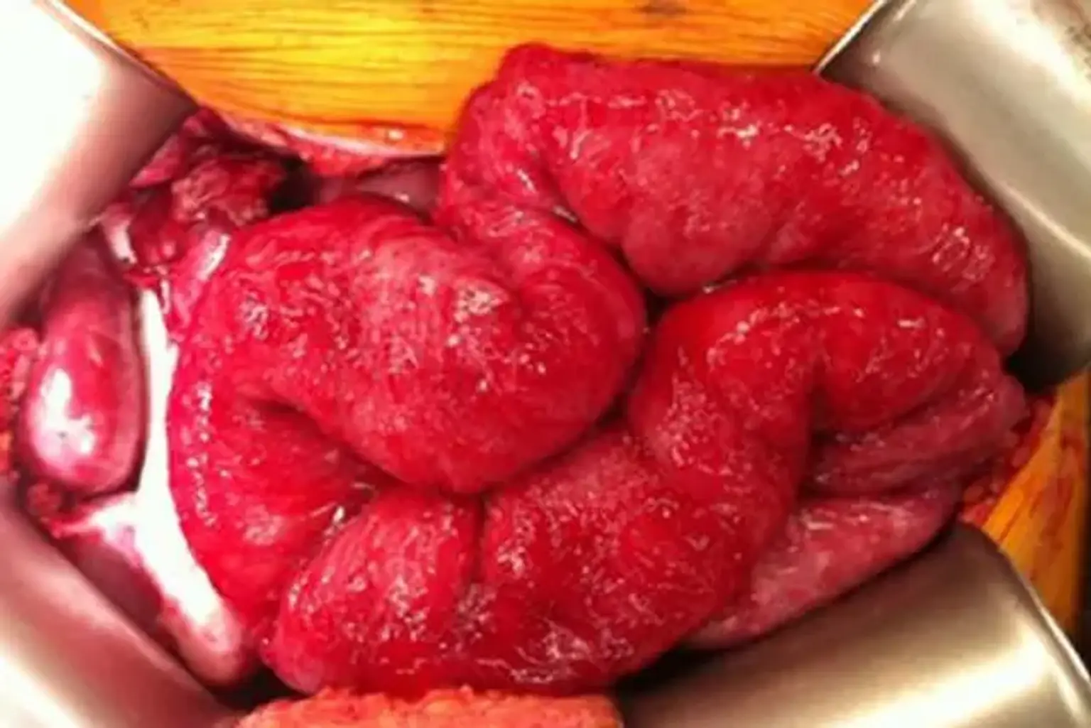
Peritoneal Carcinomatosis (PC) is a late-stage symptom of a number of gastrointestinal cancers that is distinguished by tumor deposition over the peritoneal surface. In its early stages, this can be completely asymptomatic, but as the disease advances, symptoms such as nausea, diarrhea, stomach discomfort, bloating, and weight loss may occur. When ascites or intestinal obstruction develop, the illness is commonly identified, and it is associated with a higher tumor load that is more difficult to treat. Early PC identification while the tumor load is low may improve the efficacy of existing treatment approaches.
Types of carcinomatosis
Leptomeningeal carcinomatosis
- Involvement of leptomeninges by cerebrospinal fluid seeding, which can occur either directly or indirectly through the circulation.
- It is a rare and generally late-stage cancer consequence.
- Any cancer can cause this, although adenocarcinomas are the most prevalent. One of the most prevalent reasons is breast cancer.
- The symptoms might be nonspecific (for example, headache or confusion) or focal or multifocal neurological impairments (for example, cranial nerve palsies).
- A thorough history and neurological examination, as well as CSF fluid cytology and neuroimaging, are used to make a diagnosis (preferably gadolinium-enhanced MRI).
Pulmonary lymphangitic carcinomatosis
- Diffuse pulmonary infiltration with lymphatic channel blockage.
- It may happen with a number of malignancies, the most prevalent being breast, stomach, pancreatic, lung, and prostate.
- Lymphangitic carcinomatosis can potentially impact the kidneys, causing acute renal injury (AKI).
Peritoneal carcinomatosis
- Metastases spreading into the peritoneum, most commonly from gynaecological (typically ovarian) and gastrointestinal malignancies.
- Peritoneal carcinomatosis has been proven to dramatically reduce overall survival in individuals with gastrointestinal cancer liver and/or extraperitoneal metastases.
Peritoneal Carcinomatosis
Peritoneal carcinomatosis is a rare type of cancer that affects the peritoneum, which is the thin membrane that surrounds your abdominal organs. It might be difficult to learn that you or a loved one has it, but knowing this cancer can help you feel more in control.
Peritoneal carcinomatosis often arises when existing abdominal cancers extend to the peritoneum, resulting in many new tumors on the membrane's surface.
If you have peritoneal carcinomatosis, it usually signifies that your abdominal cancer has progressed to an advanced stage. Primary peritoneal carcinomatosis, which develops in the peritoneum, is likewise quite rare. These situations are more common in women who are predisposed to ovarian cancer.
Risk Factors for Peritoneal Carcinomatosis
Because peritoneal carcinomatosis is most commonly caused by the spread of other malignancies, the greatest risk factor is having other advanced tumors, such as:
- Appendix cancer
- Colon cancer
- Rectal cancer
- Pancreatic cancer
- Gastric cancer
Primary peritoneal carcinomatosis happens almost always in women. Apart from gender, other risk factors for primary peritoneal carcinomatosis include:
- Age
- Family history of ovarian or peritoneal cancer
- BRCA genetic mutations
- Hormone replacement therapy
- Obesity
- Endometriosis
Treatments for Peritoneal Carcinomatosis
Peritoneal carcinomatosis can be difficult to treat since it is generally an advanced type of invasive cancer that has migrated from another tumor. Most peritoneal carcinomatosis tumors do not respond well, if at all, to chemotherapy. As a result, many doctors concentrate on palliative care to control symptoms, relieve discomfort, and enhance your quality of life. There are specialists that specialize in palliative care, which is available to everyone suffering from a terrible disease. If end-of-life worries become a need, hospice care is an alternative.
Depending on your particular case, other treatment options may also be available.
- Cytoreductive surgery. A surgeon removes any tumors on the peritoneum and, in some cases, nearby abdominal organs.
- Hyperthermic intraperitoneal chemotherapy. This procedure, which is frequently employed immediately following cytoreductive surgery, bathes the interior of your abdomen, where your peritoneum lies, with hot chemotherapeutic chemicals in order to destroy any leftover cancer cells.
- A peritonectomy is surgery to remove your peritoneum.
Presentation
Carcinomatosis might be a symptom of another illness. It might be the recurring presentation or the principal presenting element. The presentation will vary depending on the area impacted.
- In the lungs: shortness of breath and haemoptysis.
- In the liver: jaundice.
- In the brain there: headaches, vomiting and focal neurological features.
- In bones: pain or pathological fracture.
- Peritoneal involvement may present with varying symptoms including ascites, pain, nausea, cachexia and bowel obstruction.
Diagnosis
The purpose of investigations is to confirm the nature of the disease and to assess its severity and extent.
- Blood tests: In situations with unclear primary, FBC may indicate iron shortage, indicating gastrointestinal malignancy, microscopic haematuria, indicating occult genitourinary cancer, and occult blood, indicating a colorectal origin. FBC, U&E, creatinine, and LFTs may suggest severity in circumstances when the main is known.
- Imaging: Although an exploratory laparotomy is occasionally necessary for peritoneal carcinomatosis, modern imaging techniques such as ultrasound, CT, and MRI scanning, as well as older investigations such as CXR, give extremely useful information. As previously stated, gadolinium-enhanced MRI is the first-choice examination for suspected leptomeningeal involvement.
- Biopsy: Obtaining tissue for histology may be beneficial. A transbronchial biopsy is used to confirm the diagnosis of pulmonary lymphangitic carcinomatosis. Histological examination aids in management decision-making and is one of the aspects to examine when determining if aggressive treatment with surgery and/or chemotherapy is necessary. The advancement of molecular pathology has enabled it to make major contributions to management choices.
Imaging
Traditional imaging modalities, such as CT and MRI, are insensitive to detecting and estimating disease load in PC. Classic computed tomography (CT) symptoms of PC, such as "omental caking," omentum thickening, and peritoneal nodules, are rare radiographic findings in "early" disease stages. Several investigations aimed at determining the sensitivity and accuracy of CT scans in measuring tumor burden in PC discovered that CT scans dramatically overestimated the amount of disease present in the peritoneal cavity.
The sensitivity for CT identification of tumor nodules smaller than 0.5 cm and 1cm was found to be 11% and 25-50%, respectively. This is especially true in colorectal PC, where PCI is a key driver of full cytoreduction and long-term results. In a research focusing on CRC PC, Koh et al discovered that CT dramatically underestimated clinical PCI.
In fact, the sensitivity of small bowel involvement ranged from 8 to 17 percent in each location. Despite this, the Fifth International Workshop on Peritoneal Surface Malignancy, held in Milan, selected CT as the primary imaging modality for determining CRS appropriateness. Ultrasound also has very limited sensitivity for detecting PC nodules .
Magnetic resonance imaging (MRI), particularly diffusion weighted images, has been shown in prospective studies to improve the identification of carcinomatosis in certain locations of the abdomen. This, however, has limitations owing to peristalsis motion artifacts, expense, the necessity for radiologists skilled in their interpretation, and inter-observer variance.
Furthermore, while positron emission technology (PET) may have greater sensitivity, its limits and lack of additional therapeutic benefit sometimes restrict its use for assessing resectability. These challenges, particularly the low sensitivity, lack of meaningful clinical association, and high cost, restrict the value of non-invasive imaging in the early identification of PC. With today's technologies, laparoscopy or exploratory surgery is frequently required to confirm the diagnosis and degree of PC.
Differential diagnosis
When these symptoms appear, the question is whether they are due to a recognized disease or something else. For example, if jaundice is caused by metastatic liver cancer or gallstones.
When carcinomatosis is the presenting characteristic, imaging and histology are usually used to look for a primary tumor.
Management
Because this illness can be caused by a variety of etiologies, there are different management strategies available, some of which may be tailored to a specific malignancy. In many situations, there is no realistic prospect of curative therapy, while chemotherapy and radiation may provide some relief. Surgery might be palliative, and 'debulking' the tumor before chemotherapy could be beneficial. In a small number of cases, resection of liver metastases caused by colon cancer has been successful. There are certain patient subgroups who respond favorably to therapy.
Although the prognosis remains poor, multimodality treatment (palliation with surgery, radiation, and/or chemotherapy, delivered systemically or directly into the cerebral spinal fluid) may be employed in individuals with leptomeningeal metastases related to breast cancer. In rare circumstances, a multimodality strategy (cytoreduction surgery followed by hyperthermic intraperitoneal chemotherapy) is employed in persons with peritoneal carcinomatosis and can dramatically improve prognosis.
Chemotherapy
In certain circumstances, peritoneal carcinomatosis can be treated with intraperitoneal and/or intravenous chemotherapy. Treatment might begin postoperatively, or chemotherapy medications can be administered intravenously during or after surgery. Chemotherapy is more commonly used in ovarian cancer with peritoneal dissemination than in other primary, and the prognosis is generally greatly better.
Intrathecal trastuzumab may be a viable therapy option for HER2-positive breast cancer patients with leptomeningeal involvement in certain situations, although it is currently being studied. Methotrexate (intra-CSF) is a more well-established chemotherapeutic treatment for individuals with leptomeningeal malignancy.
Embolisation
Transcatheter arterial chemoembolisation (TACE) has been shown to be effective in patients with neuroendocrine tumors and colorectal metastases. It is increasingly being utilized in the treatment of malignant liver tumors. A microcatheter is introduced into the hepatic blood supply, and a mixture of chemotherapeutic and embolic drugs is administered. Radio-embolisation has the potential to play a growing role in the treatment and management of metastatic illness, but further studies, particularly comparisons to chemotherapy regimens, are required.
Radiotherapy
- Palliative radiotherapy can often be used to:
- Reduce or eliminate pain from bone metastases.
- Palliate brain metastases.
- Relieve spinal cord compression or compressive symptoms from visceral metastases (eg, airway or gastrointestinal obstruction).
- Control bleeding - eg, haemoptysis or haematuria.
Various ablative treatments, such as freezing, microwaves, lasers, and the use of radiofrequency alternating current, have been utilized to remove liver metastases.
Surgery
- Although palliative surgery for malignant intestinal obstruction caused by carcinomatosis can assist patients, it comes at a significant cost in terms of mortality and morbidity in comparison to the patient's remaining survival time.
- Current evidence on the efficacy of cytoreduction surgery (CRS) followed by hyperthermic intraoperative peritoneal chemotherapy (HIPEC) for peritoneal carcinomatosis indicates some improvement in survival for selected patients with colorectal metastases, but evidence for other types of cancer is limited or ambiguous.
- Surgical treatment of certain bone metastases may have a role in improving life expectancy and quality of life.
In the case of incurable patients, a candid and open dialogue is required. This may necessitate more than one session, and the ability to deliver terrible news is essential. Other factors to consider are dying at home and dyspnea in palliative care. Pain control in terminal care, as well as nausea and vomiting in palliative care, may merit consideration.
A multidisciplinary team approach is likely to be required for good palliative care. Early referral to a palliative care team is critical for specialized assistance with symptom management and emotional issues for patients and family members.
Peritoneal carcinomatosis gastric cancer
The two most critical variables influencing prognosis in gastric cancer are penetration of the gastric serosa and lymphatic dissemination (GC). When a tumor infiltrates the stomach serosa, PC becomes highly common. As a result, despite drastic surgery, up to half of patients with advanced gastric cancer (AGC) will develop peritoneal carcinomatosis, and PC is relatively frequent in gastric cancer, being present in 5% -20% of patients examined for possibly curative resection.
There are numerous approaches with varying sensitivity for identifying the presence of free peritoneal tumor cells (FPTC). FPTC in the washing might be found in up to 24% of stage IB GC patients and up to 40% of stage II or III GC patients. Furthermore, after radical resection, the peritoneum is the sole site of recurrence in 10% - 34% of patients, and one of the recurrence sites in 29% - 44% of cases.
Peritoneal carcinomatosis ovarian cancer
The treatment plan for advanced EOC is now widely acknowledged as a mix of maximum CRS and adjuvant chemotherapy, even in situations with severely peritoneal widespread disease. Grades IIIC and IV are no longer thought to be "missing." Several studies have shown that a progressively more aggressive surgical effort is related with higher disease-free and overall survival rates.
It is proposed that aggressive surgery be conducted at specialized centers with a high number of cases, which offer much lower in-hospital mortality than low volume clinics. The more radical the surgeon becomes and increases his/her surgical volume, the longer the disease-free and overall survival and the lower the in-hospital mortality. On the other hand, tumor biology and early disease dissemination have been proposed as the most essential determinants in the survival benefit of surgery.
It's still unclear how the tumor's inherent properties make intra-abdominal implants simpler to remove. Upper abdominal tumor implants, in general, indicate aggressive tumor biology. Covens and Berman were critical of CRS's position in advanced EOC. They hypothesized that tumor biology, rather than the surgeon's operating effort, determines both survival and surgical resectability.
A retrospective evaluation of data from the Scottish Randomized Trial in Ovarian Cancer found that the efficacy of optimum debulking surgery seemed to rely on the amount of disease prior to surgery in a population of 889 patients with disease stages ranging from IC to IV. A study of 456 women with advanced stage III/IV ovarian cancer found no link between nodal status and survival. Furthermore, nodal status was not a predictive factor for individuals undergoing excellent cytoreduction in advanced EOC.
Future Directions
Biomarkers for peritoneal carcinomatosis must be identified through more investigation. Distinguishing peritoneal metastases from solid organ metastases should be both physiologically viable and therapeutically effective. As we look to exosomes to give insight into the field, we must solve common problems in nanovesicle separation and validation.
For clinical application, reliable and efficient methodologies, as well as accepted standards, will need to be devised. Future clinical work in this subject should incorporate the gathering of samples in advance for retrospective analysis. This will be useful in determining clinical validity and usefulness. We are optimistic that the collaborative efforts of many will continue to deliver progress in minimizing the toll of this aggressive disease on human life.
Conclusion
Peritoneal carcinomatosis is a tremendous issue for oncologists and surgeons since it is so difficult to cure. Many surgeons and oncologists still raise the white flag when it comes to detecting them. The loco-regionality of PC, as well as the true characteristics and barrier capabilities of the peritoneum with its own lymphatic system, have yet to be thoroughly examined.
There are significant discrepancies in the treatment of different types of PC from different illnesses between different facilities and nations. As a result, several evidences in the data persist and are surely addressed. As a result, chemosurgery (the combination of chemotherapy and surgery as one entity) is not yet regarded as a definitively legitimate choice.
The many kinds of PC caused by various illnesses should no longer be treated in separate facilities. Advanced illnesses should be centralized in all nations, and chemosurgery centers should no longer treat all diseases; instead, disease-specialized facilities should begin to apply chemosurgery to the many kinds of PC.
The main risk is that the link between the PC and the primary tumor is lost: while we wait for a better understanding of peritoneal diffusion pathophysiology and try to redefine its prognostic role, it is prudent to mention the pathological classification of the primitive tumor, which is frequently overlooked.
Finally, in order to enhance knowledge and overcome actual restrictions, we must all make a concerted effort toward a multidisciplinary approach, case selection and debate, and reciprocal knowledge increase. Translational medicine should be given more attention and recognition.