Cardiac resynchronisation therapy (CRT)

    Last updated date: 11-Jun-2023

    Originally Written in English

    Cardiac Resynchronization Therapy (CRT)

    Cardiac Resynchronization Therapy (CRT)

    Overview

    Systolic heart failure is a serious concern in the United States today, with 550,000 new cases diagnosed each year, contributing to 287,000 fatalities. While pharmacologic treatment has significantly improved outcomes in individuals with systolic heart failure, hospitalizations continue to rise and remain a significant financial burden. Cardiac resynchronization treatment attempts to restore mechanical synchrony by electrically stimulating the heart in a coordinated fashion.
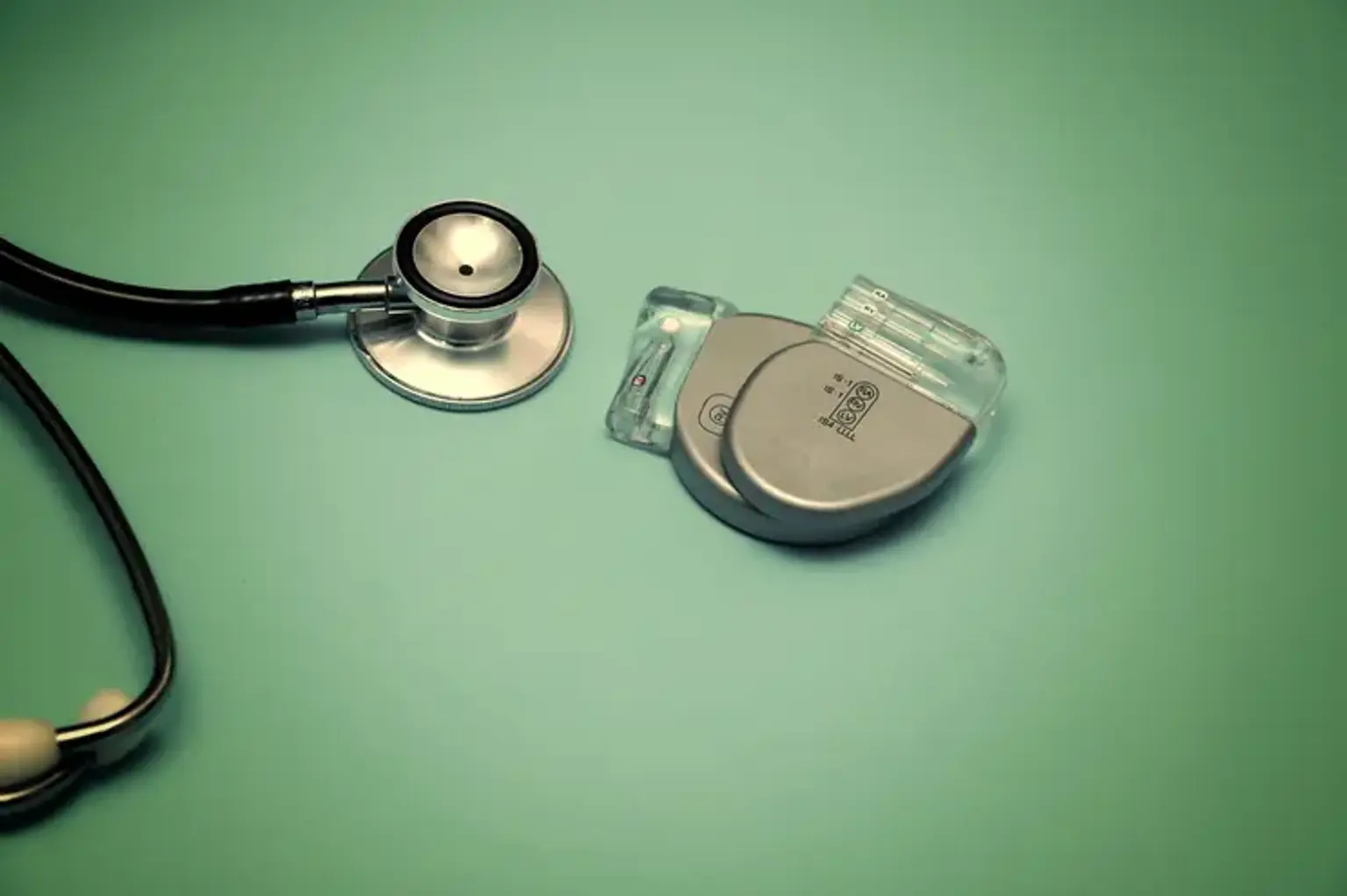
     

    What is CRT and how can it help your heart?

    CRT

    Cardiac resynchronization therapy (CRT) involves inserting electrodes into the left and right ventricles of the heart, as well as the right atrium on occasion, to treat heart failure by coordinating the function of the left and right ventricles via a pacemaker, a small device inserted into the interior chest wall.

    Cardiac resynchronization therapy (CRT) was launched in the 1990s and has transformed treatment for many patients with chronic systolic heart failure symptoms. A CRT system is divided into two components.

    • The heart gadget, which is essentially a small computer with a battery, is housed in a compact titanium metal casing the size of a pocket watch.
    • Insulated cables known as leads are inserted to transport information signals from your heart to the cardiac device as well as electrical impulses to your heart.

    CRT has had a significant influence on the management of the vast majority of patients with heart failure (HF) and an aberrant QRS duration. It was launched in the 1990s and transformed therapy for many patients with chronic systolic HF symptoms. The primary purpose of CRT is to improve mechanical functioning of the LV by restoring LV synchronization in patients with dilated cardiomyopathy and a widening QRS, which is mostly caused by left bundle branch block. This entails inserting a CS lead for LV pacing, as well as inserting a typical RV endocardial lead, with or without a (right atrial) lead.


    Normal heart rhythm

    Normal heart rhythm

    The heart is a pump that keeps blood flowing throughout the body. It consists of four compartments. The two top chambers (the right and left atrium) receive blood as it returns from the body through the veins. The bottom chambers (the right and left ventricle) are in charge of pumping blood out to the body through the arteries. The heart, like any other pump, has an electrical system that governs how it works.

    To fulfill its job (pump blood throughout the body), the heart requires a spark plug or electrical impulse to create a heartbeat. This electrical impulse normally originates in the upper right chamber of the heart (the right atrium) at a location known as the sino-atrial (SA) node. The SA node is the heart's natural pacemaker. The SA node emits electrical impulses that cause the heart to beat 60 to 100 times each minute.

    Your heart rate may be quicker if you are exercising, undertaking physical labor, or are under a lot of stress. When you rest or sleep, your heart rate slows. Certain drugs may cause your heart rate to slow. Everything is suitable.

    The electrical impulse is conveyed along the heart's conduction system from the SA node. It distributes uniformly across the right and left atria, causing them to constrict.

    When the impulse travels via the right atrium, it arrives at the atrioventricular (AV) node. This is a vital component in the heart because it provides the only electrical link between the top and bottom chambers. As a result, it is the sole means for an electrical impulse to reach the pumping chambers (the ventricles). The impulse travels via the AV node and into the heart's lower chambers or ventricles. As a result, they contract and pump blood to the lungs and body.


    Abnormal Conduction Develops in Cardiomyopathy Patients

    Cardiomyopathy

    Some cardiomyopathy patients develop a conduction system problem. A delay in normal electrical conduction via one of the normal wires - the left bundle branch - is the most prevalent anomaly. Left bundle branch block (LBBB) is characterized by a broadening of the QRS (>120ms) on the 12-lead ECG. This is present in around 40% of cardiomyopathy patients. The right ventricle is triggered a fraction of a second before the left ventricle due to the delay in conduction along the left bundle branch.

    Furthermore, LBBB causes the lateral wall of the left ventricle to activate significantly later than usual. In heart failure patients, this dyssynchrony produces an overall decline in the pump function of an already weaker heart. Patients with LBBB on ECG and symptoms of heart failure may benefit from Cardiac Resynchronization Therapy (CRT), also known as a Bi-Ventricular Pacemaker.


    Who is a candidate for CRT?

     candidate for CRT

    CRT is often used in persons who have significant heart failure symptoms and are at risk of having rapid, irregular, or life-threatening heart rhythms. Your heart will not pump enough blood if it does not contract effectively. This is referred to as a low ejection fraction. When all other alternatives for treatment have been tried, this device can aid by increasing heart function, quality of life, capacity to exercise, and survival.

    Patients with dilated cardiomyopathy, whether ischemic or nonischemic, a left ventricular ejection fraction of 0.35 or less, a QRS complex of more than 120 milliseconds, sinus rhythm, and New York Heart Association (NYHA) functional class III or IV symptoms despite maximal medical therapy for heart failure are the best candidates for CRT.

    CRT for individuals with mild heart failure has not been extensively explored and is not currently advised. The majority of patients in the CRT trials exhibited a broad QRS complex due to a left bundle branch obstruction. The advantage for patients with a left bundle branch block vs a right bundle branch block is unknown; nonetheless, the current recommendation for CRT is based on QRS length rather than QRS shape.

    Other unresolved CRT issues include the risks and benefits of left ventricular pacing without a right ventricular lead, the risks and benefits of surgical placement of a left ventricular pacing lead versus a nonthoracotomy approach, and the use of CRT for patients with NYHA class IV symptoms who are nonambulatory and reliant on intravenous inotropes for hemodynamic support.

    Early activation of the anterolateral papillary muscle, which can reduce the severity of mitral regurgitation, is another advantage of pacing from the left ventricular lateral wall. Mitral regurgitation can also be decreased over time due to reverse remodeling caused by CRT, which lowers the size of the left ventricular cavity, lowering mitral annular diameter and permitting mitral leaflet coaptation.


    Types of CRT

    Types of CRT

    The sort of CRT device you require is determined by your particular heart disease and symptoms. Following a diagnostic assessment, your doctor will review the devices with you in order to determine which one is best for you.

    • Cardiac Resynchronization Therapy Pacemaker (CRT-P)

    This CRT device, also known as a biventricular pacemaker, serves solely as a pacemaker. A CRT-P delivers a gentle electrical pulse to control the pace of ventricular contractions and restore synchronized pumping function.

    CRT-P is generally recommended for those who are not at high risk of developing life-threatening arrhythmias such as ventricular tachycardia (VT). If you simply require heartbeat pacing and not defibrillation, you may be a candidate (electric shock to restore normal heart rhythm).

    A pacemaker and a defibrillator are combined in this device. A CRT-D, like a CRT-P, synchronizes ventricular contractions. It also detects and corrects dangerously fast arrhythmias in the ventricles (known as ventricular arrhythmia)

    When there is a ventricular arrhythmia, the CRT-D delivers a greater electrical shock to the heart (defibrillation). The shock stops the aberrant rhythm and returns the heartbeat to its regular rhythm and rate. A shock to the chest may result during defibrillation.

    A CRT-D may be appropriate for those who are at high risk of VT, a form of arrhythmia that can lead to abrupt cardiac arrest or death.


    What happens prior to your procedure?

    CRT procedure

    You will get a letter from the hospital bookings clerk or the Doctor's secretary stating the date of your procedure as well as the date and time of your hospital admission.

    If you are on anti-coagulation (blood thinning) medication, such as warfarin, you must discontinue it 5 days before your treatment. After you stop taking warfarin, your doctor may arrange for you to get daily heparin injections.

    Patients undergoing the surgery at the Royal Melbourne Hospital must attend a pre-admission clinic the day before the operation. Some country patients may need to make plans to stay with relatives or friends overnight.

    A doctor will record your medical history during the pre-admission clinic. You'll also need an ECG and a blood test. The doctor will also specify the time you should be at the hospital the next day for admission.

    Before each of the procedures, you must fast for at least six hours. If your surgery is scheduled for the afternoon, you may have a light breakfast. Do not eat or drink after midnight if your proedure is in the morning, except for sips of water to help you swallow your pills.

    CRT device insertion is currently a relatively common operation. This is done under local anesthesia with sedative medicine to help you feel at ease, or under general anesthesia. This will be discussed with you by your doctor. The operation takes around two hours and is done in the cardiac catheter laboratory.

    This is a specialized room equipped with a patient table, X-Ray tube, ECG monitors, and other tools. The lab personnel will be clothed in hospital theatre attire and will wear caps and masks during the operation.

    A large number of ECG monitoring electrodes will be connected to your chest. A nurse or doctor will normally place an intravenous line in the back of your hand. This is required as a dependable method of administering drugs to you during the trial without the need for further injections. A blood-pressure cuff will also be placed to your arm and will automatically inflate at certain points throughout the process.


    What happens during the procedure?

    Chest procedure

    The device is normally implanted on the left side of the chest, just under the collarbone. A particular sterile solution is used to prepare the region, which may feel chilly. A big sterile sheet will be draped over you, partially covering your face. A nurse will be present at all times and you will be allowed to peer out from beneath the sheet to the side. You will be given oxygen to breathe through a little tube placed under your nostrils or through a mask that covers your face.

    The doctor will administer local anesthesia into the region under the collarbone where the CRT device will be implanted at the commencement of the treatment. This will hurt for a few seconds until the region becomes numb. You may feel some strong pushing in the shoulder area during the treatment, but it should not be uncomfortable. If you are in pain or discomfort, notify the nurse or doctor.

    Three leads (special cables that link the CRT device to the heart) are introduced via the vein beneath the collarbone into the heart. Under X-Ray guidance, they are maneuvered into the heart. The conventional leads are initially put, one into the right atrium and one into the right ventricle. These lines are attached to the heart wall using little hooks or a small screw. A short thin tube (called a sheath) is used to reach the coronary sinus, and the left ventricular lead is transferred through the sheath into a lateral branch of the coronary sinus. The CRT device will then be connected to all three leads.

    Due to the form of the third wire, it is not always possible to insert it into the coronary sinus. In some cases, a referral to a cardiothoracic surgeon for a second treatment to implant the lead using a restricted open surgical technique is a possibility. If the LV lead cannot be implanted successfully, this will be discussed with you.


    What happens after CRT Implantation?

    CRT Implantation

    You will be taken to the recovery room. You'll be there till the calming drug wears off. As required, your doctor will prescribe pain relievers. You will almost certainly need to spend the night in the hospital while your healthcare practitioner examines and changes the settings on your CRT device.

    It is critical that you follow all of your healthcare provider's recommendations and keep all follow-up visits after you leave the hospital.

    Here’s what you can expect once you’re home:

    • You should be able to follow your normal diet.
    • Lifting, straining, and extending with the afflicted arm (typically the left) must be avoided during the first 4 to 6 weeks. Inquire with your doctor about when you may resume these activities.
    • Keep the dressing clean and dry until your healthcare practitioner says it is OK to remove it and shower.
    • Examine your incision for symptoms of infection. Inform your doctor if you develop a fever, redness, discomfort, discharge, bleeding, or swelling.

    Here are likely long-term instructions for living with your CRT device:

    • Make sure your healthcare practitioner checks the operation of your gadget on a regular basis. This should be carried out at least once every six months.
    • Carry a CRT pacemaker identification card with you and inform all of your caregivers about your device.
    • Your pacemaker battery will last around 6-10 years. Your doctor will be able to inform you roughly 6 months before the battery dies. The replacement of the CRT pacemaker is a simple process.
    • All electrical equipment should be kept at least 6 inches away from your CRT pacemaker. They may obstruct its operation.
    • Stay clear from gadgets with high magnetic fields if suggested. These include generators and equipment such as microwaves. If you have any queries regarding what to avoid, see your healthcare professional.
    • Most X-rays and metal detectors are harmless, however metal wands used for airport inspections should be avoided. The majority of current equipment are MRI compatible.


    What are the Risks of CRT Insertion? 

    Risks of CRT Insertion

    CRT implantation is a relatively frequent and low-risk treatment, and any complications will be addressed immediately. Although the majority of people who have CRT implantation do not have any issues, you should be aware of the following risks:

    • Hematoma (large bruise) - This might happen at the pacemaker implantation site. This can be unpleasant and take many weeks to settle.
    • Pneumothorax – The pacemaker leads must be inserted into your heart through a tiny vein beneath your collarbone during the surgery. This vein is quite close to the lung, and there is a minor potential that a small hole in the lung may be formed accidently (Pneumothorax). If this happens, it normally heals on its own. However, a little tube may need to be placed on occasion to drain the air. This can be unpleasant and result in several extra days in the hospital.
    • Lead Dislodgment. – Although the pacemaker leads are carefully placed inside your heart, one of them may shift and need to be adjusted on occasion. This is generally diagnosed by checking the pacemaker during the first 24 hours following the operation.
    • Infection – There is an extremely little possibility that the pacemaker may become infected. If this happens, the pacemaker must typically be removed in order to clear the infection.
    • Cardiac perforation – Rarely, one of the leads will puncture the heart, causing blood to pool around the heart. If this happens, the problem will be addressed right away. This is an extremely unusual event.
    • Subclavian Vein Thrombosis- A blood clot in the subclavian vein is possible. This is the vein via which the leads travel to reach the heart.


    What can you expect?

    For the first 48 hours, you may experience some pain at the implant site. Inform your doctor or nurse if this continues or if there is any pain.

    The team will utilize a telemetry monitor and a holter monitor to monitor your heart rhythm while you are in the hospital. After you return home, your new gadget is monitored through a transmitter, phone monitoring, and follow-up sessions. If necessary, battery replacements can be performed as an outpatient operation.

    It takes a few weeks following implantation to resume normal activities. Consult your doctor before leaving the hospital for instructions on wound care, having a shower (typically not suggested until a few days later), moving, driving, exercise intensity, areas or appliances to avoid, and returning to work.


    Conclusion 

    CRT is a scientifically validated therapeutic option for some people suffering from heart failure. A CRT device administers tiny electrical impulses to both bottom chambers of the heart to help them beat more in sync. This may boost your heart's capacity to circulate blood and oxygen throughout your body. CRT can minimize hospitalization for heart failure and all-cause mortality in people with heart failure symptoms. In individuals with persistent atrial fibrillation, right bundle branch block, nonspecific intraventricular conduction delay, or merely slight extension of QRS duration, there is little to no benefit.