Cardiac Rhythm Disorders
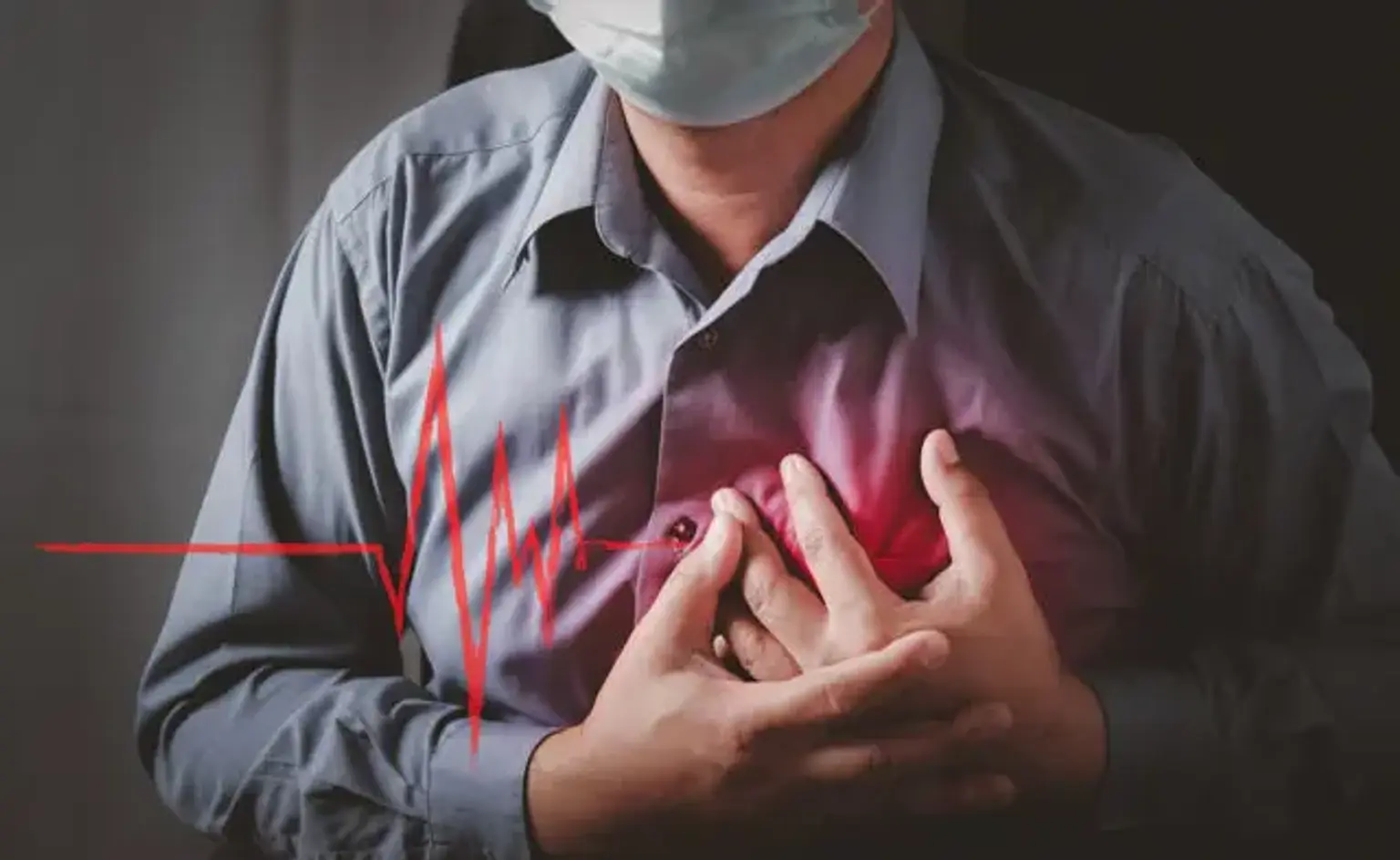
Cardiac rhythm disorders, also known as cardiac arrhythmias, are heart rhythm disorders evidenced by irregularity or unusually fast (tachycardias) or abnormally slow (bradycardias) heart rates. Palpitations, which some report as the sense of "my heart flipping over in my chest" or the awareness that their hearts are pounding rapidly or slowly, are common in patients who perceive these anomalies. Other signs and symptoms include fatigue, breathlessness, lightheadedness, dizziness, fainting (syncope), and chest discomfort. When the rate is higher, the ventricular function is worse, or the arrhythmia is coupled with anomalies in autonomic tone, the symptoms are more serious. Many people with arrhythmias, on the other hand, have no symptoms, and the disorder may only be identified during a regular exam.
Cardiomyopathy and congestive heart failure can result from a tachyarrhythmia that is fast enough and lasts long enough. Treatment of the arrhythmia in these circumstances can typically restore normal function to the ventricles. Although certain physical indications that occur during arrhythmias can aid in the diagnosis, electrocardiography (ECG) is the gold standard for detecting cardiac arrhythmias. When the arrhythmia occurs randomly, as it often does, prolonged electrocardiographic monitoring, often known as a Holter monitor, or an event recorder that the patient triggers when detecting an abnormality, may help confirm the diagnosis.
Cardiac Sinus Rhythms
The sinus node, a collection of cells at the intersection of the right atrium and the superior vena cava with the distinctive trait of automaticity shared by a few other cardiac tissues, is where the heart's normal rhythm begins. Autonomic influences through the parasympathetic nervous system, which maintains heart rates during most typical activities and at rest, and sympathetic activation, which boosts heart rates during exercise, affect the rate at which automatic cells in the sinus node release. During normal sinus rhythm, atrial activity goes from the sinus node to the atrioventricular node and from the right to the left atrium, resulting in P-waves on the electrocardiogram. In the frontal plane, this procedure results in a P wave axis that spans from around 0 to +75. The electrical activity from the atrium passes through the atrioventricular node and depolarizes the ventricles, resulting in the QRS complex on the electrocardiogram.
Adults with sinus rhythm have been categorized as having a heart rate of 60 to 100 beats per minute for decades. However, some experts believe the lower rate should be 50 beats per minute, which is common in typical athletes with a higher resting parasympathetic tone than most people. When the rate surpasses 100 beats per minute, sinus tachycardia is recognized, while sinus bradycardia is diagnosed when the rate is less than 60 beats per minute. The typical sinus rhythm in toddlers is faster than in adults. The sinus rhythm is usually a little off. Inspiration temporarily raises the rate, whereas expiration lowers it.
When the variation in the heart rhythm is extremely prominent, it is referred to as sinus arrhythmia. Sinus arrhythmia is commonly encountered in young people with healthy hearts. The sinus pacemaker may continually discharge faster than normal in a few persons, more often women than men, in the absence of any visible structural cardiac disease, additional atrial arrhythmia, or disorders such as thyrotoxicosis. If this condition causes symptoms, it may be diagnosed as inappropriate sinus tachycardia. To lower the heart rate, patients who require treatment may be given beta-blocking or calcium-blocking medications. Ablative or surgical intervention has only been used in a few cases.
Cardiac Rhythm Disorders Causes
Even if the heart is in good condition, you could develop arrhythmia. It could also occur as a result of:
- Coronary artery disease
- Electrolyte imbalance (sodium or potassium abnormalities)
- Injury to the heart or changes in the heart, such as diminished blood supply or stiffened heart muscle
- Following cardiac surgery.
- Fever or infection
- A number of drugs
- Problems with the heart's electrical impulses
- Emotional causes (stress)
- Alcohol, cigarettes, caffeine, and exercise.
Cardiac Rhythm Disorders Types
There are two general types of cardiac rhythm disorders: tachyarrhythmia and bradyarrhythmia. Supraventricular tachycardia, atrial tachycardia, and ventricular tachycardia are subtypes of tachyarrhythmia while sick sinus syndrome and atrioventricular heart block are the subtypes of bradyarrhythmia.
Supraventricular Tachycardia (SVT)
In young people, this is the most prevalent form of aberrant tachycardia. The rapid heart rate, which can reach above 150 beats per minute, begins in the upper chambers of the heart or in the electrical conduction system. Palpitations, chest aches, an upset stomach, a loss of appetite, lightheadedness, and weakness are some of the signs and symptoms. Slowing down one's heart rate can be learned by some. Straining, such as closing one's mouth and nose and attempting to exhale (Valsalva Maneuver), may help. When the heart rate returns to normal, medication therapy can usually avoid recurrent instances. To fix the condition, patients frequently require an electrophysiologic study along with radiofrequency ablation.
Atrial Tachycardia
Atrial tachycardia is a kind of SVT that is also known as atrial flutter or atrial fibrillation. This is a rapid heart rate that begins in the upper chambers of the heart and travels to the lower chambers. It's prevalent after atria (upper chambers) surgery, particularly the Mustard, Senning, and Fontan surgeries, as well as in disorders that cause the atria to grow (most commonly from leak or obstruction or the mitral or tricuspid valves inside the heart). Other symptoms include weariness, dizziness, lightheadedness, and fainting, in addition to a fast heart rate. Medications or radiofrequency ablation are frequently used to treat it.
Ventricular Tachycardia
This is a rapid heart rate that begins in the lower chambers of the heart. It is frequently caused by severe cardiac disease and necessitates immediate or emergency treatment. The symptoms can be modest, but they are usually severe. Dizziness, lightheadedness, and fainting are some of the symptoms. Medication, radiofrequency ablation, and inserting a device (defibrillator) that shocks the heart into a normal rhythm, as well as surgery, are all alternatives for treatment.
Sinus Node Dysfunction
The heartbeat begins at the sinus node. Sick sinus syndrome can occur if this is destroyed, which commonly happens following surgery. The heartbeat is slow and may not accelerate in a timely manner as a result of exercise. Patients may experience no symptoms at all, or they may experience weariness, exercise intolerance, dizziness, or fainting. If treatment is required, an artificial pacemaker can be installed.
Atrioventricular Heart Block (AV Block)
When an electrical signal cannot travel normally from the upper to lower chambers of the heart, it is called a heart block. Heart block can occur if the A-V node is destroyed during surgery. Heart block can develop without surgery in some cases. An artificial pacemaker can help you get your heart rate and rhythm back to normal. It is divided into three categories, first, second, and third.
First-Degree Heart Block
- Atrial conduction is slowed
- The passage of the impulse through the AV node to the ventricles is prolonged, resulting in a longer P-R interval.
- Every P wave has a QRS complex, and the rhythm is regular.
- Delayed conduction time is common in athletes and the elderly.
Second-Degree Heart Block
There are two forms of second-degree heart blocks: Mobitz type 1 and Mobitz type 2.
1. Mobitz type 1
- Occurs when there is a gradual increase in conduction time over a number of beats.
- The phenomenon repeats with a gradual lengthening of the PR interval over 4-6 beats until an impulse is completely blocked and a P wave occurs without an accompanying QRS.
- QRS is erratic and frequently slow.
- Usually confined to the AV node
2. Mobitz type 2
- Mobitz Type 2 is characterized by the occurrence of a dropped QRS complex without a prior P-R interval lengthening.
- Dropped beats can happen on a regular or irregular basis.
Third-Degree (Complete) Heart Block
Characterized by a complete or permanent halt of AV conduction, obstructing all impulses from above the AV junction.
In most situations, a focus below the block takes over the pacemaker function, and the heartbeat is subsequently supported by impulses from the area around the AV node or the ventricles.
Nodal rhythm has a rate of 40-60 beats per minute, whereas ventricular rhythm has a pace of 30-40 beats per minute.
Ventricular standstill will ensue if this escape rhythm does not form, and if not managed, it will be deadly.
The atrial and ventricular rates may be normal and regular, but there is no impulse conduction between the atria and ventricles.
On an ECG, a P wave and a QRS complex occur separately.
Other Conditions Associated with Cardiac Rhythm Disorders
Hypertrophic cardiomyopathy (HCM) is a rare but common cardiac hereditary disorder in which the heart muscle thickens. In young individuals and athletes, it is the most frequent cause of sudden cardiac death. Atherosclerosis puts those with HCM at danger for atrial fibrillation and heart failure.
Another cause of sudden cardiac mortality in athletes and others is arrhythmogenic right ventricular dysplasia (ARVD). Hypertrophic cardiomyopathy is more common, but it is less common. ARVD patients are at risk of developing heart failure.
Long QT syndrome increases a person's chance of fast, erratic heartbeats, which can cause fainting and be fatal. Though the syndrome is frequently hereditary, it can also be brought on by drugs or other medical disorders.
Cardiac Rhythm Disorders Diagnosis
- Electrocardiogram (ECG). An ECG is a test that captures the heart's electrical activity. For the fast, painless test at the doctor's office, you wear small electrode patches on the chest, arms, and legs.
- Holter monitor. This is a portable ECG (also known as an ambulatory electrocardiogram) the size of a postcard or a digital camera that you can use for up to 2 weeks. The study determines how electrical signals or waves go through the heart. These impulses direct the heart to squeeze (contract) and pump blood. Electrodes will be attached to the skin. It's painless, though the tape used to adhere the electrodes to the chest can cause slight skin irritation in some individuals. While wearing the electrodes, you can do anything except shower or bathe. You will return to the doctor after the test period has ended. They will get the data by downloading it.
- Event monitor. If the symptoms aren't frequent, the doctor may recommend that you try one of these for a month. When you press a button, the heart's electrical impulses are recorded and stored for a few minutes. When you see symptoms, try to acquire a reading. The results will be interpreted by the doctor.
- Implantable loop recorder. This device is implanted under the skin and continuously captures the electrical activity of your heart. It has the ability to send data to the doctor's office.
- Stress test. There are various types of stress testing. The idea is to determine how much stress the heart can withstand before developing a rhythm disorder or failing to receive enough blood. You will walk on a treadmill or peddle a stationary bike while getting an ECG and having the heart rate and blood pressure recorded in the most frequent form of stress test. Technicians gradually increase the intensity of your workout.
- Echocardiogram. Ultrasound is used to examine the heart muscle and valves.
- Cardiac catheterization. The doctor will place a catheter, a long, flexible tube, into a blood artery in the arm or leg. They will use a special X-ray scanner to direct it to the heart. Then, using the catheter, they will administer dye to aid in the creation of X-ray videos of the heart valves, coronary arteries, and chambers.
- Electrophysiologic study. This test monitors the electrical activities and pathways of the heart. It can assist you in determining what is causing your cardiac rhythm disorders and determining the best therapy option for you. The doctor will safely stimulate the abnormal heart rhythm during the test. Then they may prescribe medications to see which one works best for you, or they may recommend a treatment or device to treat it.
Cardiac Rhythm Disorders Treatment
Medical Treatment
Medication can help with a variety of rhythm abnormalities, including tachycardias. There are several medications on the market, and more are being investigated. These won't cure an arrhythmia, but they can help with symptoms by stopping episodes from occurring, lowering the heart rate during an episode, or reducing the duration of an episode.
Several medications may need to be attempted before the proper one is discovered. All medications, including those used to treat arrhythmias, have negative effects. Many adverse effects aren't dangerous and go away once the drug is stopped or the dose is altered. Some of the negative effects are dangerous.
To begin the medication, you may need to be hospitalized at the hospital. A slow heart rate is a typical adverse effect of many anti-arrhythmic drugs. A pacemaker may be required if the medication is required to avoid high heart rate problems. Because of the potential adverse effects, it's critical to follow the doctor's instructions properly. It may be required to keep track of the drug's concentration in the blood. The doctor will determine whether these blood tests are necessary.
Blood clots developing in the heart, particularly in the upper chambers, is one risk of chronic cardiac rhythm disorders. A clot that breaks off might travel to other regions of the body, such as the lungs or the brain, and cause major issues. To avoid this, anticoagulants (drugs that help prevent blood clots) are prescribed.
Cardioversion Therapy
Cardioversion is preferable in individuals with atrial arrhythmia who are hemodynamically unstable or whose rate control has failed. In a young patient with no significant comorbidities, it is also preferable. Cardioversion can be done within 36 hours of the onset of atrial fibrillation, but if the onset is delayed or the length is uncertain, transesophageal echocardiography is needed to confirm the absence of thrombi. If echocardiography reveals a thrombus, the patient will require anticoagulation for at least three weeks before cardioversion. After cardioversion, the patient must be on an anticoagulant for at least four weeks. Cardioversion therapy can take the form of synchronized electrical cardioversion or chemical cardioversion using drugs such as flecainide, propafenone, amiodarone, or dronedarone. The Maze procedure is usually limited to patients who are undergoing other types of heart surgery.
Radiofrequency Ablation
During an intracardiac electrophysiologic technique, radiofrequency ablation is performed. A catheter is placed immediately above the tachycardia-causing region. The catheters' tip is then heated with radiofrequency waves to change a small portion of the heart. This blocks the passage of electrical charge through the tissue. To treat the arrhythmia, this treatment may need to be repeated. Its success is directly tied to the underlying etiology of the arrhythmia and the complexity of the congenital cardiac condition. These treatments should be conducted in centers with special experience in the majority of patients with congenital cardiac disease. For supraventricular tachycardia, the success rate can be as high as 92%.
Surgical Treatment
Surgical intervention is occasionally required to stop aberrant electrical conduction. This operation is frequently performed in conjunction with surgery to correct other heart problems.
Implanted Defibrillators and Pacemakers
Artificial pacemakers can be used to treat a wide range of rhythm problems. The most prevalent causes for using a pacemaker are slow heart rates (bradycardias). A pacemaker is made up of a generator, which is a simple device that is hidden beneath the skin and attached to the heart by one or two thin wires. To make the heartbeat, it provides little, harmless quantities of energy to it. Pacemakers are implanted during a quick procedure. The pacemaker is put beneath the skin right below the clavicle, and the wires are introduced into the heart through veins in the shoulder.
The pacemaker generator is sometimes put in the abdomen and the wires must be placed on the outside of the heart through minor surgery. Once you've had a pacemaker, you'll need regular examinations to ensure the battery is charged and the wires are in good working order. Pacemaker batteries often last a long time. The majority of persons with pacemakers are able to participate in routine activities. An implanted cardioverter-defibrillator (ICD) may be required for patients who have very fast cardiac rhythms and significant symptoms such as fainting. This gadget is worn in the belly or slightly beneath the clavicle. With one or two thin wires, it is attached to the heart. When the gadget identifies a rapid cardiac rhythm, it shocks the heart to reset the rate.
After you've received an ICD, you'll need to get it checked on a regular basis to ensure that the battery and cables are in good working order. It's perfectly safe to be near microwave ovens and other common household electrical equipment. The doctor can advise you on whether or not you should avoid using other electrical devices and how to reduce your risk. For persons who have pacemakers or defibrillators, certain medical procedures, such as magnetic resonance imaging (MRI) or treatments that involve electrical pulses, aren't indicated. Newer gadgets and MRI machines, on the other hand, are being designed that are compatible.
Conclusion
Arrhythmias can be difficult to diagnose. Even for the same arrhythmia, the patient's presentation can vary, necessitating a different therapy approach dependent on the patient's medical problem and features. While a clinical history, medical examination, and ECG readings may disclose certain types of arrhythmia, forming an effective therapy strategy without a professional opinion might be difficult at times. While an internist is always involved in the care of patients with arrhythmia, a multidisciplinary team of professionals, including cardiologists, electrophysiologists, and interventionists, is required. The patient may require more testing, such as electrophysiology or ischemic workup, to determine the origin of the arrhythmia, which will then be addressed with cardiac catheterization or ablation treatment.