Cardiac SPECT
A heart is a major essential body organ that defines a person’s general health and life. It can, however, be susceptible to a wide range of diseases and disorders that can impact the quality of life. If the doctor suspects a heart condition, they often recommend nuclear medicine imaging test to assess the problem further. In most cases, they can suggest PET or SPECT scan methods.
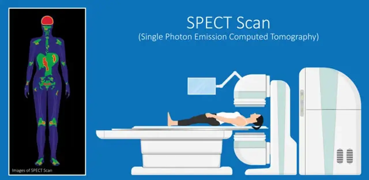
Single-photon emission computed tomography (SPECT) is a type of nuclear medicine imaging test. It’s a non-invasive technique that utilizes radioactive content (tracers) injectable through the vein to generate detailed heart images. With these images, the doctor can easily identify a problem within the heart and come up with possible solutions. Cardiac SPECT is thus an effective method of identifying any underlying heart disease and malfunction.
Objectives Cardiac SPECT Scans
The primary objective of SPECT scans is to obtain images of the heart structure and surrounding areas. This provides important information to the healthcare provider regarding the health state of the heart. Furthermore, it also helps determine the following;
- Areas within the heart that have limited blood flow during exercise and when at rest
- Presence of coronary artery disease
- How well the cardiac procedures such as bypass surgery are working
- If you have initially suffered a heart attack and the scar tissue areas, exist
- If you have a heart attack and require additional urgent tests, including percutaneous coronary intervention (PCI) or cardiac catheterization
- Whether you are at a high risk of suffering a heart attack
How Cardiac SPECT Scan Works
An injectable radioactive tracer that is normally injected into the bloodstream facilitates cardiac SPECT procedure. Once in the body, the tracer emits gamma rays, a type of radiation energy. The gamma camera detects tracer signals, which are then converted by a computer into images of blood flow via the heart. The pictures of thin slices taken through the heart can be generated from a variety of angles and directions.
These pictures are then analyzed to determine the tracer's locations. With the thin-slice images, computer graphics are applied to produce a 3-dimensional view of your heart. Regions of the heart with healthy blood flow will appear bright on the images. On the other hand, areas of inadequate blood circulation will be darker. Multiple SPECT scans generate colored pictures. The distinct colors reflect varying degrees of tracer absorption.
Areas of the heart with strong blood flow will be bright on the images, while regions of poor blood flow will be dark. Several SPECT scans generate color images. The numerous colors reflect differing degrees of tracer absorption.
SPECT scans can be performed when you are resting and in the process of an exercise stress examination known as a nuclear stress test. This stress examination provides the doctor with a clear understanding of how well the heart responds to stress. When you are unable to exercise, the doctor can give you medication. This helps stimulate blood flow through the heart, just like how it is during exercise. This is referred to as a pharmacologic or chemical stress test.
With thecardiac SPECT procedure, the doctor can notice the following;
The flow of blood via the coronary vessels is normal if the test is normal in both the exercise and at rest.
At rest, the test can demonstrate the normal flow of blood or perfusion, but not after exercise. This may be caused by an obstruction of one or more heart or coronary arteries. Such blockage may result in a perfusion defect or a region with slight to no tracer uptake.
While exercising and when at rest, the test could be irregular. The tracer would not be apparent in this situation. This thus means that there isn't sufficient blood currently circulating to that part of the heart.
Lack of tracer normally indicates that the cells within that region are dead as a result of a previous heart attack. It could also mean that they have turned into scar tissue.
SPECT scans might also demonstrate how well the left ventricle of the heart or lower pumping chamber is functioning. This is by using special radioactive tracers optimized for this test.
How to Prepare For Cardiac SPECT Scan
Before the procedure, the doctor will schedule a consultation visit first. This aims at assessing your overall health state to determine if you are eligible for the scan. During the visit, ensure that you inform the doctor of any drugs you are taking. This includes over-the-counter medications, dietary supplements, and vitamins. The physician might ask you to stop taking them a few days before the procedure. However, you should not stop using the medication unless the doctor instructs you to.
Sometimes, the doctor can as well ask you to abstain from certain foods like drinks containing caffeine. This can be for 24 hours prior to the examination. However, if the test could be delayed or canceled in case you take caffeine beverage within 24 hours.
Lastly, you should avoid eating anything for at least four to six hours before the examination. Instead, you can only drink water. Putting on loose, comfortable clothes and shoes is essential when undergoing an exercise stress examination.
What to Expect During Cardiac SPECT Test
The nuclear medicine technician and the medical provider often conduct the SPECT scan in a clinic or hospital setting. They also use a specialized tool for this purpose.
The doctor will begin by placing tiny metal disks known as electrodes on the arms, legs, and chest. These disks consist of wires that attach to a device that tracks the electrocardiogram (ECG). During the exam, the ECG will keep track of the heart pulse. This can be used to notify the camera when to capture an image. You will be required to put on a brace around the arm in order to measure the blood pressure.
The doctor will insert an intravenous (IV) line into the arm and inject the tracer through it. When performing a resting scan, you will be asked to lie down on a table. A gamma camera will rotate around the chest during this period, converting the tracer's signals into images.
You can either walk on a treadmill or ride a stationary bicycle during a nuclear stress test. After that, you will lay down on the table for the device to take more images. If you are unable to exercise, you will take a drug referred to as chemical stress via IV. This increases the rate of blood flow in the heart, the same as what happens during physical activity. Adenosine, dobutamine, and dipyridamole (Persantine) are examples of such chemical stress medications. The entire scan test can take about two to three hours.
What to Expect After Cardiac SPECT Test
Immediately after the examination, you can resume your normal day-to-day activities. It's also essential to take plenty of water for at least three to four days following the test. This helps flush out the radioactive substance from the body system.
After several days, you and the doctor will meet to discuss the scan results as well as the next step. If the results show that you have a heart disorder, the doctor will use the information to develop a suitable treatment plan. This will commence immediately since early treatment prevents additional complications and enhances successful recovery.
Risk of Cardiac SPECT
A specialist will closely monitor you during the stress test. He or she will also assess your blood pressure level, breathing, and electrocardiogram (ECG). Nonetheless, there is a chance of some unfavorable problems happening during the procedure, regardless of the precautions to maintain safety. These can include disorders such as;
- Irregular heartbeat
- Cardiac arrest
- Heart attack
- Death in the event of the cardiac stress test
In addition, you will experience considerable pain involved with injecting the needle that would be used to deliver the radioactive tracer into the vein. It’s also possible to have a very slight chance of bleeding from the venipuncture area.
The degree of radiation the body can get is approximately six times that of normal radiation exposure in the atmosphere in a year. Besides, the nuclear cardiac stress test is a common routine analysis that has been applied for over 20 years.
The Cardiac SPECT test is generally considered safe. The level of radiation exposure is minimal. Besides, the body can eliminate it via the kidneys in an average of 24 to 72 hours.
It’s essential to inform the doctor if you are pregnant or suspect you might be pregnant before the test. Also, if you are a nursing mother, consult first with the doctor before undergoing this examination. This is because it could endanger the child.
Conclusion
SPECT scan is similar to positron emission tomography (PET) scan. It’s a nuclear imaging test that involves the injection of radioactive tracer into the bloodstream. Doctors use cardiac SPECT imaging to check for coronary artery diseases and determine if a heart attack has initially occurred. It also indicates how well the blood circulates in the heart and how the heart functions.
For the best cardiac SPECT procedure, you can consider the CloudHospital healthcare platform. It consists of medical experts and nuclear medicine technicians who utilize state-of-the-art equipment to perform the test. Using the test results also helps develop an effective treatment plan.
