What is Cardiac SPECT Imaging?
Cardiac SPECT imaging (Single Photon Emission Computed Tomography) is an advanced diagnostic tool used to evaluate the health of the heart. It provides detailed, 3D images of the heart's blood flow and helps doctors assess areas where blood flow may be reduced due to blockages or damage. This imaging technique uses a small amount of radiation to capture nuclear images, which are then used to evaluate myocardial perfusion—how well oxygen-rich blood flows through the heart muscle.
Cardiac SPECT is particularly helpful for diagnosing heart conditions such as coronary artery disease, heart failure, and myocardial ischemia (reduced blood flow). By visualizing areas with poor blood flow, SPECT imaging helps doctors identify the extent of damage and make informed decisions about treatment.
Why Cardiac SPECT Imaging Is Essential for Heart Health
Cardiac SPECT imaging is an invaluable tool for diagnosing heart conditions early, before they become serious. This non-invasive procedure helps doctors assess heart function and detect abnormalities in blood flow, which is crucial for conditions like coronary artery disease. Unlike traditional imaging techniques like X-rays or echocardiograms, SPECT imaging provides detailed, real-time views of how blood flows through the heart muscle, revealing any blockages or issues with perfusion that could lead to heart attacks or other serious conditions.
The ability to detect heart disease early can significantly improve treatment outcomes. For patients with risk factors such as high blood pressure, diabetes, or a family history of heart disease, regular SPECT scans allow for early intervention and monitoring of heart health over time. This helps doctors manage risks and make timely decisions regarding further diagnostic tests or treatments, like angioplasty or coronary artery bypass surgery.
Understanding Nuclear Medicine for Heart Health
Nuclear medicine plays a pivotal role in cardiac diagnostics, and cardiac SPECT imaging is one of the most widely used techniques in this field. Unlike traditional X-rays, nuclear medicine uses small amounts of radiation to create images of the body’s organs and tissues. In the case of cardiac SPECT imaging, the focus is on assessing blood flow to the heart muscle and identifying areas that may be lacking oxygen-rich blood.
In nuclear imaging, the radiotracer used in the test emits gamma rays, which are detected by the gamma camera to create 3D images. This enables doctors to visualize the heart’s structure and function with high precision, allowing for more accurate assessments of coronary artery disease, heart muscle damage, and perfusion issues.
This imaging technique is highly valued for its accuracy, efficiency, and non-invasive nature, making it a preferred choice for patients with known risk factors for heart disease. It provides essential information that can guide treatment plans, whether it’s for medications, angioplasty, or more advanced procedures.
Diagnosing Coronary Artery Disease with SPECT Imaging
Cardiac SPECT imaging is a crucial tool in diagnosing coronary artery disease (CAD). CAD occurs when plaque builds up inside the coronary arteries, reducing blood flow to the heart muscle. This condition can lead to heart attacks, angina, and heart failure.
SPECT myocardial perfusion imaging helps doctors assess the extent and severity of CAD by showing areas of the heart that receive insufficient blood flow. The radiotracer used during the scan highlights regions with reduced perfusion—a clear indication of blockages or narrowing in the coronary arteries. This allows doctors to identify not only the presence of CAD but also its impact on the heart’s functionality.
By detecting coronary artery disease early, doctors can recommend appropriate treatments, such as angioplasty, stent placement, or bypass surgery, to prevent heart attacks and other life-threatening complications.
The Role of Cardiac SPECT Imaging in Myocardial Ischemia Detection
Myocardial ischemia occurs when the heart muscle is not getting enough oxygen due to reduced blood flow, often caused by narrowed arteries or blockages. Cardiac SPECT imaging is one of the most effective methods for diagnosing this condition. It allows doctors to observe how blood flows through the heart during rest and stress, revealing areas where blood flow is restricted.
SPECT myocardial perfusion imaging identifies ischemic regions of the heart that are not receiving adequate blood supply, which could eventually lead to heart muscle damage. Detecting myocardial ischemia early helps doctors intervene before it causes significant harm, allowing them to provide preventive treatments like lifestyle changes, medications, or surgical procedures such as angioplasty.
By visualizing these areas of reduced blood flow, SPECT imaging plays a key role in preventing heart attacks and improving long-term heart health.
Cardiac Stress Testing with SPECT Imaging
Cardiac stress testing is an essential part of heart disease diagnosis, particularly when assessing how the heart responds to physical or emotional stress. Traditionally, patients undergo a treadmill stress test, during which their heart rate and blood pressure are monitored while they exercise. However, cardiac SPECT imaging adds an important layer by evaluating myocardial perfusion before and after stress.
During the SPECT stress test, a radiotracer is injected into the bloodstream while the patient is exercising (or under stress-inducing medication if unable to exercise). The SPECT scan captures images of the heart, showing how blood is flowing through the heart muscle. If areas of the heart show reduced blood flow during stress, it may indicate the presence of blockages or coronary artery disease.
The combination of SPECT imaging and stress testing provides a comprehensive look at heart health, allowing doctors to assess both resting heart function and stress-induced issues, enabling earlier diagnosis and more accurate treatment recommendations.
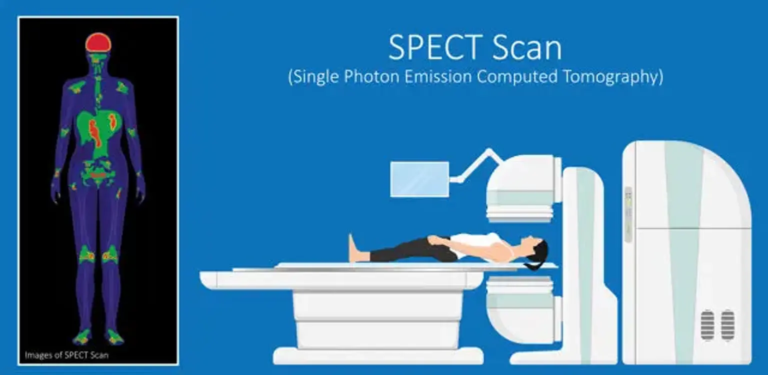
How Cardiac SPECT Imaging Works
The process of a cardiac SPECT scan involves the use of a radiotracer—a small amount of radioactive material that is injected into a vein, typically in the arm. The radiotracer travels through the bloodstream, and because it emits energy, it can be captured by a special camera called a gamma camera. The camera rotates around the patient and records how the radiotracer moves through the heart, creating detailed images of the heart’s blood flow.
Before the scan, patients may be asked to exercise on a treadmill or take medication that simulates exercise to stress the heart. This helps the doctor observe how the heart performs under stress, which is crucial for diagnosing coronary artery disease or myocardial ischemia. The SPECT imaging then highlights areas of the heart muscle that may not be receiving enough blood flow, which can indicate a blockage or previous damage.
The procedure is non-invasive and generally takes about 30-60 minutes to complete, with very little discomfort. There is a small risk from the radiation used, but it is minimal and carefully controlled to ensure patient safety.
Cardiac SPECT Imaging for Monitoring Heart Function in Aging Patients
As people age, the risk of developing heart disease increases, making regular heart health assessments essential. Cardiac SPECT imaging is especially important for aging patients, as it helps monitor heart function, detect early signs of heart disease, and assess the severity of existing conditions.
SPECT imaging allows doctors to track blood flow in the heart muscle and evaluate how well the heart is pumping oxygen-rich blood. This is particularly useful for elderly patients with conditions like coronary artery disease or heart failure, as it can identify areas of reduced perfusion that may not yet show symptoms.
Regular SPECT scans can detect early cardiac issues, allowing doctors to intervene early with treatments like medications, lifestyle changes, or surgical interventions. For patients with existing heart disease, SPECT imaging helps doctors monitor how well the heart is responding to treatment and adjust plans accordingly.
How SPECT Imaging Aids in Early Diagnosis of Heart Disease
One of the most significant advantages of cardiac SPECT imaging is its ability to detect heart disease early, often before symptoms become apparent. Early diagnosis is crucial in preventing the progression of coronary artery disease (CAD), heart failure, and other heart conditions.
SPECT scans can identify areas of the heart that may not be receiving sufficient blood flow due to blockages or narrowing of the arteries. By capturing real-time images of the heart’s blood flow, doctors can detect subtle changes in perfusion, even in the early stages of heart disease. Early intervention, whether through medications, lifestyle changes, or surgical procedures, can prevent these conditions from worsening and reduce the risk of heart attacks or other complications.
Regular SPECT imaging helps monitor heart health, ensuring that any signs of heart disease are detected and addressed as early as possible, improving long-term outcomes for patients.
Advanced Cardiac Diagnostics with SPECT Imaging
SPECT imaging is at the forefront of advanced cardiac diagnostics, offering more detailed insights into heart health than traditional methods. This non-invasive imaging technology helps doctors assess the blood flow and oxygen supply to the heart muscle, providing a clearer picture of how the heart is functioning.
Unlike basic tests like ECGs or echocardiograms, SPECT scans provide dynamic, three-dimensional images of the heart, allowing for a more accurate diagnosis. SPECT myocardial perfusion imaging shows how well blood is reaching the heart muscle at rest and under stress, which is crucial for detecting issues like coronary artery disease, myocardial ischemia, and heart failure.
With advanced cardiac diagnostics, SPECT imaging enables doctors to diagnose complex heart conditions early, guide treatment decisions, and provide better care for patients, especially those with high cardiovascular risk.
Cardiac SPECT Imaging in Korea: A Leading Destination for Heart Health
South Korea is recognized as a global leader in cardiac care, offering cutting-edge medical technology, including SPECT imaging for heart disease diagnosis. Many international patients seek cardiac SPECT imaging in Korea due to its advanced technology, highly trained specialists, and world-class healthcare infrastructure.
Korean hospitals and clinics are known for their expertise in heart disease diagnosis, and SPECT myocardial perfusion imaging is an integral part of the diagnostic process. For patients dealing with heart disease, seeking expert care in Korea can provide accurate, timely diagnoses and access to advanced treatments.
In addition to traditional heart care, Korea also excels in combining cardiac SPECT imaging with cosmetic surgery assessments, helping international patients monitor heart function during recovery from cosmetic treatments or volume restoration procedures.
How SPECT Imaging Improves Early Diagnosis of Heart Diseases in Korea
SPECT imaging plays a crucial role in improving early diagnosis of heart diseases in Korea. The country’s healthcare system is equipped with state-of-the-art technology that allows doctors to perform high-quality SPECT scans to assess heart function and detect coronary artery disease, heart failure, and other cardiovascular issues.
By identifying heart problems in their earliest stages, SPECT imaging allows for earlier and more effective interventions. Korean doctors are known for their expertise in using SPECT myocardial perfusion imaging to create personalized treatment plans that can prevent serious cardiovascular events like heart attacks and stroke.
In addition, the non-invasive nature of SPECT imaging makes it a highly effective tool for continuous monitoring, helping patients stay ahead of potential health issues and receive ongoing care tailored to their needs.
Cardiac SPECT Imaging for Monitoring Heart Function in Aging Patients
As people age, the risk of developing heart disease increases, making heart health monitoring especially important for seniors. Cardiac SPECT imaging is a valuable tool for monitoring heart function in aging patients, as it provides detailed insights into blood flow and oxygen delivery to the heart muscle.
For elderly patients with a history of coronary artery disease, heart failure, or hypertension, SPECT imaging helps doctors assess how well the heart is functioning, even if the patient is asymptomatic. It can also identify areas of the heart that might not be receiving enough blood supply, which could lead to further complications like myocardial ischemia or heart attacks.
By using SPECT scans, healthcare providers can detect early signs of heart disease, enabling early intervention and more personalized treatment plans. This is crucial for preventing worsening conditions and improving the quality of life for aging patients.
The Role of Cardiac SPECT Imaging in Preventing Heart Disease
Cardiac SPECT imaging is an important tool in the prevention of heart disease. By providing detailed images of the heart’s blood flow, it helps identify early signs of coronary artery disease and other heart conditions that could lead to heart attacks or heart failure. Early detection of these issues allows doctors to implement treatment strategies before the condition becomes life-threatening.
Patients at risk for heart disease, such as those with a family history, high blood pressure, high cholesterol, or diabetes, can benefit from regular SPECT scans. These scans allow for proactive heart health management, offering insights into the heart’s perfusion and the potential need for lifestyle changes, medications, or more intensive interventions. In some cases, SPECT imaging can also detect vascular blockages that might otherwise go unnoticed.
By using SPECT imaging for early diagnosis, doctors can help prevent serious cardiac events, ensuring better outcomes for patients and reducing the long-term costs of heart disease treatment.
Non-Invasive Cardiac Tests for Evaluating Heart Health in Patients Considering Cosmetic Surgery
Before undergoing cosmetic surgery, especially volume restoration procedures like fat grafting, it’s essential to assess heart health, particularly for patients with pre-existing heart conditions. Non-invasive tests like SPECT imaging provide detailed information on blood flow to the heart, ensuring that the patient is fit for surgery.
SPECT scans allow doctors to evaluate cardiac function without the need for invasive procedures. By identifying any potential issues, SPECT imaging helps ensure that patients are physically ready for cosmetic treatments, minimizing the risk of complications. This non-invasive heart test is a critical step in making informed decisions about cosmetic surgery and overall health.
Cardiac SPECT Imaging for Detecting Heart Failure and Blockages
Heart failure and blockages in the coronary arteries are serious health concerns, often linked to coronary artery disease (CAD). Cardiac SPECT imaging plays a vital role in detecting these conditions early, providing detailed, real-time images of how blood flows to the heart muscle.
In cases of heart failure, SPECT scans can show areas of the heart that are weakened or not pumping effectively. For patients with CAD, SPECT imaging highlights narrowed arteries or blockages that prevent adequate blood flow to the heart muscle. By identifying these issues, doctors can develop a treatment plan tailored to the patient’s condition, whether that includes medication, angioplasty, or more invasive procedures like bypass surgery.
Early detection through SPECT imaging is critical for managing heart failure and preventing heart attacks or other serious complications that arise from blocked arteries.
SPECT Imaging for Heart Disease Prevention and Its Role in Heart Health Management
Preventing heart disease requires early diagnosis and regular monitoring, both of which are facilitated by cardiac SPECT imaging. SPECT scans are instrumental in detecting blockages, reduced blood flow, and areas of the heart muscle that may be at risk of further damage. This non-invasive imaging technique helps healthcare providers stay ahead of potential issues by identifying heart conditions early and offering timely interventions.
SPECT imaging is often used as part of a comprehensive heart health management plan, particularly for patients with risk factors such as high blood pressure, high cholesterol, diabetes, or a family history of heart disease. By using SPECT scans as a diagnostic tool, doctors can monitor changes in heart function over time, allowing for early intervention and continuous care.
By offering valuable insights into myocardial perfusion, SPECT imaging supports heart disease prevention and enables doctors to implement personalized treatment plans, from lifestyle changes to surgical interventions, depending on the severity of the condition.
The Role of Cardiac SPECT Imaging in Post-Cosmetic Procedure Heart Assessments
After undergoing cosmetic procedures, especially those involving volume restoration or fat grafting, it’s essential to monitor heart health for patients with a history of cardiovascular disease. Cardiac SPECT imaging is a valuable tool in ensuring that patients maintain healthy heart function during recovery.
By performing SPECT scans, doctors can assess blood flow to the heart and detect any irregularities that may be aggravated by the stress of surgery or recovery. Monitoring heart function post-surgery is particularly important for patients at risk of heart disease or those who have undergone extensive cosmetic treatments.
Using SPECT Imaging to Monitor Heart Health in Patients with Volume Restoration Treatments
Patients undergoing non-surgical volume restoration treatments, such as fat grafting, often need to monitor their heart health to avoid complications during recovery. SPECT imaging offers an effective, non-invasive method for assessing heart function and blood flow, ensuring that patients are not at risk from underlying heart conditions.
By using SPECT scans, healthcare providers can assess whether the heart is operating efficiently and address any issues before they progress. This is particularly useful for aging patients or those with pre-existing heart conditions who might be considering cosmetic procedures.
Benefits of SPECT Imaging in Early Detection and Prevention of Heart Disease in Korea
SPECT imaging plays a vital role in the early detection and prevention of heart disease in Korea, where advanced cardiac diagnostics are readily available. SPECT myocardial perfusion imaging helps doctors identify areas of the heart with reduced blood flow, allowing for early intervention and treatment.
In Korea, SPECT scans are used widely to assess heart function, especially for patients at high risk of coronary artery disease, heart failure, or myocardial ischemia. By identifying heart problems early, patients can receive personalized treatment plans, including lifestyle changes, medications, or surgeries, to reduce the risk of severe heart events.
By detecting heart disease in its early stages, SPECT imaging allows for more effective prevention and long-term health management, reducing complications and improving quality of life.
Conclusion
Cardiac SPECT imaging is an essential tool for diagnosing and managing heart disease. It helps detect issues like coronary artery disease, myocardial ischemia, and heart failure early, allowing for timely interventions and better outcomes. By providing detailed, non-invasive images of the heart, SPECT scans offer valuable insights into heart function and blood flow, which are crucial for both heart disease prevention and cosmetic surgery assessments.
In Korea, SPECT imaging is widely used, offering advanced diagnostic capabilities for both local and international patients. Whether used for pre-surgical evaluations or monitoring recovery post-cosmetic treatments, SPECT scans provide a safe, effective way to ensure heart health.
With its ability to detect heart issues early and monitor ongoing conditions, SPECT imaging plays a critical role in improving patient outcomes and maintaining heart health throughout life.