Cardiovascular toxicities of cancer therapy

    Last updated date: 29-Aug-2023

    Originally Written in English

    Cardiovascular toxicities of cancer therapy

    Cardiovascular toxicities of cancer therapy

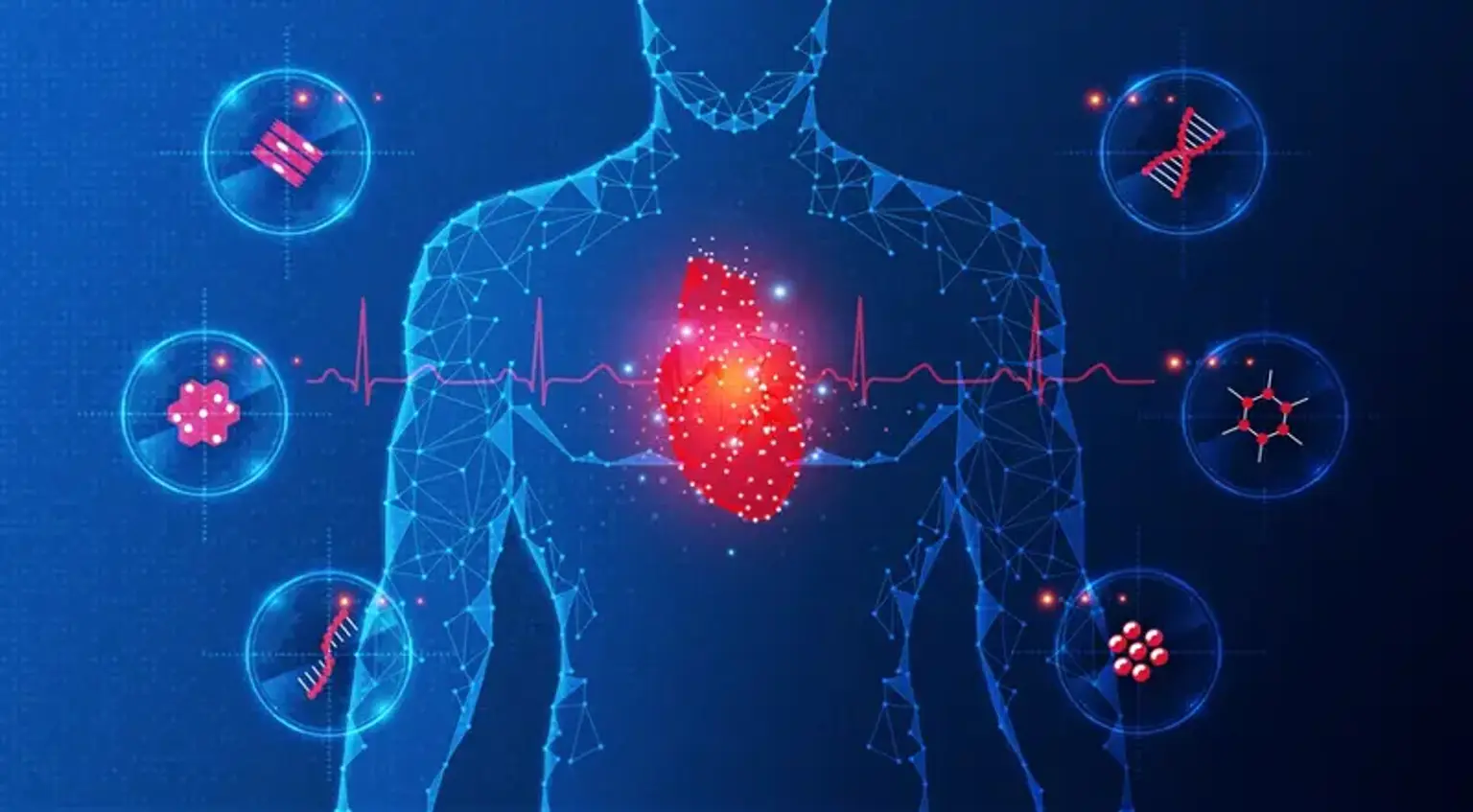
    What is Cardiotoxicity?

    Cardiotoxicity refers to cardiac damage caused by some cancer therapies or medications. It can appear years after cancer treatment, especially in people who were treated as children. Certain cancer therapies are more likely to cause cardiotoxicity.

    Cancer medications and therapies can cause direct cardiac damage. This is referred to as cardiotoxicity. Patients used to be focused on healing from cancer, but as cancer survival has grown, so has the prevalence of cardiac problems connected with chemotherapy and radiation.

    Cardiotoxicity can make it difficult for your heart to flow blood throughout your body as efficiently as it should. It can progress to cardiomyopathy, a heart muscle problem that makes it difficult for your heart to pump blood.

    During or after cancer therapy, damage to the heart (cardiotoxicity) or blood arteries (cardiovascular toxicity) can occur. As therapies have improved, more individuals are living longer following a cancer diagnosis than ever before. The quality of life of these survivors is an ongoing study topic that now covers the health difficulties around cardiotoxicity, both who is at risk and how to lessen that risk, which has to be investigated further.

    Cardiotoxicity can occur in anyone who has had cancer therapy. It is more frequent in those who have taken particular medicines or received chest radiation therapy. It's also rather prevalent among people who were treated for cancer as children. The exact rate of cardiotoxicity in people who had cancer therapy as adults is difficult to ascertain. However, some experts believe that up to 20% of this population will have cardiac issues, with up to 7% to 10% developing cardiomyopathy or heart failure.


    What are the effects of Cardiovascular toxicity of cancer therapy?

    Cardiovascular toxicity

    Although cancer therapies have considerably improved in the last decade, they can still induce adverse effects and raise the risk of heart disease. Cardiovascular disease is the primary cause of death among cancer survivors, surpassing the risk of death from cancer itself. Cancer survivors had a 42% greater chance of having cardiovascular disease and a 59% higher risk of acquiring heart failure than those without past cancer, according to a new study.

    Certain cancer therapies can harm the heart and circulatory system. Chemotherapy and radiation therapy, as well as newer kinds of cancer treatment such as targeted treatments and immunotherapies, can induce or worsen these adverse effects, which include high blood pressure, irregular cardiac rhythms, and heart failure.

    Cardiotoxicity can result in a variety of cardiac issues, including:


    What are the symptoms of cardiotoxicity of cancer therapy?

    symptoms of cardiotoxicity of cancer therapy

    Cardiotoxicity can develop years after cancer therapy has been completed. Children who have received chemotherapy or radiation treatment are more likely to have cardiac problems later in life. In fact, children with cancer have a 5-7 percent probability of developing cardiac problems as adults, including heart disease and hypertension.

    Symptoms of a potential cardiac problem caused by cardiotoxicity include:

    • Shortness of breath
    • Chest Pain
    • Heart palpitations
    • Swelling and fluid retention (edema) in the legs.
    • Distention of the stomach
    • Dizziness

    Many cancer medications can induce heart failure, high blood pressure, low blood pressure, heart attacks, irregular pulse, slow heart rhythm, or fluid around the heart. One-third of cancer patients who get chemotherapy medications including trastuzumab (Herceptin) and anthrycyclines will have cardiac cell damage.

    Radiation therapy to the chest can induce fibrosis (tissue thickening or scarring), which can lead to heart valve problems, heart attack, and thickening of the pericardium (the lining of the heart), resulting in constrictive pericarditis. Patients being treated for illnesses such as leukemia, left-sided breast cancer, or anybody receiving direct radiation to the left chest region may be affected.


    What drugs can cause cardiovascular toxicity?

    cardiovascular toxicity drugs

    Cardiotoxicity may be caused by some cancer therapies, including:

    • Anthracyclines. Chemotherapy drugs, such as doxorubicin (Adriamycin), are frequently used to treat leukemia, lymphoma, breast cancer, sarcoma, and multiple myeloma.
    • Trastuzumab (Herceptin), A targeted treatment medication widely used to treat breast cancer, stomach cancer, or cancer of the gastroesophageal junction, which is the connection between your food pipe (esophagus) and your stomach. If coupled with an anthracycline medicine, it is most likely to induce cardiomyopathy.
    • Radiation therapy to the chest, commonly used in the treatment of breast cancer or leukemia


    How cancer treatment can affect the heart?

    cancer treatment

    Drug-induced cardiotoxicity, most typically shown as cardiac muscle dysfunction that can develop to heart failure, is a significant side effect of certain regularly used classical antineoplastic drugs, e.g., anthracyclines, cyclophosphamide, 5-fluorouracil, taxanes, as well as newer agents such as biological monoclonal antibodies, e.g., trastuzumab, bevacizumab, and nivolumab; tyrosine kinase inhibitors, e.g., sunitinib and nilotinib; antiretroviral drugs, e.g., zidovudine; antidiabetics, e.g., rosiglitazone; as well as some illicit drugs such as alcohol, cocaine, methamphetamine, ecstasy, and synthetic cannabinoids. Most of the affected patients had no prior manifestation of the disease.

    Some forms of chemotherapy (particularly those in the anthracycline class of medications) damage the cardiac muscle due to calcium accumulation and other chemical processes in the body that create damaging free radicals. Thus, cardiomyopathy (enlargement) or congestive heart failure are examples of chemotherapy adverse effects. Chemotherapy, on the other hand, does not enhance the risk of a heart attack since it typically has no effect on blood flow to the arteries.

    Radiation therapy to the chest region, for example, for breast or lung cancer, can cause thickening of the blood vessels and heart valves, inflammation, and arterial obstructions. Radiation-induced heart issues frequently affect young people as well.


    Who is at greatest risk of heart problems from cancer treatment?

     greatest risk of heart problems

    Chemotherapy patients are at a higher risk of having cardiovascular issues, and the risk is increased by 30% if there is a history of heart disease or concurrent radiation, increasing the frequency of events by 30% compared to the general population.

    The time course of cardiotoxicity varies depending on several factors, including the patient's age at the time of exposure and the class effect of chemotherapy drugs, with childhood cancer survivors experiencing an exponentially rising risk of delayed cardiovascular events, whereas adult cardiovascular risk manifests earlier and is dependent on the number of conventional coexisting cardiac risk factors, particularly hypertension.

    Patients who have a moderate to high risk of developing or are suspected of having cardiotoxicities based on their medical history or abnormal imaging and biomarker levels may benefit from risk factor management, alternative cancer treatment choices, and cardioprotectant delivery.

    The following are risk factors for drug-induced cardiotoxicity:

    • Patient age at time of exposure (below 4 years and old age)
    • Female gender
    • Black ethnicity
    • Class of chemotherapeutic agent
    • Total cumulative dose
    • Concomitant radiotherapy/cardiac irradiation
    • Abnormal cardiac imaging or biomarkers levels
    • History of heart disease or left ventricular dysfunction
    • Hypertension
    • Obesity
    • Diabetes
    • Dyslipidemia
    • Physical inactivity
    • Smoking
    • Genetic predisposition

    One of the primary goals for cardiologists and oncologists in personalizing cancer therapy and arranging early preventative measures is to identify individuals at risk of drug-induced cardiotoxicity. It is critical to optimize and standardize the care of these patients in a multidisciplinary manner; an integrated strategy based on genetic, imaging, and clinical data may enable for the identification of individuals at risk of developing chemotherapy-related cardiotoxicity.


    How is drug-induced cardiotoxicity diagnosed?

    drug-induced cardiotoxicity

    Cardiotoxicity can be diagnosed by monitoring your heart's pumping performance using left ventricular ejection fraction (LVEF) and analyzing the function of your heart valves. When your heart contracts, LVEF measures how much blood flows out of your lower left heart chamber (left ventricle). Doctors employ a variety of tests to assess the heart's pumping and valve function, including:


    Radionuclide ventriculography (RVG):

    The gold standard for cardiotoxicity screening has long been radionuclide ventriculography. However, a more recent meta-analysis found that LVEF evaluated with RVG is insufficient for predicting heart failure since RVG frequently overestimates LVEF, resulting in volume measurement errors. Furthermore, the approach is costly, has little temporal and geographical resolution, and a significant risk of irradiation. As a result, RVG cannot be employed in contemporary medical practice.


    Positron emission tomography (PET):

    PET's application in cancer patients was centered on the identification of metastatic lesions and the response to treatment. Furthermore, this approach is capable of assessing pericardial metastases. However, PET has limited use in monitoring cardiac dysfunction in cancer patients after chemotherapy.


    Cardiac magnetic resonance (CMR):

    The ACC/AHA guideline now recognizes CMR as a tool for screening chemotherapy-induced cardiotoxicity, providing comprehensive information on myocardial function, valve and pericardial involvement. Even though this method has several advantages, particularly in obese patients with poor image quality, and it has been considered the gold standard for measuring LVEF, with the unique ability to demonstrate myocardial edema seen in acute myocardial injury, it is less used for routine screening and monitoring of cardiotoxicity, likely due to the widespread availability of echocardiography.


    Echocardiography:

    Echocardiography, with repeated measurements of the LVEF or LVSF, is perhaps the most widely accessible approach for assessing cardiotoxicity. Furthermore, it can offer additional information about numerous cardiac consequences of chemotherapy, such as valve and pericardial involvement, and it can be utilized to detect early, subclinical myocardial damage using the most advanced echocardiographic techniques.


    Other imaging tests:

    Cardiac stress test: This test assesses how your heart reacts to strenuous activities. While attached to a machine that analyzes your heart rate and blood pressure, you can walk on a treadmill or ride a stationary cycle.

    Multi-gated acquisition scan (MUGA): This test determines how well your heart's ventricles are working. It employs a safe radioactive tracer that is visible on an imaging scan. Your provider measures your ejection percent based on how the tracer flows through your circulation.

    Cardiac CT scan: CT scans employ a large number of X-rays from various angles to create a comprehensive image of your heart. This test may be particularly effective for those who have suspected cardiotoxicity following chest radiation treatment.


    Biological markers:

    Other non-invasive approaches for monitoring cardiotoxicity, such as biological markers, have been examined. Biomarkers may give valuable information on the cause of cardiac dysfunction and may identify people at risk of developing irreversible heart failure; hence, they can be employed in the diagnosis of chemotherapy-induced cardiotoxicity and in the monitoring of cancer therapy. Several biological markers, such as troponin isoforms, can detect cardiomyocyte damage and may be effective in identifying acute cardiotoxicity caused by cell death-inducing medicinal drugs.

    On the other hand, cardiac natriuretic peptides, which are markers of hemodynamic overload and increased wall stress, have been used as biomarkers in the detection of cardiotoxicity, but definitive evidence for a diagnostic or prognostic role in predicting chemotherapy-induced cardiotoxicity is still lacking. Other biomarkers used to evaluate cardiovascular damage, such as myeloperoxidase, should also be validated for use in cardio-oncology clinical trials. In the future, genomics, proteomics, and/or newly discovered oligoclonal B-cell repertoires may give genomic profiles and serological biomarkers for assessing cardiotoxicity.


    What is the management of cardiovascular toxicity of cancer therapy?

    Conventional chemotherapeutic

    Conventional chemotherapeutic drugs are frequently regarded as gold-standard therapies for a variety of cancers due to their familiarity and decades of credible evidence supporting their usage. However, because of their nonspecific cytotoxic effects on cardiac cells, several drugs in this class are well recognized to produce chemotherapy-induced cardiotoxicity. Anthracyclines, 5-FU, and capecitabine are the most common of these agents.

    Cardiotoxicity, toxicity mechanisms, treatment, and possible consequences differ between agents. Fundamentally, each chemotherapeutic class slows tumor development by a cytotoxic mechanism; however, this is not the same mechanism that causes cardiotoxicity.

    There are no precise guidelines for the treatment of chemotherapy-induced cardiotoxicity at the moment, although a few modest studies suggest the use of neurohormonal antagonists in the treatment and prevention of this condition.

    In addition to lowering the total dose of anthracyclines, the American College of Cardiology/American Heart Association (ACC/AHA) and Heart Failure Society of America (HFSA) recommendations for treating cardiac failure offer various techniques that may lessen the risk of cardiac cell death. Administering anthracyclines as infusions rather than boluses, or encapsulating doxorubicin in liposomes, are all approaches that may assist reduce cardiac toxicity.

    In combination with doxorubicin or epirubicin, dexrazoxane, an EDTA-like chelator, may minimize the risk of cardiotoxicity. Its usage, however, is restricted to individuals who have received a cumulative dosage of doxorubicin more than 300 mg/m2. Carvedilol, a beta-blocker with antioxidant characteristics, on the other hand, may lessen the risk of anthracycline-induced cardiotoxicity.

    Cardiotoxicity with 5-fluorouracil occurs at a rate of around 20%. Cardiotoxicity is caused by this drug by a multifactorial mechanism associated to its administration. When 5-FU is given as a bolus, the risk of cardiotoxicity is lower than when it is given as a continuous infusion. Patients frequently approach with immediate or severe symptoms ranging from chest discomfort and acute HF to myocardial infarction.

    It is critical to inform patients that they may have chest discomfort, sweating, and nausea while undergoing 5-FU infusion; however, these side effects are reversible as the infusion is stopped. If a patient continues to have cardiac symptoms after secondary exposure, the risk of cardiac side effects increases, especially if the patient has a history of coronary artery disease. To lessen the risk of cardiac consequences, beta-blockers, calcium channel blockers, or nitrates may be started in rare circumstances.

    The treatment of trastuzumab-induced left ventricular dysfunction (LVD) remains debatable. The left ventricular ejection fraction (LVEF) should be measured before starting trastuzumab and at frequent intervals throughout treatment. In the case of a 16% absolute decline in LVEF from pretreatment values, LVEF below institutional boundaries of normal, or a 10% absolute decrease in LVEF from pretreatment values, trastuzumab delivery should be delayed for at least 4 weeks.

    Trastuzumab may be restarted if LVEF recovers to normal limits within 4 to 8 weeks and the absolute drop from baseline is 15%. Tratuzumab should be completely discontinued if there is a prolonged decline in LVEF (>8 weeks) or if administration has been halted more than three times due to cardiotoxicity.

    Overall, once the diagnosis of chemotherapy-induced cardiotoxicity is confirmed, the oncologist and cardiologist should assess the patient's prognosis while considering the risks of quitting the cardiotoxic drug. The commencement of regular heart failure treatment, as well as the termination of the cardiotoxic drug, will hasten the recovery of left ventricular function.


    How to prevent cardiovascular toxicity of cancer therapy?

    cardiovascular toxicity of cancer therapy

    Identifying individuals who are at risk of cardiotoxicity is an important part of the therapy regimen. Patients with preexisting cardiac risk factors such as diabetes, hypertension, or a history of heart disease are among those included in this group. Catching these patients early, educating them on risk factor modification and living a heart-healthy lifestyle, and changing medicines and therapies can frequently avert problems and lower the long-term risk of cardiovascular disease.

    The use of advanced imaging to check for cardiac problems during cancer therapy is another important component of the treatment. Early detection of toxicity allows for the beginning of remedies, which are frequently drugs that assist minimize or reverse the toxicity of therapy. The objective is for patients to finish their cancer treatments safely, with as few interruptions as possible while avoiding cardiac problems that may occur during or after treatment.

    Prior to the administration of chemotherapeutic drugs, all patients should have their hearts monitored. Patients taking anthracyclines with trastuzumab should undergo cardiac function testing at baseline, three, six, and nine months into treatment, and twelve and eighteen months after treatment begins.

    Patients undergoing cancer treatment should be advised on the benefits of lowering cardiovascular risk by controlling blood pressure, quitting smoking, lowering lipids, and other lifestyle changes. Most people should aim for a blood pressure goal of 140/90 mmHg; those with diabetes or chronic renal disease should aim for 130/80 mmHg.

    Patients who undergo toxic cancer therapy, particularly pediatric cancer survivors, have a lifelong risk of heart disease. Long after cancer treatment is completed, these individuals should be watched for evidence of cardiovascular problems.


    Conclusion

    Cardiovascular toxicities of cancer therapy

    Cardiotoxicity is a significant side effect of several standard chemotherapy drugs. Cardiotoxicities come in numerous forms, including reversible, irreversible, acute, chronic, and late-onset. Early diagnosis of cardiotoxicity requires knowledge of the consequences of cardiotoxicity, its management, and dose changes for chemotherapeutic drugs such as anthracyclines, fluorouracil, taxanes, monoclonal antibodies, and tyrosine kinase inhibitors.

    Chemotherapy-induced cardiotoxicity is a significant consequence that endangers life and restricts the clinical use of several chemotherapeutic drugs, notably anthracyclines. Understanding the molecular underpinnings of chemotherapy-induced cardiotoxicity is essential for developing efficient preventative treatments and a more successful chemotherapy regimen. Despite the lack of a proven and efficient preventative medication, multiple studies show that chemotherapy-induced cardiotoxicity includes the production of reactive oxygen species (ROS).

    Drug-induced cardiotoxicity is a serious side effect that has been observed with various therapeutically relevant medications, particularly antineoplastic therapies. This toxicity has already resulted in the post-marketing removal of a number of pharmacologically active medications, as well as limiting the efficacy of other therapeutically valuable treatments. Currently, analyzing the cardiotoxicity potential is an important aspect in drug development, and several models have been developed to help forecast it in order to minimize such toxicity.

    Depending on your existing drug regimen, your healthcare professional may advise you to discontinue or reduce the dosage of certain medications. Your doctor may also prescribe drugs to help your heart function better, such as:

    • ACE inhibitors, such as lisinopril or fosinopril sodium, to open your arteries and improve blood flow.
    • Beta-blockers, such as metoprolol or atenolol, to increase blood flow and slow your heart rate.
    • Digoxin, also called digitalis, to slow your heart rate and help it beat more efficiently.
    • Diuretics, such as furosemide (Lasix), to rid your body of excess fluid.
    • Vasodilators, such as isosorbide dinitrate, to open up (dilate) your blood vessels so that blood flows more efficiently.

    Cardiotoxicity might be reversable. Cardiotoxicity caused by trastuzumab may be reversible, according to research. Cardiotoxicity caused by anthracycline usage is frequently irreversible and necessitates long-term therapy. Cardiotoxicity caused by chest radiation is very difficult to cure and may need long-term therapy, including surgery.