Cerebral aneurysm
Last updated date: 20-Apr-2023
Originally Written in English
Cerebral aneurysm
Overview
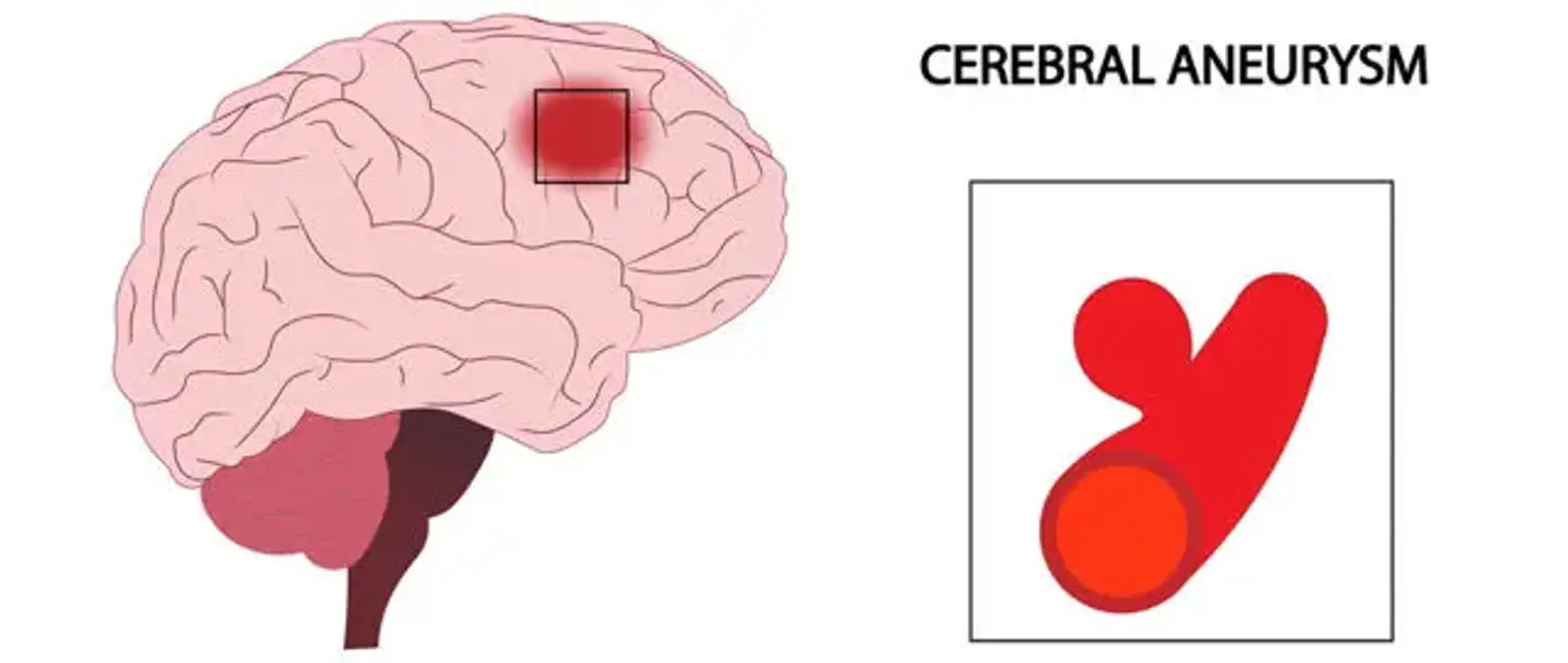
A cerebral aneurysm is a ballooning that occurs as a result of a weak spot in the wall of a blood artery in the brain. If the brain aneurysm grows in size and the blood artery wall gets too thin, the aneurysm will rupture and leak into the brain space.
What is cerebral aneurysm?
Cerebral aneurysms are dilations that form at weak sites in the cerebral artery circulation. They can range in size (small less than 0.5 mm, medium 6 to 25 mm, and large greater than 25 mm). The majority are saccular (berries), with a thin or nonexistent tunica medium and an absent or badly fragmented internal elastic lamina.
Fusiform (circumferential) and mycotic (infectious) aneurysms, on the other hand, are seen in a limited number of instances. The majority of brain aneurysms are quiet and may be discovered by chance on neuroimaging or at autopsy. Approximately 85 percent of aneurysms are found in the anterior circulation, primarily at junctions or bifurcations in the Willis circle. Subarachnoid hemorrhage (SAH) is generally caused by a rupture and is linked with a significant morbidity and fatality rate.
Epidemiology
The global prevalence of cerebral aneurysms is around 3.2 percent, with a mean age of 50 and a gender ratio of 1:1. After the age of 50, this ratio changes dramatically, with a rising female predominance nearing 2:1, which is assumed to be due to decreasing circulating estrogen, which causes a decrease in the collagen composition of the vascular tissue. The rate of rupture resulting in SAH is around 10 per 100,000.
This is more prevalent among some groups, such as the Finnish and Japanese. This is not because these populations have a higher rate of aneurysms. Overall mortality from aneurysmal SAH is estimated to be 0.4 to 0.6 percent of all-cause fatalities, with an estimated 20% mortality and an additional 30% to 40% morbidity in patients with known rupture.
Etiology
The majority of cerebral aneurysms are acquired lesions, having a higher prevalence in individuals with risk factors such as advanced age, hypertension, smoking, alcohol misuse, and atherosclerosis. Cocaine use, tumors, trauma, and some embolic-forming illnesses, such as endocarditis, are other causes.
There is also a substantial hereditary component, with the incidence being much higher in people with a strong family history of aneurysms (in other words, more than one family member affected). Certain genetic disorders are linked to a greater rate of occurrence.
Autosomal dominant polycystic kidney disease, Ehlers-Danlos syndrome, fibromuscular dysplasia, tuberous sclerosis, arteriovenous malformations (AVM), and aortic coarctation are all examples of this.
Pathophysiology
An aneurysm is a blood-filled outpouching of a blood vessel wall. Aneurysms form at a weak spot in the vessel wall. This might be due to an acquired illness or to inherited causes. The repetitive stress of blood flow against the vessel wall presses against the site of weakness, causing the aneurysm to grow in size.
According to Young-law, Laplace's increasing the area raises stress against the aneurysmal walls, resulting in enlargement. A combination of computational fluid dynamics and morphological indicators has also been presented as a good predictor of cerebral aneurysm rupture.
Flowing blood with both high and low wall shear stress can produce aneurysms and rupture. The mechanism of action, however, is still unclear. It is thought that low shear stress causes big aneurysms to develop and rupture by an inflammatory reaction, whereas high shear stress causes tiny aneurysms to grow and burst via a mural response .
Cigarette smoking, hypertension, feminine gender, family history of cerebral aneurysm, infection, and trauma are all risk factors for aneurysm development. Shear stress damage to the structural integrity of the artery wall triggers an inflammatory response that includes the recruitment of T cells, macrophages, and mast cells.
Smooth muscle cells from the tunica media layer of the artery, on the other hand, migrated into the tunica intima, where their role changed from contractile to pro-inflammatory. This results in arterial wall fibrosis, with a decrease in the number of smooth muscle cells and aberrant collagen production, culminating in thinning of the artery wall and the development of aneurysms and rupture. There are no identifiable gene loci that have been linked to cerebral aneurysms.
Aneurysms greater than 7 mm in diameter should generally be treated since they are prone to rupture. Meanwhile, aneurysms smaller than 7 mm in diameter form in the anterior and posterior connecting arteries and are more readily burst than aneurysms formed in other places.
Classification
Cerebral aneurysms are categorised according to their size and form. Small aneurysms are smaller than 15 mm in diameter. Large (15 to 25 mm), gigantic (25 to 50 mm), and super-giant aneurysms are the largest aneurysms (over 50 mm).
Berry (saccular) aneurysms
Saccular aneurysms, also known as berry aneurysms, are the most frequent type of cerebral aneurysm and look as a spherical outpouching. Connective tissue disorders, polycystic kidney disease, arteriovenous malformations, untreated hypertension, tobacco smoking, cocaine, and amphetamines, intravenous drug abuse (can cause infectious mycotic aneurysms), alcoholism, heavy caffeine consumption, head trauma, and bacteremia infection in the arterial wall are all possible causes (mycotic aneurysms).
Fusiform aneurysms
Fusiform dolichoectatic aneurysms are a widening of a portion of an artery that extends around the entire blood vessel rather than only originating from one side of the artery's wall. They have an estimated yearly rupture risk of 1.6–1.9 %.
Microaneurysms
Microaneurysms, also known as Charcot–Bouchard aneurysms, are small blood vessel aneurysms (less than 300 micrometres in diameter), most commonly in the lenticulostriate arteries of the basal ganglia, and are linked with persistent hypertension. Aneurysms of the Charcot–Bouchard type are a prevalent source of cerebral bleeding.
Brain aneurysm symptoms
Unruptured cerebral aneurysms are asymptomatic and so cannot be discovered based only on history and physical exam. When they burst, however, they frequently appear with a quick onset, severe headache. This is commonly referred to as a "thunderclap headache" or the "worst headache of my life." The discomfort is lateralized to the side of the aneurysm in 30% of individuals.
A headache may be followed by a momentary loss of consciousness, meningismus, nausea and vomiting, or other symptoms. Seizures are uncommon, occurring in fewer than 10% of patients. In 10 to 15% of patients, sudden death may occur. Surprisingly, 30 to 50% of individuals with serious SAH describe a sudden and intense headache 6 to 20 days before the event. This is known as a "sentinel headache," because it indicates a tiny hemorrhage or "warning leak."
Elevated blood pressure, dilated pupils, visual field and/or cranial nerve deficits, mental state changes such as sleepiness, photophobia, motor or sensory deficits, neck stiffness, and lower back discomfort with neck flexion are all possible physical exam results. Clinicians can utilize the Hunt and Hess grading system to predict outcomes based on baseline neurologic state. There are five classes based on the severity of symptoms, which correspond with the total fatality rate.
Grade 1 symptoms include a minor headache and slight nuchal stiffness. A strong headache with a stiff neck but no neurologic damage other than cranial nerve palsy is assigned a grade of 2. The patient is tired or disoriented with a minor focal impairment in grade 3. The patient is stuporous and has moderate to severe hemiparesis in grade 4. Finally, a coma with decerebrate posture is included in grade 5.
Diagnosis
Laboratory Studies
Lab studies that may be helpful for diagnosis include the following:
- CBC with platelets: Monitor for infection, evaluate anemia, and identify bleeding risk.
- Prothrombin time (PT)/activated partial thromboplastin time (aPTT): Identify a coagulopathy that increases bleeding risk.
- Serum chemistries, including electrolytes and osmolarity: Obtain baseline studies to monitor hyponatremia, address arrhythmogenic abnormalities, assess blood glucose, and monitor hyperosmolar therapy for elevated intracranial pressure.
- Liver function tests: Identify hepatic dysfunction that may complicate clinical course.
- Arterial blood gases: Assess blood oxygenation.
Imaging Studies
Most unruptured cerebral aneurysms are discovered by chance when a patient undergoes neuroimaging for another reason. Individuals at high risk, on the other hand, may be tested using magnetic resonance imaging (MRI), computed tomographic angiography (CTA), or traditional angiography.
A non-contrast CT of the brain, with or without a lumbar puncture, is commonly used to make the diagnosis of a suspected rupture producing SAH (LP). When the patient appears early, CT alone is regarded extremely sensitive for SAH, although sensitivity falls over time. According to some research, CT is 100 percent sensitive if conducted within 6 hours after the beginning of symptoms, but lowers to 92 percent after 24 hours and 58 percent after 5 days (14 to 18).
If the CT scan is negative but there is still a clinical suspicion of SAH, an LP should be performed. The characteristic LP findings include an increased opening pressure and an increased red blood cell count that does not decrease from tube 1 to tube 4. The presence of xanthochromia, a pink or yellow tinge to the cerebrospinal fluid (CSF) caused by hemoglobin breakdown products, strongly suggests SAH.
Xanthochromia can be diagnosed visually or through spectrophotometry, which is more than 95% sensitive when conducted at least 12 hours following the commencement of the bleed. Once SAH has been diagnosed, the cause of the bleeding should be determined. This can be accomplished with the use of CTA, MRA, or digital subtraction angiography (DSA). DSA entails introducing a catheter into the arterial circulation and injecting contrast under fluoroscopic guidance. This is regarded as the "gold standard" for detecting aneurysmal SAH.
Angiography
Traditional angiography is the gold standard for detecting and characterizing brain aneurysms. This technique can assess aneurysm location, size, and shape in both acute and chronic settings.
Digital subtraction angiography with biplanar magnification views gives information that may aid in the identification of an acutely ruptured aneurysm. Acute rupture may be accompanied by aneurysmal irregularity, the existence of a daughter loculus, or localized spasm. Vasospasm may be consistently represented, and collateral circulation can be demonstrated.
Perform a four-vessel angiography to rule out distant vasospasm and the existence of numerous aneurysms. Acute angiography occasionally produces negative findings (for example, due to thrombosis or vasospasm), in which case the procedure should be repeated 1-3 weeks later. However, given the risks and costs involved, this approach may not be appropriate for screening high-risk people.
In roughly 5-10% of individuals, a junctional dilation of the terminal carotid artery near the origin of the PCoA is seen. Less than 3 mm infundibula or conical enlargements are unlikely to expand or rupture. Overt aneurysms at the confluence of the terminal carotid artery and a persistent PCoA structure, on the other hand, may be more prone to rupture.
Following the recent development of 3-dimensional rotational angiography, more advancements in the characterization of cerebral aneurysms are predicted. Recent research suggests that this approach may provide greater resolution and sensitivity for detecting tiny aneurysms.
Brain aneurysm treatment
The choice to treat is complex and is influenced by the patient's size, location, age, and comorbidities, as well as the presence or absence of a rupture. There are two types of treatment: surgical and endovascular. Under general anesthesia in the operating room, a small metal clip is placed across the neck of an aneurysm, stopping blood from entering the aneurysmal sac and therefore eliminating the danger of bleeding.
Using a microscope, the aneurysm is accessible by temporarily removing a tiny part of the skull, dissecting the dura, and separating it from other blood arteries. After carefully applying the clip, the skull is held in place with tiny metal plates and screws, and the incision is closed. The aneurysm will decrease and scar with time, but the clip will likely stay for life.
Endovascular coiling
Endovascular coiling is a less invasive procedure that can be performed in some cases. Thromboembolism and intraprocedural aneurysm rupture are also potential risks. The operation is carried out by putting a catheter into the femoral artery and up into the artery harboring the aneurysm.
Following that, a second microcatheter with the platinum coil is placed via the first catheter. To remove the coil from the catheter within the aneurysm lumen, an electric current is employed. This stimulates the production of local clots and the obliteration of the aneurysmal sac.
In the event of a rupture, care also involves the treatment of SAH consequences. This is frequently done in an intensive care unit (ICU) to look for indicators of clinical worsening such as rebleeding, symptomatic vasospasm, hydrocephalus, seizures, and hyponatremia. Nimodipine, a calcium channel blocker, is widely used to avoid cerebral ischemia after SAH due to vasospasm.
Differential Diagnosis
- Arteriovenous malformations
- Cavernous sinus syndromes
- Carotid/vertebral artery dissection
- Cerebral venous thrombosis
- Fibromuscular dysplasia
- Migraine and cluster headaches
- Moyamoya disease
- Pituitary apoplexy
- Stroke - ischemic or hemorrhagic
- Vein of Galen malformation
Prognosis
The size of the aneurysm influences the outcome. Small aneurysms (less than 7 mm in diameter) have a minimal risk of rupture and grow slowly. For aneurysms of this size, the probability of rupture is less than 1%.
The prognosis for a ruptured cerebral aneurysm is determined by the aneurysm's size and location, as well as the patient's age, general health, and neurological status. Some people who have a burst brain aneurysm die as a result of the first hemorrhage. Other people who have had a brain aneurysm recover with little or no neurological damage. The Hunt and Hess grade, as well as age, are the most important determinants in predicting result.
Individuals with Hunt and Hess grade I and II hemorrhage on admission to the emergency room, as well as patients younger than the normal age range of susceptibility, should generally expect a satisfactory prognosis, with no death or lasting impairment. Patients above the age of 65, as well as those with lower Hunt and Hess scores at the time of admission, have a bad prognosis. Approximately two-thirds of patients have a negative result, such as death or lifelong disability.
The increased availability and accessibility of medical imaging has resulted in an increase in the frequency of asymptomatic, unruptured cerebral aneurysms detected incidentally during medical imaging studies. This might result in surgery, including endovascular surgery, or simply monitoring. Aneurysm height, aspect ratio, height-to-width ratio, inflow angle, deviations from ideal spherical or elliptical shapes, and radiomics morphological traits have recently been studied in their capacity to predict aneurysm rupture status.
Brain aneurysm how to prevent
You can't always prevent brain aneurysms, but you can lower your risk by not smoking and reducing high blood pressure.
Smoking
If you smoke, quitting can lower your chance of having a brain aneurysm greatly. If you're determined to quit smoking but don't want to be sent to a stop smoking program, your doctor should be able to prescribe medicinal medication to alleviate any withdrawal symptoms you may have after stopping.
High blood pressure
High blood pressure might also raise your chances of having a brain aneurysm.
You can help reduce high blood pressure by:
- Eating a healthy diet - specifically, limiting salt intake and eating lots of fruits and vegetables
- Limiting your alcohol consumption - both men and women are recommended not to consume more than 14 units per week on a regular basis.
- Maintaining a healthy weight - even dropping a few pounds may have a significant impact on your blood pressure and general health.
- Exercising on a regular basis — Being physically active and engaging in regular exercise reduces blood pressure by maintaining your heart and blood vessels in excellent working order.
- Caffeine reduction — It's good to take tea, coffee, and other caffeine-rich drinks as part of a balanced diet, but these drinks shouldn't be your primary source of fluid.
Complications
- Recurrent bleeding
- Vasospasm
- Seizures
- Syndrome of inappropriate antidiuretic hormone secretion
- Hydrocephalus
- Arrhythmias. congestive cardiac failure
- Gastrointestinal bleeding
- Deep vein thrombosis
- Neurogenic pulmonary edema
Conclusion
An intracranial aneurysm, also known as a brain aneurysm, is a cerebrovascular condition that produces a localized dilatation or ballooning of a cerebral artery or vein. Aneurysms in the posterior circulation (basilar, vertebral, and posterior connecting arteries) are more likely to rupture. Although basilar artery aneurysms account for approximately 3–5% of all cerebral aneurysms, they are the most prevalent aneurysms in the posterior circulation.