Chest Wall Tumors

    Last updated date: 12-Oct-2022

    Originally Written in English

    Chest Wall Tumors

    Chest Wall Tumors

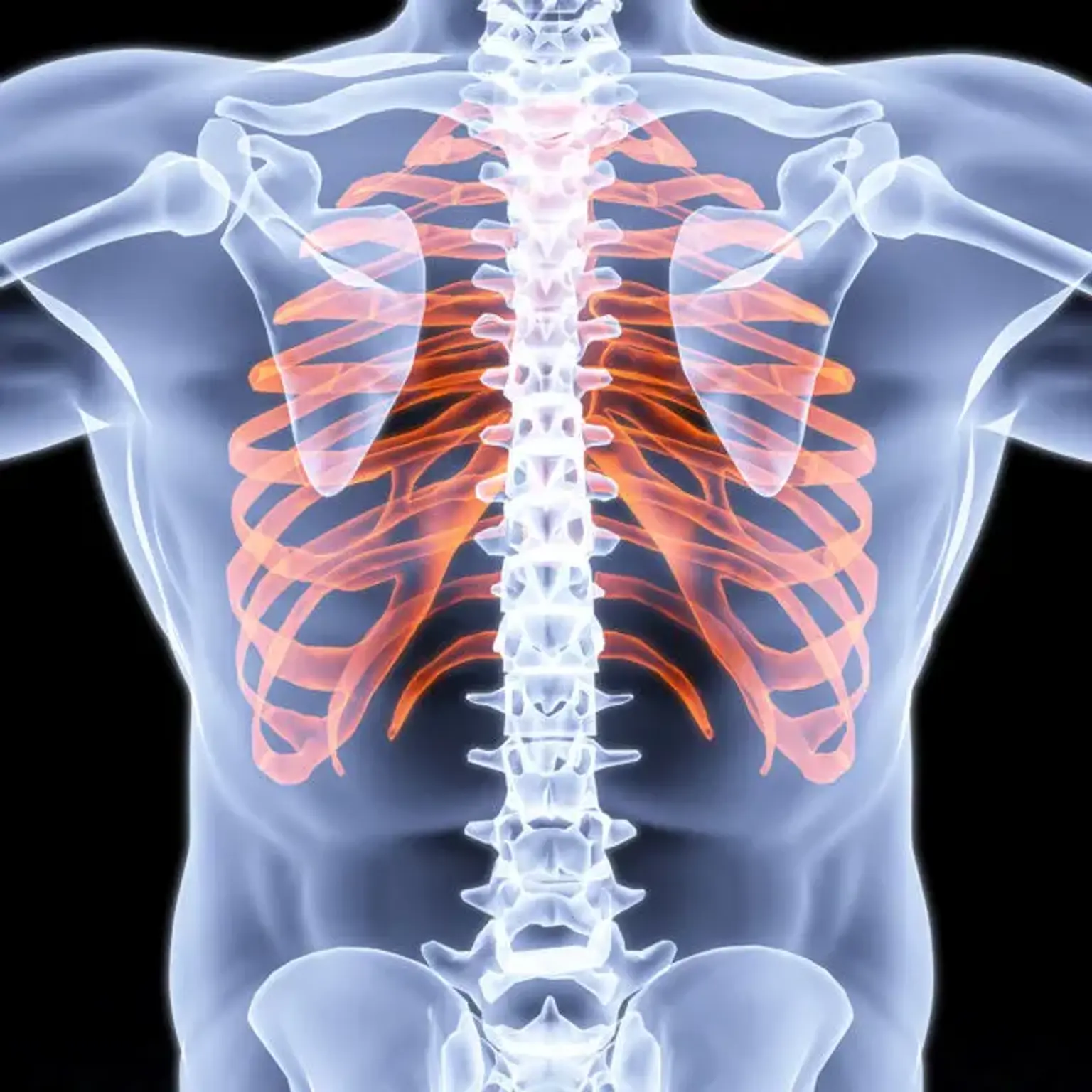
    The chest cavity is a space that encloses the ribs, breast bone, and spine. It also houses the heart, lungs, esophagus, thoracic aorta, and other essential organs. On the other hand, the chest wall comprises the diaphragm and the rib cage. It’s usually strong enough to shield the organs within the thoracic cavity and flexible to move in and outwards when breathing. 

    However, just like any body structure, the chest wall is also susceptible to diseases and disorders, including tumor growth. This condition is associated with abnormal cell growth, either cancerous (malignant) or non-cancerous (benign). Chest wall tumors can highly affect pulmonary functions; hence early diagnosis and treatment are crucial. 


    Types of Chest Wall Tumors 

    Chest wall cancers can develop in the soft tissues, cartilage, or the bones of the chest cavity. The most prevalent types of chest wall tumors include; 

    Chondrosarcoma: This is the most prevalent type of primary chest wall tumor. It develops in the cartilage and can be described as a gradually enlarging and sore mass.

    Osteosarcoma: This condition develops in the chest bone, especially during the stage of rapid growth and development, such as adolescence. Osteosarcoma can be described as a rapidly growing, enlarging, and painful mass.

    Ewing’s sarcoma: This type of tumor usually begins within the chest bone. It mainly affects young children and adolescents. Ewing’s sarcoma can be described as a painful mass associated with extreme fatigue fever.

    Plasmacytoma: This tumor is an isolated type of multiple myeloma and usually originates from plasma cells. Patients with plasmacytoma can experience pain but no visible mass or growth. Typically, it’s one of the few types of tumor that is rarely addressed via surgery.

    Malignant fibrous histiocytoma: This is the most prevalent type of soft tissue sarcoma that mainly affects adults. Malignant fibrous histiocytoma usually develops slowly and is characterized by a painless mass.

    Rhabdomyosarcoma: This type of tumor develops in the striated muscle and is more common among young children and adolescents. Rhabdomyosarcoma can be described as a fast-growing and painless growth.


    Causes of Chest Wall Tumors 

    There are no specific chest wall tumors causes. However, medical experts believe that certain factors can trigger chest wall cancers. These factors include; 

    • Hereditary or genetic factors 
    • Diet 
    • Overall lifestyle choices 


    Signs and Symptoms of Chest Wall Tumors 

    The signs and symptoms of chest wall tumors vary from one person to another, depending on various factors. It can be the types of cancer, the extent, age, and overall health of the patient. 

    The common symptoms associated with non-cancerous and cancerous chest wall tumors can include; 

    • Chest pain 
    • Muscle atrophy 
    • Swelling in the chest 
    • Impaired mobility 
    • A lump or mass protruding in the chest 

    At times, chest wall cancers don’t show any signs or symptoms, and approximately 20% are usually detected on chest x-rays. Due to this, you should immediately see a doctor if you suspect a tumor or notice some irregularities. 


    Risk Factors of Chest Wall Cancer 

    Although chest wall cancer is a rare health condition, factors that increase the risks of developing this condition include; 

    • Previous chest radiation 
    • Old age (certain types of chest wall tumors are more common among older people)
    • Medical history of previous tumor or cancer 
    • Residing in some parts of the world


    Diagnosing Chest Wall Tumors 

    When it comes to diagnosing chest wall tumors, the doctor will first review your medical history and perform a physical examination. The purpose of this is to find out if you have initially had a similar condition. It also helps analyze the symptoms you are currently experiencing. 

    Depending on the nature of your health condition, they can recommend other additional tests such as; 

    Imaging tests: 

    Examples of imaging tests that doctors will use to diagnose chest wall cancer include; 

    • Chest x-ray 

    Chest x-ray

    This is an imaging procedure that uses X-ray technology to visualize the chest organs and structures. It allows the doctor to check how well the heart and lungs are functioning. This is because certain heart-related problems can result in changes in the lungs. On the other hand, certain conditions can trigger changes in the lungs or heart structure. 

    Chest X-rays provide effective results regarding the shape, size, and position of the chest wall tumors. It can also display surrounding organs such as; 

    • The lungs 
    • Heart 
    • Aorta
    • Bronchi 
    • Pulmonary arteries 
    • Chest bones 
    • Middle chest sections 


    • Computerized tomography (CT) scan 

    This imaging test uses x-ray and computer technology to create detailed images of the structures and organs within the chest. Unlike the regular x-rays, the pictures of the CT scan are more precise. This provides more information regarding the location of the tumor, the size, shape, and extent. 

    • Magnetic resonance imaging (MRI)

    This method utilizes radio waves, magnetic fields, and computer technology to create in depth images of the chest organs and structures. It enables doctors to assess chest wall tumors and abnormal masses. It also helps them determine the size, shape, the extent of its spread to other organs or structures. 

    • Positron emission tomography (PET) scan 

    PET scan is a form of nuclear medicine imaging technique that uses a radioactive tracer to show certain body organs or tissue. Doctors often use this method to check and examine the body tissues so as to determine certain chest wall tumors. It can be by checking the blood flow rate, oxygen use, and metabolism. 

    Biopsy: 

    A biopsy is an alternative diagnostic procedure to assess chest wall tumors. It involves taking samples of the growths or lumps in the organs and tissues within the chest. It can as well include the lungs lining and the lymph nodes. The retrieved samples are then brought to the lab for further assessment under a microscope. 


    Other additional diagnostic tests that doctors can opt for to check for chest wall tumors include; 

    Endoscopic ultrasound (EUS): This is a minimally invasive technique that doctors use to check growths and masses in the chest wall. It involves the use of high-frequency sound waves to create detailed pictures of the chest lining. This makes it easier to identify a tumor. 

    Bone density test: This is a test to measure bone density and mineral content. 

    Endobronchial ultrasound (EBUS): This is a diagnostic test that enables the doctor to view the inside of the bronchi. This is the air path that leads to the lungs. It also allows them to conduct a biopsy procedure successfully. 


    Treating Chest Wall Tumors 

    The treatment options for chest wall tumors usually depend on factors like the type of cancer, the size, location, and extent. Some of the common forms of treatment include; 

    • Surgery 

    The primary objective of a surgical procedure is to eliminate all the visible tumors and masses. Surgeons often conduct chest wall tumors surgery with the aim of retaining the normal functionality of the chest and arms. This enables the patients to keep on enjoying various essential things and live a normal life. Examples of the surgical alternatives to address the chest wall tumors include:

    Resection and reconstruction of the chest wall: This is the major surgical option that doctors use to address chest wall tumors. It involves removing one or more ribs as a way of eliminating cancer. After this, the surgeon will reconstruct and restore the initial appearance and form of the region. They can use prosthetic materials or rotate the muscle flaps to accomplish this. 

    Video-assisted thoracic surgery (VATS): This is a minimally invasive technique that surgeons perform via small incisions created on the chest. Through the incisions, they will insert thoracoscopes attached with tiny cameras to facilitate viewing. The surgical instruments will then be inserted through another incision to take out the tumor. 

    • Chemotherapy

    Chemotherapy

    Chemotherapy is a treatment alternative to address chest wall tumors. It uses certain medications or sometimes a combination of various drugs to treat cancer. Doctors can administer chemotherapy drugs orally in the form of pills or intravenously through injection. Once the drug reaches the bloodstream, it moves towards the affected area and destroys the cancerous cells. Chemotherapy treatment is more effective for treating the type of cancers or tumors that have spread or metastasized to other body parts. 

    • Radiation therapy 

    Doctors can sometimes recommend radiation therapy to help manage chest wall tumors and reduce associated risks. According to medical research, radiation therapy to the chest wall minimizes the possibility of cancer returning. Furthermore, it boosts the survival rate by a small percentage. In a case where chemotherapy treatment is required, the doctor will administer radiation therapy once the initial treatment is complete. 


    Conclusion 

    Chest wall tumors comprise malignant and benign tumors that develop within the chest cavity and alter pulmonary functions. It can also affect organs within the chest, including the heart, lungs, esophagus, thoracic aorta, and other important organs. Overall, chest wall cancers are chronic conditions that require immediate treatment to prevent complications and fatalities. 

    CloudHospital is dedicated to helping patients with all types of chest wall cancers. It works with a number of medical experts with skills and experience in handling such health conditions. They also provide comprehensive diagnostic services that will eventually help develop a proper treatment plan.