Chiari malformation

Chiari malformation

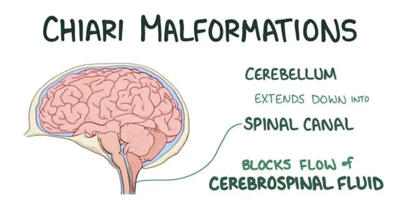
Chiari malformation refers to the structural problems within the skull and the cerebellum base. The cerebellum and the brain stem areas normally sit over a hole in the skull through which the spinal cord passes. It's known as Chiari malformation, when a portion of the cerebellum extends into the upper spinal canal below the foramen magnum. 

When a portion of the skull is small or malformed, it presses on the brain. It also forces the cerebellum down into the spinal canal, causing Chiari malformations. Pressure on the brain stem and cerebellum may disrupt normal functions regulated by these regions and obstruct the cerebrospinal fluid (CSF) flow. 


Types of Chiari Malformation 

The following are the major types of Chiari malformations; 

Chiari malformation Type I: 

Generally, this is the most frequent type among children. The lower section of the cerebellum, excluding the brain stem, stretches into a hole (foramen magnum) in the skull base in this form. Only the spinal cord normally travels via this hole. This is the only Chiari malformation type that is acquired. 

Chiari malformation Type II: 

This is exclusively observed in children who were born with spina bifida. The inadequate spinal cord development and the shielding covering is the spina bifida. Type II Chiari malformation is also known as Arnold-Chiari malformation or "classic" Chiari malformation. Both the brain stem and cerebellum stretch into the foramen magnum in this Chiari malformation type. 

Chiari malformation Type III: 

Basically, this is the most severe kind of Chiari malformation yet a very uncommon type. It’s characterized by the protrusion or herniation of the cerebellum and brain stem via the foramen magnum into the spinal cord. Normally, this results in significant neurological problems. 

Chiari malformation Type IV:

This usually occurs due to an underdeveloped or incomplete cerebellum. At times, it’s linked to sections of the skull and spinal cord that are exposed. Nonetheless, type IV Chiari malformation is a very uncommon type.


Signs and Symptoms of Chiari Malformation 

Some children and adults with Chiari malformation don't exhibit any signs or symptoms. But based on the tissue and nerves compression, as well as the CSF pressure accumulation, the symptoms might alter in some people. 

The following are the common signs and symptoms of Chiari malformation; 

  • Breathing difficulties
  • Buzzing and ringing in the ears (tinnitus)
  • Depression
  • Dizziness
  • Feeding problems and an incapability to gain weight
  • Gagging, drooling, or vomiting in excess
  • Hand coordination and fine motor skills issues
  • Headache, particularly after abrupt sneezing, coughing, or straining
  • Insomnia
  • Neck ache
  • Numbness or muscle weakness
  • Problems with hearing or balancing
  • Swallowing or speaking difficulties
  • Swallowing problems
  • The spine curvature (scoliosis)
  • Vomiting


Causes of Chiari Malformation 

There are several causes of Chiari malformation. A structural problem in the brain and spinal cord which can occur while the infant is still growing in the womb results in Chiari malformation.

Also, some researchers believe that a structural deficiency within the skull (a smaller-than-standard size in the region where the cerebellum rests) causes pressure and crowding on the brain. This pushes it to pass through the foramen magnum, which connects the brain and spinal cord. 

In certain circumstances, Chiari malformations may be hereditary (passed down through generations). This is because they can occur in more than one member of the family. In addition, a shortage of vital minerals during pregnancy, according to some researchers, may contribute to the disorder.

Chiari malformations are virtually always present at birth; however, the symptoms might not appear till later. Chiari malformations can also occur in people who were not born with the condition. Tumors, abnormalities in the spine, and hematomas may cause the spinal cord or the skull to change shape in these circumstances. 


Chiari Malformation Diagnosis 

Chiari Malformation Diagnosis 

Chiari malformation can sometimes be detected on prenatal ultrasounds even before a child is born. Ultrasound imaging is a procedure that employs sound waves to create images of soft tissues. 

Neurologists also conduct a thorough physical examination to look for Chiari malformation. He or she will examine your hands and feet for movement, sensation, and balance. Memory impairments, cognitive deficits, and learning problems are also examined. 

Your doctor can order the following diagnostic tests to check a comprehensive image of the spinal cord and the brain; 

  • Magnetic resonance imaging (MRI): MRI can be used to view soft tissues as well as bones. It also helps examine the spinal cord, brain, and cerebrospinal fluid. In other cases, this imaging test can reveal the extent to which the cerebellum extends into the spinal canal. 
  • Computerized tomography (CT) scan: Doctors can use this method to look for any disorders in the bones at the skull base and in the spinal column. It can also show conditions such as brain damage, brain tumors, and blood vessels defects. 
  • X-ray: Alternatively, the medical provider can suggest the x-ray technique to look for abnormalities in the head and neck. 


Chiari Malformation Treatment 

Chiari malformation that does not cause any symptoms and doesn’t affect a person's daily activities may just require routine monitoring and diagnostic imaging like Chiari malformation MRI and CT. To relieve associated headaches, pain, and discomfort, the physician may give you medications. 

In most cases, surgical procedure is the only option for alleviating symptoms or stopping the advancement of the central nervous system damage. It can also help to stabilize and enhance the symptoms. However, the disorder may necessitate more than one operation to manage successfully. These surgeries include; 

  • Posterior fossa decompression

This is usually the most prevalent method for treating Chiari malformation. It involves removing a portion of the rear of the skull known as the posterior fossa to reduce pressure within the brain and gives it enough room. 

  • Laminectomy

The surgeon might have to take out a small section of the bones that covers the spinal column to give greater room for the spinal cord and restore the cerebrospinal fluid flow. This, however, depends on the severity of the Chiari malformation. 

  • Electrocautery

Surgeons can use a small percentage of electricity to reduce a small area of the brain known as the cerebellar tonsils. This makes more space and enables the cerebrospinal fluid to drain. The tonsils in the cerebellum retract without resulting in any neurologic injury or damage.

  • Duraplasty

This type of decompression surgery involves opening the dura mater, a membrane located outside the brain. The surgeon will then stitch in a patch to expand the membrane and give the brain more space. This technique expands the brain's surface area and reduces pressure. 


Complications of Chiari Malformation 

Complications of Chiari Malformation 

Chiari malformation may be a progressive condition for some individuals, leading to significant risks and complications. In certain cases, there might be no symptoms at all, and treatment is not required. 

The following are some of the complications linked with Chiari malformation; 

  • Hydrocephalus

This is an accumulation of excess CSF in the brain. A Chiari malformation can obstruct the natural flow of the fluid, resulting in pressure inside the skull. This can cause mental disability or a larger or deformed skull. If left untreated, severe hydrocephalus can be life-threatening. Hydrocephalus can develop with any form of Chiari malformation, although Type II is the most prevalent. 

  • Syringomyelia 

This is a condition whereby the CSF-filled tubular cyst known as a syrinx develops in the central canal of the spinal cord. The expanding syrinx damages the core of the spinal cord and puts pressure on the nerves. As a result, it causes weakness, pain, stiffness, and even incapacity to sense extremes of cold or hot, particularly around the hands.

  • Spina bifida 

Spina bifida is the deficient closure of the backbone and the membranes surrounding the spinal cord. Myelomeningocele is common in people with Type II Chiari malformation. Also, the spinal cord of the child stays open in one part of the back as well as the lower spine. The spinal cord and membranes protrude via the opening within the spine, forming a pouch on the child's back. This can result in neurological problems, including paralysis, muscle weakness, or scoliosis. 

  • Tethered cord syndrome

This develops when the spinal cord becomes improperly attached to the tissues surrounding the bottom of the spine. Hence, it prevents the spinal cord from rotating freely in the spinal canal. The problem aggravates as the child grows. It can thus lead to irreversible damage to the nerves regulating the muscles in the legs and around the lower body. Minors with myelomeningocele are more likely to develop a tethered cord in the future. 

  • Spinal curvature 

People who have syringomyelia or Chiari malformation Type I have a high rate of developing spinal curvature. The spinal cord can bend to the left or right, scoliosis, or bends forward (kyphosis).  


Conclusion 

A structural abnormality in the skull makes the portion of the brain press against the spinal canal, resulting in Chiari malformation. Chiari malformations are virtually always present at birth, even if symptoms may not appear until later in life. The most prevalent symptom is a headache.

The form, severity, and related symptoms can influence the treatment of Chiari malformation. The common treatment options include routine monitoring, medicines, and surgical procedure. In other circumstances, treatment is not necessary.