Chronic Venous Insufficiency
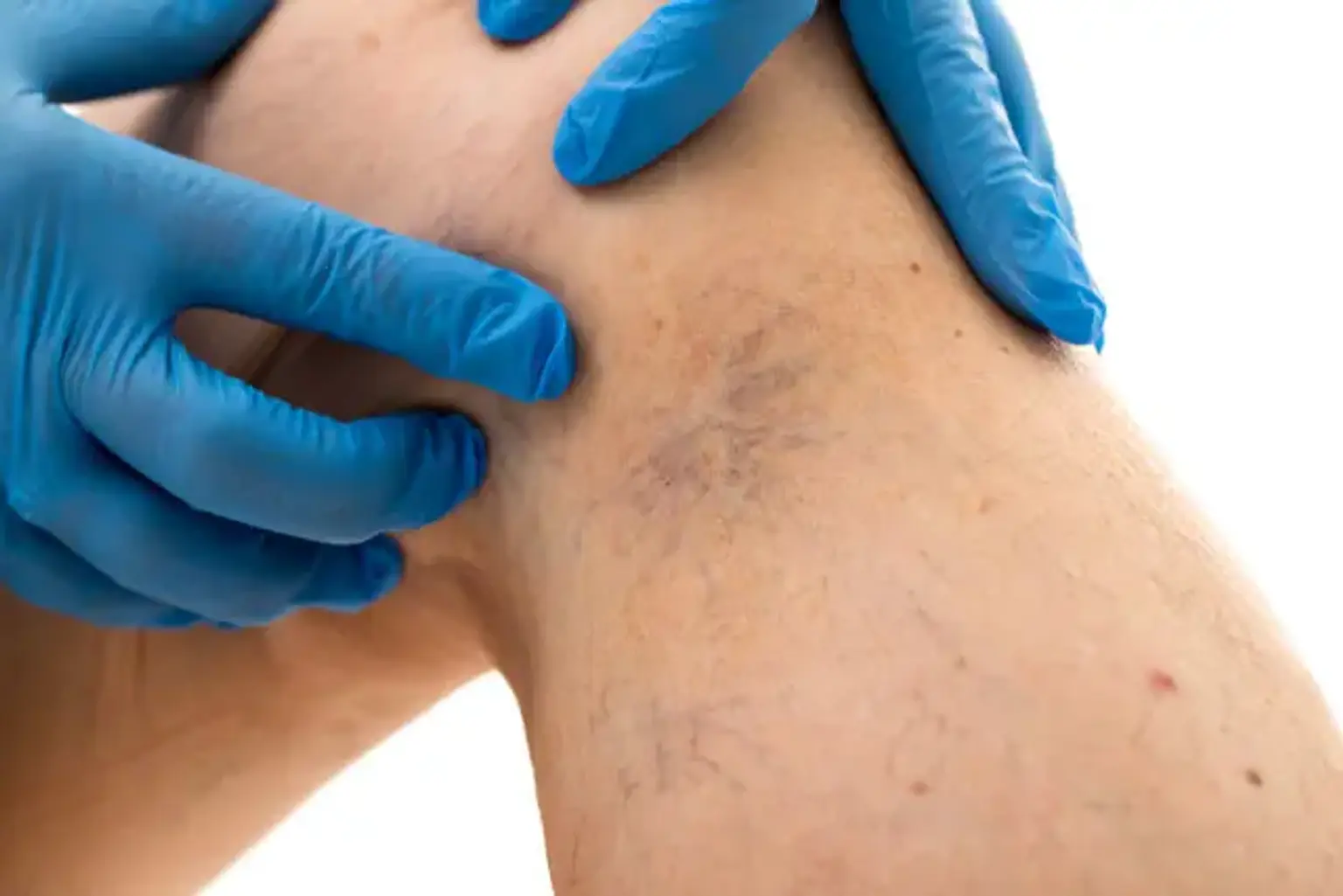
Chronic venous insufficiency (CVI) of the lower limbs can cause a wide range of symptoms, from asymptomatic cosmetic issues to severe complaints. Telangiectasias, reticular veins, varicose veins, edema, hyperpigmentation and/or dermatitis, lipodermatosclerosis, atrophie blanche, and venous ulceration are all examples of this.
Because of an underappreciation of the magnitude and significance of the condition, as well as insufficient recognition of the many presenting signs of primary and secondary venous problems, CVI is a rather common medical problem that is often neglected by healthcare practitioners. Up to 55% of people have abnormal venous circulation in their lower limbs, while the reported prevalence of CVI completely depends on demographic studies.
CVI is associated with increasing age, family history, extended standing, obesity, smoking, sedentary lifestyle, lower extremity injuries, past venous thrombosis, the existence of an arteriovenous shunt, high estrogen situations, and pregnancy. According to a multiethnic cross-sectional study, the prevalence of CVI in the Asian population is much lower than in non-Hispanic whites, but the prevalence of CVI in South Korea is projected to rise due to likely underdiagnosis of CVI, an increase in obesity, and an older population.
Chronic Venous Insufficiency Epidemiology
In the United States, CVI is a major public health issue. It is estimated that 3-5 percent of all Americans suffer from CVI-related symptoms. The prevalence of varicose veins in the adult population has been estimated to range from 9% to 62%, with most studies showing clinical varicose reflux in about 45% of the population. Approximately 500,000 persons are affected by venous stasis ulcers. The average rate of CVI hospitalization is 92 per 100,000 patients.
Venous insufficiency is thought to be more common in Westernized and industrialized countries than in undeveloped countries, owing to variations in lifestyle and activity.
It is estimated that 2-3 percent of the adult population suffers from lower-limb ulceration, with 75-90 percent of these ulcers having CVI.
Chronic Venous Insufficiency Pathophysiology
Ambulatory venous hypertension, which is produced by venous valve reflux, venous flow blockage, or both, is the most common pathophysiological cause of CVI of the lower extremities. In a stationary posture with no skeletal muscle contraction, the venous pressure in the foot vein can reach 85 to 90 mmHg. During ambulation, this pressure drops to less than 30 mmHg in a patient with healthy venous valves. The fall in venous pressure with leg movements is reduced in a patient with CVI. High pressures created in the deep veins by calf-muscle contraction can be conveyed to the superficial system and skin microcirculation if valves in the perforator veins are ineffective. Ambulatory venous hypertension is the medical term for this condition. Due to the persisting blockage of the venous flow and valvular reflux due to valve damage, post-thrombotic syndrome after deep vein thrombosis (DVT) also produces venous hypertension and valve reflux.
Chronic Venous Insufficiency Causes
A valve can be damaged by a blood clot in a deep vein in your leg (deep vein thrombosis). CVI can also be caused by a lack of activity. Long periods of sitting or standing might also be damaging. This increases the pressure in the veins, perhaps weakening the valves.
CVI is more frequent in females than in males. You may also have a higher probability if you are:
- Obese
- Over the age of 50
- Pregnant or have previously been pregnant
- Coming from a family with a CVI history
- Someone who has had blood clots in the past
- A person who smokes
Chronic Venous Insufficiency Symptoms
Discomfort, edema, varicose veins, and skin problems or ulceration are all symptoms of CVI. After prolonged standing, venous leg discomfort is commonly described as a dull soreness, pounding or heaviness, or pressure sensation that is eased by any treatment that lowers venous pressure, such as elevating the leg, wearing compression stockings, or walking. Leg discomfort, on the other hand, is missing in roughly 25% of patients with other CVI clinical symptoms, whereas it is the only clinical feature in approximately 12% of patients. Tenderness may be seen in people with varicose veins due to venous distension. Venous claudication can occur in people who have a deep vein blockage.
CVI is characterized by leg edema. It's generally pitting and varies a lot depending on the time of day and whether or not you have orthostasis. It starts in the peri-malleolar area and works its way up the leg. Congestive heart failure, hypoalbuminemia due to nephrotic syndrome or severe hepatic impairment, myxedema due to hypothyroidism, and medications such as calcium channel blockers and thiazolidinediones can all produce bilateral leg edema. Lipedema, or non-pitting leg edema produced by fat accumulation, should also be evaluated. Lipedema does not include the feet. Clinically, it might be difficult to distinguish from lymphedema. One of the clinical symptoms of lymphedema is the Stemmer's sign. Furthermore, up to one-third of CVI instances result in secondary lymphedema; however, if the underlying CVI is treated, the secondary lymphedema may resolve.
Varicose veins are distended, bulging, and superficial veins that become convoluted and larger with time. Patients with varicose veins are frequently asymptomatic but are concerned about their legs' look. If superficial thrombophlebitis occurs, they cause pain and might lead to continuous bleeding.
Skin darkening, stasis dermatitis, and ulceration are examples of skin alterations. Hemosiderin deposition causes hyperpigmentation. Non-venous hyperpigmentation, such as acanthosis nigricans or hemosiderosis, is more widespread and affects various parts of the body. Psoriasis, periarteritis nodosa, and allergic dermatitis should all be distinguished from stasis dermatitis. Lipodermatosclerosis is a kind of subcutaneous fat inflammation. A venous ulcer differs from an ischemic ulcer in that ischemic ulcers are deeper and often contain gangrenous margins or a gangrenous center.
Chronic Venous Insufficiency Diagnosis
For an accurate diagnosis of CVI, a thorough medical history and physical examination are required. The physical examination should be performed standing up to allow for maximum vein distension. Diagnostic testing, both non-invasive and invasive, is required to aid in the diagnosis.
Brodie-Trendelenburg Test
The Brodie-Trendelenburg test is useful for determining whether reflux is deep or superficial. To drain the veins, the patient lies down with the leg elevated. After the patient is instructed to stand, a tourniquet or manual compression is applied over the superficial veins, and the veins are examined. If the varicose veins fill up in less than 20 seconds, they are produced by superficial venous insufficiency. In the presence of profound (or mixed) venous insufficiency, on the other hand, varicose veins widen rapidly.
Plethysmography
Plethysmography is a non-invasive venous test that assesses each probable component of CVI pathophysiology, such as reflux, blockage, and muscle pump dysfunction. It is possible to determine venous volume, venous refilling times, maximal venous outflow, segmental venous capacitance, and ejection fraction. Impedance plethysmography, strain gauge plethysmography, photoplethysmography, and air plethysmography are the four primary types of plethysmography. This technique is only used in academic or hospital settings when DUS does not provide clear information on the pathogenesis of CVI due to its complexity.
Computed Tomography and Magnetic Resonance Venography
Although advances in computed tomography (CT) and magnetic resonance (MR) imaging have improved the examination of venous pathology, these techniques are rarely used to diagnose CVI and plan treatment. To obtain excellent images and eliminate artifacts in a specific venous system, proper image acquisition timing based on venous filling time is essential. Furthermore, these methods do not give functional data. These approaches, on the other hand, are particularly beneficial for evaluating isolated or complex lesions at proximal veins and their surrounding structures, as well as determining whether or not there is intrinsic or extrinsic obstruction.
Venous Duplex Ultrasonography
DUS is the most widely used and effective diagnostic method for CVI, providing both etiological and anatomical information. DUS detects venous blockage and venous reflux in superficial and deep veins using a combination of B-mode imaging and spectral Doppler. The use of color-assisted DUS to visualize venous flow patterns is beneficial.
Venous Obstruction Diagnosis
The absence of flow, decreased augmentation, the presence of an echogenic thrombus within the vein, or the inability of the vein to collapse after compression can all be signs of venous occlusion. At resting, large veins including the inferior vena cava, iliac, femoral, and popliteal vessels exhibit spontaneous blood flow. Respiratory changes are reflected in this flow. Because of the increased intra-abdominal pressure during inspiration, normal flow pauses during inspiration and resumes after expiration. Because of their small size, small veins, such as calf veins, rarely demonstrate spontaneous flow. A restriction proximal or distal to the area of evaluation may cause a lack of spontaneous flow. Furthermore, a proximal stenosis is indicated by essentially constant high velocity flow without any respiratory variations. Not in a standing position, but in a supine or modest reverse Trendelenburg position, should spontaneous flow be assessed. In normal veins, augmentation can be examined by exerting a relatively tough squeeze over the calf to augment flow going centrally. It's ideal to squeeze and hold for around 0.3 second before releasing while conducting a compression. This procedure is performed to confirm the vein segment's patency. The increase has been blunted, which indicates occlusion. The fluctuation of the compression force, on the other hand, is a key disadvantage of this operation. The most reliable method of detecting an intraluminal thrombus is compressibility, which is done in a short-axis view. The higher blood velocity from DUS at the iliac vein indicates non-thrombotic stenosis.
Venous Reflux Diagnosis
The direction of flow is used to detect venous reflux. Venous reflux is defined as any considerable reverse flow toward the foot. In the reverse Trendelenburg posture, venous reflux is measured. Although reverse flow can be seen without the provocation maneuver, venous reflux can be confirmed via the Valsalva maneuver or augmentation by compressing the calf. Intra-abdominal pressure is raised by the Valsalva technique. The major purpose of this test is to assess the central vessels' flow characteristics and valve functioning. Downward pressure is carried down and through the defective valves until it reaches the working valve. Venous reflux is indicated by prolonged reversal flow after augmentation. However, using a cuff inflation-deflation approach with rapid cuff deflation in the standing posture is the preferable provocation procedure. For detecting reflux in the superficial and deep veins of the leg, this approach delivers more uniform measurable data. The term "reflux time" refers to the length of time that reflux lasts. It's regarded normal to have a lot of venous reflux. Pathologic reflux is defined as a time interval of more than 1.0 second in the femoral or popliteal veins, more than 0.5 second in the saphenous systems, and more than 0.35 in the perforators. The duration of reflux does not correspond with clinical signs, despite the fact that it represents the severity of the disease.
In individuals with CVI, a DUS assessment should reveal both structural patterns of veins and anomalies in venous blood flow in the limbs. The following information should be gathered: (1) the number, site, diameter, and function of incompetent perforating veins; (2) the degree of reflux in the saphenous veins of the thighs and legs; (3) other related veins that demonstrate reflux; (4) the origin of filling of all superficial varices if not from the veins already described; (5) veins that are hypoplastic, atretic, missing, or have been removed; and (6) the condition of the saphenous junctions.
Intravascular Ultrasound
Because venous intravascular ultrasound (IVUS) is better than conventional venography for morphologic diagnostic testing of iliac venous outflow blockage and provides great assistance in the accurate placement of venous stents, IVUS is quickly gaining acceptance in the management of chronic venous iliofemoral disease.
Chronic Venous Insufficiency Treatment
All patients with CVI signs and/or symptoms should get conservative treatment at first. Conservative management relies mostly on the use of compression stockings. Reducing risk, such as weight loss in an obese patient, frequent walking activity, and quitting smoking, should be supported as conservative management in the patient. Eberhardt and Raffetto provide a comprehensive summary of CVI diagnosis and treatment.
Compression Stockings
Compression stockings are designed to give graded external compression to the leg and counteract the hydrostatic pressures that cause venous hypertension. Gradually graduated compression stockings are favored over non-graded compression stockings. For the prophylaxis of DVT in surgical patients, there is no difference between knee-length and thigh-length graduated compression stockings. Most patients, especially the elderly, tolerate knee-length stockings better.
Patients with varicose veins with or without edema should wear compression stockings with a compression pressure of 20 to 30 mmHg. Patients with extensive venous skin change or an ulcer should wear stockings with a pressure of 30 to 40 mmHg. Stockings with a pressure of 40 to 50 mmHg are recommended for people with recurring ulcers.
Compression therapy with moderate pressure (20 to 30 mmHg) is recommended for individuals with symptomatic varicose veins who are not suitable for saphenous vein ablation, according to current clinical practice standards. Furthermore, compression therapy is indicated as the standard treatment for venous ulcer healing and as an adjunct to superficial vein ablation for ulcer recurrence prevention.
Despite compression stockings' clinical efficiency, they have a number of drawbacks, including application difficulty, physical limitations (obesity), and concurrent vascular insufficiency. Many reports state that about half of patients are unable to maintain compression therapy for a variety of reasons, including tightness and warmth.
Medical Treatment
Symptomatic varicose veins, ankle edema, and venous ulcers can all be treated with venoactive medications. Saponins (e.g., horse chestnut seed extract), gamma-benzopyrenes (flavonoids), the micronized purified flavonoid fraction (MPFF), and other plant extracts have all been tested with varied degrees of success. Although the goal of utilizing these venoactive medications is to increase venous tone and capillary permeability, the exact mechanism of action is uncertain. According to a Cochrane meta-analysis, there is inadequate evidence to justify the widespread use of venoactive medications to treat CVI.
Pentoxifylline is a microcirculatory medication that inhibits inflammatory cytokine release, leukocyte stimulation, and platelet aggregation. Pentoxifylline in combination with compression was related with increased healing rates of venous ulcers in a meta-analysis of five trials relative to compression alone, albeit the magnitude of the benefit appears to be small and its significance in patient treatment is uncertain. Pentoxifylline at a greater dose was more beneficial than at a lower dose, however the higher amount caused more gastrointestinal disturbance.
To help alleviate pain and swelling caused by CVI, current clinical practice guidelines recommend using venoactive drugs such as flavonoids, MPFF, and horse chestnut seed extract, as well as using pentoxifylline or MPFF in combination with compression to speed venous ulcer healing.
Surgical Treatment
Open surgical therapy of varicose veins with high ligation and stripping of the GSV combined with the excision of large varicose veins has been the standard of care for more than a century. This therapy is performed in the following sequence: incisions are made in the groin and upper calf; the GSV is ligated (high ligation) below the SFJ, and a wire is inserted into the GSV and advanced distally; the proximal part of the GSV is secured to the wire and retrieved (stripping) via the calf incision. Stripping of the GSV below the knee and stripping of the SSV are not usually performed because of the high risk of nerve injury. Complications of ligation and stripping include DVT, bleeding, hematoma, infection, and nerve injury.
Endovenous ablation therapy has mainly replaced traditional ligation and stripping over the last decade. Patients with a large distended and tortuous saphenous vein located immediately beneath the skin or aneurysmal enlargement at the SFJ, patients with prior thrombophlebitis of the GSV or SSV where percutaneous placement of the laser fiber or radiofrequency catheter may not be feasible, and patients for whom open techniques must be used for vein removal have been the only indications for this procedure.
Stab phlebectomy entails the removal or avulsion of varicose veins by minor stab wounds or a puncture hole produced with a larger needle. In the past, this technique was done in accordance with saphenous vein ligation and stripping.
Sclerotherapy
Sclerotherapy is the least intrusive percutaneous procedure for closing undesirable veins with chemical irritants. Detergents (e.g., sodium morrhuate), osmotic agents (e.g., hypertonic saline), and chemical agents (e.g., sodium tetradecyl sulfate, sodium tetrade (e.g., poly-iodinated iodine). The sclerosing substance is given as a liquid or as a foam by mixing it with air or CO2/O2. In individuals with CVI, sclerotherapy can be done alone or in combination with a surgical operation. Sclerotherapy can be used to treat telangiectasias, reticular veins, minor varicose veins, and venous segments with reflux.
Endovenous Thermal Ablation Therapy
Thermal ablation therapy is divided into two types: EVLA and RFA. Both are guided ultrasounds. A heat generator induces local thermal damage to the vein wall, resulting in thrombosis and fibrosis, according to the mechanism.
Both are commonly used for GSV reflux and have replaced surgery due to shorter convalescence and pain while providing similar efficacy. A meta-analysis found that EVLA and RFA were equally safe and effective in terms of quality of life, blockage, thrombophlebitis, hematoma, and recanalization after one year. This operation necessitates the use of tumescent anesthetic. Tumescent anesthesia is a technique for administering a large amount of anesthetic with a low dose.
Bruising is the most common consequence, occurring in up to 80% of patients undergoing ablation therapy. Slight venous thrombosis, DVT, skin burn, pigmentation, and nerve damage are all possible but rare effects. After perforator ablation, an arteriovenous fistula has been documented.
Stent Implantation and Valvuloplasty
Some patients with chronic iliac vein occlusions who have significant CVI symptoms and have not responded to conventional treatments may be candidates for catheter-based procedures such as stent placement or surgical bypass.
Valvular incompetence is treated with surgical reconstruction of the valves, which involves tightening the valve by commissural apposition of the deep veins, and valve transfer procedures, which involve inserting a segment of the patent valve from the brachial or axillary vein into the vein with an incompetent valve.
Conclusion
CVI of the lower limbs is a rather common medical condition that is usually underdiagnosed. It's crucial to approach patients with suspicion of the disease because it's linked to such a wide range of symptoms. To adequately evaluate and diagnose the pathophysiology of CVI, a thorough understanding of normal venous architecture and function is required. Although structural imaging with Computed tomography or magnetic resonance imaging may be adequate to diagnose a patient with lower extremity arterial disease, functional imaging with DUS is required to diagnose patients with CVI. Compression stockings are the gold standard for conservative treatment, yet low compliance is a serious challenge. In symptomatic patients, venous ablation therapy is recommended earlier. The iliac vein stenting technique can considerably reduce symptoms in patients with severe symptomatic iliac vein compression or stenosis.