Colonoscopy
Overview
A colonoscopy is an important tool in today's medicine. Because of its adaptability and utility, it is a critical life-saving technique in both the short and long term. It can be used to treat sigmoid volvulus, gastrointestinal hemorrhage, and colonic impactions, among other oncological and non-oncological disorders. Colonoscopy screening is critical for detecting and treating early colorectal cancers. It is used to guide steps in oncologic treatment, such as surgical intervention planning.
Colonoscopy definition
Colonoscopy is a common procedure for examining the colon. Colonoscopy is a diagnostic and therapeutic treatment that examines the large intestine (colon, rectum, and anus) as well as the small intestine's distal section (terminal ileum). Flexible colonoscopy can access any part of the large intestine for diagnostic and therapeutic purposes.
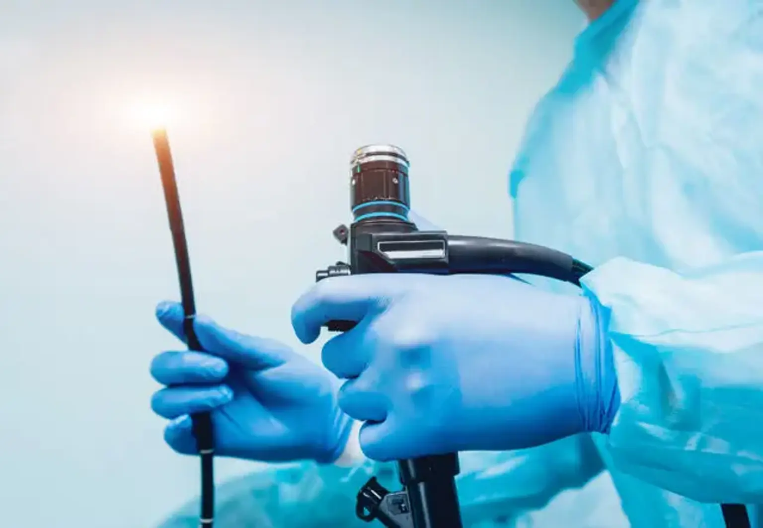
It is done with a colonoscope, a hand-held flexible tube-like device with a high-definition camera positioned at the tip and accessory channels that allow the introduction of equipment and fluids to clean the colonoscope lense and the intestinal mucosa.
The visual data that the camera provides to the screen aids in the detection of anomalies such as colonic wall overgrowth, allowing us to analyze, biopsy, and remove mucosal lesions utilizing various types of biopsy instruments via these supplementary channels. Colonoscopy has been at the forefront of making colorectal cancer an easily avoidable and early identified disease during the last few decades due to its great value.
Anatomy and Physiology
The foregut, midgut, and hindgut are the three parts of the gastrointestinal system. The foregut is made up of the mouth to the second portion of the duodenum (part of the small intestine), the midgut is made up of the remaining duodenum (third and fourth portions) to about two-thirds of the distance of the transverse colon, and the hindgut is made up of the distal one-third colon to the anus.
This is crucial to know when it comes to lesions and disease in the colon, as it will aid in future surgical planning, particularly for the colon. The small and large intestines are the parts of our gastrointestinal system that follow the stomach. The small intestine is separated into the duodenum, jejunum, and ileum as it moves from proximal to distal.
The ileum comes to an end at the ileocecal valve, which leads to the initial part of the large intestine. The cecum is a pouch-like structure that leads to the ascending colon, which is followed by the transverse, descending, and sigmoid colons. The colon enters the rectum, which leads to the anal canal. The entire distance between the anus and the cecum varies from male to female, although it is roughly 120 cm to 160 cm in most cases.
The right colon is made up of the ascending colon, hepatic flexure, and transverse colon, whereas the left colon is made up of the descending colon, splenic flexure, and sigmoid. The diameters of the cecum, transverse colon, and descending colon are 9 cm, 6 cm, and 3 cm, respectively, as a general rule. When going through a colonoscopy and determining the position within the colon, it's crucial to keep this in mind.
The superior mesenteric artery (SMA) and the inferior mesenteric artery (IMA) are the two main arteries that deliver blood to the colon (IMA). The SMA and IMA branch out into a number of additional arteries that nourish various parts of the colon (Media 1 and 2). The SMA provides nutrition to the small intestines, cecum, ascending colon, and proximal two-thirds of the transverse colon, whereas the IMA provides nutrition to the distal one-third, descending colon, sigmoid colon, and proximal rectum.
It's crucial to understand whether surgical intervention is required and appropriate anatomic resections are taken into account.
The colon, like the small intestine, is made up of various layers (from intraluminal to extraluminal):
- Mucosa
- Submucosa
- Muscularis
- Serosa
While there are several noteworthy changes between the large and small intestines, the organization of the muscularis layers is one of the most significant. Circular muscle fibers and longitudinal muscle fibers make up the muscularis layer.
The longitudinal layer is organized into three bandlike formations termed the taenia coli, unlike the small intestine, where these fibers run throughout the intestinal wall. The colon also has haustra, which are outpouchings. When doing a colonoscopy, keep this in mind because these haustra create folds throughout the colon that can conceal polyps if not correctly observed.
The length and diameter of the colon are important when it comes to colonoscopies. The estimated lengths are listed below:
- Anal canal: 4 cm
- Rectum: 15 cm
- Sigmoid colon: 50 cm
- Descending colon: 10 cm
- Transverse colon: 50 cm
- Ascending colon 10 cm
- Cecum: 5 cm
Despite generalized length estimations, different landmarks and variances in colonic diameter, color tones, vascular, and morphology aid in directing location, as the colonoscope might loop on itself and offer an inaccurate length measurement.
Indications
Colonoscopy can be done for a variety of reasons. There are two types of indications: diagnostic and therapeutic. Screening vs. elective diagnostic indications are two types of diagnostic indications. Colorectal cancer screening colonoscopies are performed to assess a patient's risk for colorectal cancer (average vs. high).
As long as the colonoscopy results were unremarkable or no pathology was detected that would put the patient at a higher risk, average risk screening begins at the age of 50 and continues for at least 10 years. At 10-year intervals, patients will have another colonoscopy to check for colorectal cancer or pre-malignant lesions.
Surveillance colonoscopies are the colonoscopies that follow. On the basis of the outcomes of the first (index) procedure, earlier surveillance is carried out.
The screening technique is performed on patients who have a high risk of developing colorectal cancer before they reach the age of 50, and it is repeated every 1, 2, or 5 years depending on the primary risk and findings during the procedure. A history of inflammatory bowel disease, a family history of colorectal cancer before the age of 60, hereditary polyposis (such as Peutz Jegher syndrome and Familial Adenomatous Polyposis, caused by an APC gene mutation) and non-polyposis syndromes (LYNCH I and II), and surveillance after colorectal cancer resection are all examples of high-risk populations.
Individuals with first-degree relatives who have been diagnosed with colon cancer should have their first colonoscopy at the age of 40, or 10 years before the relative was diagnosed, whichever comes first. For reasons such as known or occult gastrointestinal bleeding or stool positive for occult blood, unexplained changes in bowel habits or patterns, iron deficiency anemia or weight loss in elderly patients, persistent abdominal pain, suspected inflammatory or infectious colitis, and barium enema showing radiographic structural abnormalities, an elective colonoscopy is performed.
Excision and ablation of lesions, treatment of bleeding lesions, dilatation of stenosis or strictures, foreign body removal, decompression of colonic volvulus or megacolon, and palliative therapy of known neoplasms are all therapeutic indications for colonoscopy.
United States Preventative Services Task Force is a group that makes recommendations for medical treatments and procedures. Patients between the ages of 50 and 75 who are at an average risk for colorectal cancer should get a colonoscopy every ten years, according to recommendations.
Other screening methods, such as yearly fecal occult blood tests (FOBT) with flexible sigmoidoscopy every 5 years and fecal immunochemical testing, are also available, but they are beyond the focus of this study and are normally reserved for people at average risk of colorectal cancer. In high-risk populations, these screening methods are not recommended.
While colonoscopy is most commonly used to diagnose colorectal cancer, it's also used to diagnose, treat, and plan surgical procedures for a variety of inflammatory, mechanical, and anatomic problems. Crohn's disease, ulcerative colitis, Ogilvie syndrome, diverticulitis, and sigmoid volvulus are only a few examples.
Contraindications
While colonoscopy is used to diagnose acute or chronic pathology, there are various contraindications that should be considered before proceeding with the procedure. Naturally, in order to proceed with any operation, especially a colonoscopy, a consenting patient is required. If the patient has little desire to proceed, the bowel preparation, which is detailed elsewhere in this article, can be unpleasant and difficult to tolerate. In addition, when selecting whether or not to have a colonoscopy, active inflammation should be taken into account. Inflammation from toxic megacolon, fulminant colitis, ulcerative colitis, Crohn's flares, diverticulitis, and other conditions can cause this.
While colonoscopy is used to diagnose acute or chronic pathology, there are various contraindications that should be taken into account before proceeding with the procedure. To proceed with any surgery, particularly a colonoscopy, a consenting patient is required. If the patient has little desire to continue, the bowel preparation, which is detailed elsewhere in this article, can be painful and difficult to tolerate.
Furthermore, when selecting whether or not to have a colonoscopy, active inflammation should be considered. Toxic megacolon, fulminant colitis, ulcerative colitis, Crohn's flares, diverticulitis, and other conditions can all cause inflammation.
The procedure
Equipment
A colonoscopy does necessitate the use of specific devices as well as a team of people who are familiar with the process. High-definition monitors, an insufflation device that creates positive pressure within the intestinal lumen, a suction device, several different gripping devices, and equipment for administering irrigation to the gut will all be included in a colonoscopy suite.
A colonoscope is a long cylindrical tube that can be maneuvered by a single person and is flexible, robust, and cleanable. Endoscopes are available in both pediatric and adult sizes. The scope's length varies between 160 and 180 cm, with a diameter ranging from 1.0 to 1.2 cm, depending on the manufacturer and scope size. There are several important functional pieces at the end of the colonoscope.
Two to three lenses are used to help offer depth and clarity to images, two light-emitting diodes (LED) are used to provide suitable illumination of the colonic mucosa, and two small holes serve as working ports for passing irrigant and tools through.
The colonoscope's handle is designed to be gripped with the left hand, while the right hand manipulates the scope and advances/retracts it near the anus. Depending on the scope brand, there is a dial at the base of the handle that can be used to raise or decrease the scope's stiffness. This enables the colonoscope to be maneuvered around tight corners and to be pulled and pushed with varying degrees of stress.
Too much rigidity can cause discomfort for the patient and increase the risk of perforation, therefore take caution when increasing it during the colonoscopy. Several functioning devices are also attached to the handle, allowing for manipulation of the scope's tip, image quality adjustment, and control of numerous videography choices throughout the colonoscopy.
The wheels to the right of the handle are two noticeable controls on the scope. These are designed to be operated by the left hand's thumb and index finger, though the right hand can help if necessary. The bigger wheel, placed proximal to the handle, allows the scope to move upward and downward, while the smaller wheel, located distal to the handle, allows the scope to move left and right.
These, together with the colonoscope's rotational aspect, provide for a wide range of motion and movement, allowing for improved colon viewing. Depending on the scope model and manufacturer, the head of the scope has a viewing angle of 140 degrees to 170 degrees while it is not moving. This, combined with the mobility of the scope's end, gives you a nearly 360-degree vision in any direction.
It's also worth noting that two switches, proximal and distal to the two wheels, are employed to lock the scope's up/down, left/right characteristics. Before beginning the surgery, make sure these controls are turned off and that the scope's tip slides freely before inserting it into the anus. Failure to do so may cause discomfort to the patient as well as harm to the anus' sensitive tissue.
Furthermore, this will complicate the procedure by making it more difficult to guide the scope through tight junctions and turns, increasing the danger of unintentional perforation. For a visual representation of a colonoscopy.
As previously stated, the two small holes allow for the passage of equipment and irrigation. These functional ports are essential for a colonoscopy's good viewing and therapeutic benefits. The working port can accommodate long wires and equipment. Grippers, needles, snares, and clip appliers are all examples of this.
Stopping a bleeding vessel, removing a suspicious polyp, tattooing a mass for future surgical planning, or cauterizing worrying and bleeding tissue are all possible treatments.
Personnel
It's critical for patient safety that the right people are in the room, aiding an experienced physician with the surgery. At a minimum, this would comprise a nurse to administer medications and a technician to assist with device setup, patient positioning, and troubleshooting. With an extra set of hands, it's easier to manipulate the patient and apply counterpressure to the abdomen as necessary. It has been proven that having an expert assistance improves patient happiness and colonoscopy success.
Preparation
Preparation for a colonoscopy is the most common complaint among patients regarding the procedure, and it is a major cause for screening colonoscopies not being completed. Appropriate intestinal preparation is required for a colonoscopy to properly detect malignant or pre-malignant lesions and reduce the need for repeat colonoscopies.
Failure to have enough intestinal preparation can be a contraindication to undergoing the surgery, as it raises the risk of perforation and false-negative results. Several studies have shown that using an appropriately cleansed colon before a colonoscopy improves patient care and early cancer detection.
This, along with a number of other parameters such as the time to the cecum, ileocecal intubation rate, and withdrawal time, are indicators of the success of an adequate colonoscopy.
The foundation of proper bowel preparation is straightforward: the colon should be free of stool and the colonic mucosa should be visible. While the theory is straightforward, the method for obtaining it can be a challenging aspect of colonoscopy and a source of discomfort for people.
Polyethylene glycol, magnesium citrate, magnesium hydroxide, bisacodyl, and sodium picosulfate are just a few of the drugs and oral laxatives that can be used in a bowel preparation routine. The best bowel cleansing regimens have been determined through several investigations. Each doctor has their own routine that they've devised and discovered to be the most effective in their practice. When deciding on a bowel cleansing routine, however, various aspects must be taken into account.
While the colon must be clean, stool will never be completely removed. Patients should be informed that they will continue to produce stool, which may or may not be colored. The key to the bowel cleanse is that the feces is liquid and can be cleansed rapidly with a colonoscope's irrigation function. There is no perfect cleansing program that is painless to date. The day before the surgery, patients should follow a clear liquid diet.
This, together with the chemical cleansing agent, gives the colonoscopy the best chance of success. Patients who cannot endure a 24-hour cleanse may be able to follow a clear liquid diet for up to three days before to the procedure, along with a light laxative regimen, to aid with colon cleansing.
Enemas can be used before a colonoscopy, but they do not provide adequate preparation or a thorough bowel cleaning on their own. This is owing to an enema's limitations in terms of reaching the transverse colon, ascending colon, and cecum. They may give a last cleanse for the rectum and sigmoid colon, but their application is limited.
Colonoscopy antibiotic prophylaxis is a hotly debated topic. While there are no clear evidence that preventive antibiotics should be used in colonoscopies, adjusting antibiotic use based on patient risk factors should be considered. Peritonitis after colonoscopy and polypectomy may be more common in immunocompromised patients, those who undergo peritoneal dialysis, and those who have diabetes. These patients should be taken into account, as they may benefit. Antibiotics are unlikely to be necessary in the general population.
Any decisions and procedures should be properly explained to the patient, and any questions should be answered before starting. It is especially beneficial to sit with and discuss the procedure steps and the required bowel preparation prior to a colonoscopy.
According to studies, this form of education results in more effective bowel preparation than merely distributing a brochure or leaflet. Colonoscopies with incomplete bowel preparations have been recorded in as many as 10% to 20% of cases. By explaining the bowel preparation timeframe and patient expectations in person, you can improve patient compliance and ensure a successful colonoscopy.
Technique
Colonoscopy is a difficult skill to learn and requires a lot of practice. While it may appear straightforward to observe an experienced doctor perform a colonoscopy, the process takes time, patience, and a lot of practice. It's not easy to navigate inside a cylindrical tube that can flex, dilate, contract, and move. In addition, when preparing for a colonoscopy, patient factors such as obesity, superfluous colonic tissue, surgical history, bowel preparation compliance, and underweight persons must all be taken into account.
It takes several hours on a simulator and proctored colonoscopies with an experienced surgeon or gastrointestinal specialist for a physician to become proficient in colonoscopy.
Before beginning the colonoscopy, it is critical that the clinician is familiar with the equipment and has tested it. The colonoscope, as previously stated, has numerous components. Manipulation of all scope elements (up, down, right, left, clockwise, and counterclockwise rotation) adds to the procedure's complexity.
The patient should be in the lateral decubitus posture on the left side. If circumstances require, certain professionals may prefer the patient to be on their back or right side. To aid prevent injury to the bony prominence and maintain position in the left-sided position, the patient's legs should be flexed and pillows should be positioned around their back, head, and between their knees. The technician or nurse's job is to keep the patient stable and prevent him or her from rolling forward or backward.
They're also there to aid the endoscopist navigate corners and turns by applying counter pressure to the abdomen. The puborectalis and pubococcygeus muscles are relaxed when the legs are flexed toward the chest. This makes it easy to enter and traverse through the sacral prominence's angle. Navigation past the rectum at the level of the sacrum becomes more difficult if this position is not achieved.
Behind the patient, the endoscopist should be. A digital rectal exam (DRE) with a water-soluble lubricant is required before inserting the scope. The clinician should feel for any lumps, bulges, rectocele, or masses at this time. They should also record the rectal tone, but they must also consider the level of sedation the patient has obtained, since this may impact the anus tone.
Following the DRE, the physician should hold the scope in his or her left hand and the endoscope in his or her right hand, around 10 cm to 20 cm from the scope's working end or lense. To aid prevent harm to the sensitive tissue of the anus, a substantial amount of water-soluble lubricant should be applied once more.
The clinician can use certain landmarks to help them figure out where they are in the colon. While broad anatomy distances are valuable, using the scope to measure them is not always accurate. Keep in mind that the scope can twist and turn on itself and will not offer an exact measurement further into the colon.
Complications
There are inherent complications with any invasive operation that should be known and described to the patient before beginning. Generic problems include anesthetic reactions like stroke, heart attack, respiratory distress, and death are always connected with sedating operations and should be discussed during consent. Patients should be informed that colonoscopies have the risk of rectal rips, bleeding, pain, bloating, and infection.
Intestinal perforation is a more significant consequence that can occur, albeit it is uncommon. Perforation risk has been estimated to be around 0.14 percent (or 1 in 1000 colonoscopies). In general, the risk of perforation associated with colonoscopy is modest, and the operation is relatively safe. As a result, it is a great screening technique for colon cancer with a low risk.
Perforation can happen at any point in the colon or terminal ileum. The most common site of perforation, however, has been described numerous times as the sigmoid colon. Shear injury by pushing and pulling on the colonic wall, overdistention from too much insufflation against a weaker colonic wall, and perforation via mechanical polypectomy and electrocautery are the most common causes of perforation.
Each turn or corner in the colon serves as a focal point against which the scope can press to proceed through the colon. Manipulation methods involving the assistant pressing against the abdominal wall can help reduce push and pull forces and prevent damage. As the scope proceeds to the cecum, the sigmoid colon, as the first corner or turn in the large intestine, has the largest risk of injury since it receives the most force.
Conclusion
Colonoscopy is an endoscopic examination of the large intestine and the distal portion of the small bowel using a camera or fiber optic camera on a flexible tube inserted via the anus. It can offer a visual diagnosis (e.g., ulceration, polyps) and allows for the biopsy or excision of colorectal cancer lesions.
GGI bleeding, unexpected changes in bowel habits, and suspicion of cancer are all reasons for a colonoscopy. Colonoscopies are commonly used to detect colon cancer, but they are also used to detect inflammatory bowel disease. An unexplained decline in hematocrit (one symptom of anemia) in older individuals (and occasionally even younger ones) warrants a colonoscopy, generally in conjunction with an esophagogastroduodenoscopy (EGD), even if no visible blood is detected in the stool.