Colposcopy

Last updated date: 08-Jun-2023

Originally Written in English

Colposcopy

Cervical cancer was diagnosed in 527,500 people globally in 2012, and 265,500 people died from it, making it the fourth most commonly diagnosed cancer and the fourth greatest cause of cancer mortality among women. It is the second most frequent cancer in developing countries and the third leading cause of mortality after breast and lung cancers. In high-income nations with good cervical cancer screening systems, however, the incidence of cervical cancer has dropped by >82 percent during the last 40 years. This decline can be linked to the widespread use of Pap cytology and colposcopy to diagnose and treat premalignant cervical lesions. Cervical cancer was the seventh most prevalent cancer among women in the Republic of Korea in 2014, with 3,550 women diagnosed, representing an annual percent change of -3.8 percent in cervical cancer occurrence since 2000. When abnormal Pap test results are discovered, cervical conization was routinely performed in the past, but in recent years, the focus has shifted to choosing the best treatment option based on the biopsy findings via colposcopy. Cervical conization has become less indiscriminate as a result of this.


What is a Colposcopy?

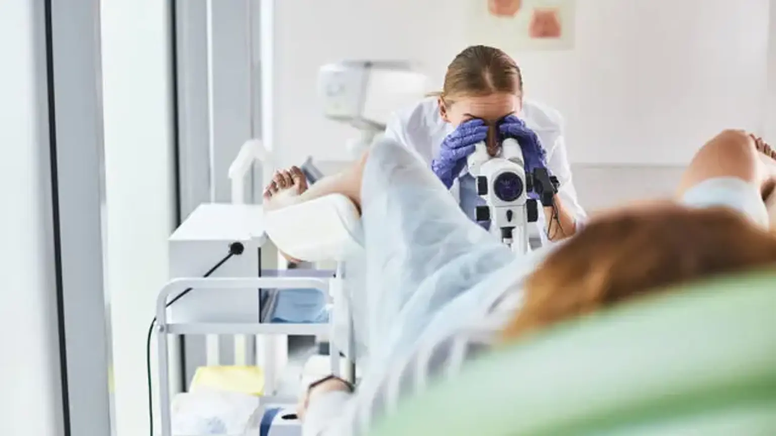
Colposcopy is a practice in which the cervix, vagina, and vulva are examined using a lit, magnifying device called a colposcope. Colposcopy was first described as a cervical cancer screening method by Hans Hinselmen of Germany in the1920s. It is a diagnostic procedure used to assess women who have had an abnormal Papanicolaou (Pap) test, women who have had a visual inspection with acetic acid (VIA), women who have tested positive for high-risk human papillomavirus (HPV) DNA, or women who have a suspicious-looking cervix even if the PAP test is normal. It's also used to monitor intraepithelial and invasive carcinoma patients after treatment.

Advanced practice physicians, family medicine practitioners, gynecologists, gynecological oncologists, and some internal medicine doctors use colposcopy. This process has a lack of uniformity, as well as training and development based on daily, monthly, or less regularly conducted procedures. Colposcopy is well known for its high variability and low reliability among colposcopists. To address these and other concerns, the ASCCP (American Society for Colposcopy and Cervical Pathology) issued colposcopy standards in 2017. The uniformity of nomenclature was adopted to make things easier and to ensure that every meeting included a thorough colposcopic examination.


Colposcopy Indications

Colposcopy Indications

The need for a colposcopy is depending on the danger involved. Based on their cytological findings, HPV screening if it was done, and personal history of cervical dysplasia, women referred for colposcopy have a spectrum of underlying risks for cervical pre-cancer. Each can be triaged appropriately, but when colposcopy is required, it is utilized to determine whether dysplasia is present and the severity of the dysplasia.

Colposcopy is indicated when:

  • Women who have had a Pap test that is abnormal.
  • Women who have a positive test for high-risk HPV DNA.
  • VIA-positive women.
  • Even if the Pap smear is normal, a suspicious-looking cervix and postcoital/postmenopausal bleeding should be evaluated.
  • Bleeding in the lower genital tract that is unexplained.
  • Cervical cytology that persists inflammatory or unsatisfactory despite treatment, especially in patients with high-risk characteristics for cervix carcinoma.
  • Examining chronic abnormal vaginal discharge or vulvae pruritus.
  • Identification and treatment of papillomavirus infection in the subclinical stage.
  • Diethylstilbestrol (DES) exposure during pregnancy
  • Intraepithelial neoplasia is treated with caution.
  • Cervical neoplasia that has spread to the vaginal region to be identified and managed.
  • Follow-up after treatment.
  • After intraepithelial and invasive carcinomas have been treated.
  • Follow-up after radiotherapy.

As a result, while many Pap tests require a colposcopy, not all of them do. Low-risk Pap tests, such as those that reveal a low-grade squamous intraepithelial lesion (LSIL) or atypical squamous cells of undetermined significance (ASCUS) with no HPV, are less likely to reveal major colposcopic abnormalities that lead to severe dysplasia. As a result, urgent colposcopy is not necessary, and the patient can have a repeat Pap test a year later.


Colposcopy Contraindications

If the Pap test continues abnormal the next year, with LSIL or ASCUS positive for HPV, colposcopy is suggested. Some Pap test results are more tightly correlated to severe cervical dysplasia than others. High-grade squamous intraepithelial lesions (HSIL) and atypical squamous cells (ASC-H) are among them; nonetheless, high-grade intraepithelial lesions cannot be ruled out. When high-grade lesions are suspected, there is a chance that invasive cervical cancer is involved. In patients with initial Pap test findings that are more closely associated with severe cervical dysplasia, immediate colposcopy is recommended.


Colposcopy Equipment

Colposcopy Equipment

Except for an ongoing or unresolved cervical or vaginal infection, there are no definite contraindications to a colposcopy. Certain steps of the colposcopy technique are not performed if a patient is pregnant. Due to the risk of harmful effects on the pregnancy and the lack of benefit, the endocervical curettage aspect is not performed. Furthermore, because cervical excisional procedures are prohibited during pregnancy, pregnancy can limit therapeutic options. A vaginal speculum, a colposcope, 5 percent acetic acid, Lugol's solution, biopsy forceps, an endocervical speculum, a Kevorkian curette or endocervical brush, and a solution or means to stop bleeding are all required for a proper colposcopy. A dissecting microscope that can magnify cervical, vaginal, or vulvar tissue is known as a colposcope. Colposcopes come with a variety of lens options, computer-generated pictures, light filters, and even cameras to capture pictures or videos. For evaluating a lesion, colposcopes should have two settings: low power and high-power magnification.

The magnifications of 10x and 18x are interchangeable in most scopes. In order to discover vascular patterns that are difficult to perceive with white light, the scope should contain both normal light and a green light filter. A cotton ball soaked in 5% acetic acid is placed on the cervix and left to soak for 2 to 3 minutes. When dysplastic cells are exposed to acetic acid, they dehydrate and turn acetowhite. When first applied, this may cause very mild discomfort to the patient. The cervix and upper vaginal tissue should be thoroughly examined in all directions. Some colposcopists will use Lugol's solution to accentuate the dysplastic area, which will turn yellow due to the brown solution's lack of absorption. This is known as Schiller's test. A region that does not stain with iodine is a Schiller positive test. In order to properly inspect the cervical os, an endocervical speculum may be required. Tischler cervical biopsy punch forceps, Burke biopsy forceps, or some variant of these are the most common kind of biopsy forceps used for cervical biopsy.

There are several ways to control bleeding following a biopsy, including utilizing Monsel's solution, silver nitrate, or even Bovie cautery. There's also a new technology called digital video colposcopy, which uses an integrated camera and a powerful light source to offer magnification and lighting. The colposcopic image is projected on a high-resolution video monitor, and no binocular eyepieces are needed. It offers various advantages, including ease of manipulation and simultaneous co-visualization of images by several observers, including trainers and the patient. It produces a permanent record of the findings in the form of a copy of the image that the examiner is viewing.


Personnel

It is critical to consult with a skilled colposcopist. Having a helper to handle the tools and specimen containers during the procedure is beneficial, although they can also be handled independently. Because this is an invasive practice, there should always be a chaperone present.


Prepare for a Colposcopy

The patient does not need to prepare for the colposcopy; but, due to obscuring blood, it may be difficult to conduct if she is on her menstrual cycle. The patient's visit will be shorter if the appropriate equipment is easily available in the room.


Colposcopy with Biopsy

Colposcopy with Biopsy

Standardization criteria for colposcopy have been released by the ASCCP (American Society for Colposcopy and Cervical Pathology). The ASCCP gives recommendations for both extensive and minimal colposcopy requirements. The vulva, vagina, and cervix should be examined grossly in their natural form as well as after the addition of 5% acetic acid. For adequacy, the complete cervix and SCJ (squamocolumnar junction) must be shown. To detect any lesions, the visual field should be lighted with both white light and a red-free filter.

Each aberrant finding should be subjected to targeted biopsies. The visibility extent, size, position, and description of each lesion, presence or absence of acetowhitening, complete or incomplete visibility of the SCJ, documentation of biopsies and sites, if an endocervical curettage was conducted, and finally the colposcopy impression (benign-normal/low grade/high grade/cancer) should all be documented in a minimum of text format. After the colposcopy has been performed and all biopsies have been obtained, Monsel's solution or silver nitrate should be used.


Colposcopy Results

Document the cytological abnormality's grade, colposcopic adequacy, visibility, and SCJ type. The lesion's site, size, and extent, as well as any endocervical or vaginal extension, should all be noted. Colposcopic observations should be documented in detail by region, and a colposcopic impression should be given in terms of a low-grade or high-grade lesion, as well as Reid's/ Swede score. Colposcopy alone should never be used to make a histopathologic diagnosis.


Abnormal Colposcopy

Abnormal Colposcopy

You may not need to take any action right away if the doctor or nurse discovers abnormal cells. You may require additional tests or treatments at times. It depends on the degree to which the cervical cells are aberrant.

The doctor or nurse may advise you to wait and see if the cells heal on their own. You will undergo another Pap test and possibly other tests to monitor the cells in this case.

The biopsy may also serve as a treatment. This is because, during the biopsy, the doctor may be able to remove all of the problematic cells. You won't need any more treatment if this is the case. You will begin undergoing pelvic exams, Pap tests, and/or HPV tests on a regular basis. The doctor or nurse will advise you on how frequently you should get these.

You may possibly require additional treatment. There are four treatments for eliminating aberrant cells and avoiding cervical cancer that are very effective.

  • Cryotherapy. It is the freezing of aberrant cells.
  • Loop Electrosurgical Excision Procedure (LEEP). In which aberrant cells are eliminated by passing an electrical current through a narrow wire loop.
  • Laser therapy. A laser is used to eradicate unhealthy cells.
  • Cone biopsy. A cone-shaped wedge is carved from the cervix to remove the abnormal cells during a cone biopsy.

It's critical to continue undergoing regular pelvic examinations after you have been treated for abnormal cells, regardless of the treatment you get. Even though these techniques are very efficient, aberrant cells do occasionally reappear.


Colposcopy Side Effects

Colposcopy Side Effects

Based on the patient's history, colposcopy complications are likely to be attributable to an obscured visual field, significant atrophy, or scarring. Significant hemorrhage, infection, and long-term morbidity are all minimal procedure risks. Anxiety and patient discomfort are bad side effects of the operation that should not be overlooked, although it can be hard to ascertain if the negative emotions are connected to the concept of HPV infection or the procedure itself. Colposcopy performed by an inexperienced doctor has the potential to cause injury.

Competency in colposcopy requires ongoing training and experience. Colposcopy has a false-negative rate of 14 percent to 70 percent (missed high-grade squamous intraepithelial/invasive carcinoma). With cytology, HPV molecular testing, and risk-based evaluations, there are now better screening techniques. As a result, diagnostic testing is less necessary with colposcopy, necessitating the engagement of a more experienced and professional colposcopist.


Sources of Errors in Colposcopy

Every colposcopic image reflects a unique tissue pattern formed by the interaction of surface epithelium and stroma. The most prevalent error in colposcopy is a misinterpretation of patterns. A flat, mild acetowhite grade 1 lesion that mimics immature or active metaplastic epithelium in young women, regenerative epithelium, subclinical HPV infection, and congenital transformation zone is more probably to be over-diagnosed. These lesions must be biopsied if there is any uncertainty. Colposcopy should be avoided while the epithelium is regenerating after CO2 laser ablation, cryosurgery, or trauma. In cases where the cervix is hidden from view by an endocervical polyp or large retention cyst, or there is a stenotic internal os, or in cases of an incomplete view of the squamocolumnar junction colposcopist has difficulty in making the diagnosis.

Colposcopy in postmenopausal women can be challenging, with 26% of women having an unsatisfactory colposcopy due to insufficient visibility of the squamocolumnar junction and vaginal atrophy. Because of the physiological and morphological changes that occur during pregnancy, errors are possible. During pregnancy, vasodilation and congestion cause exaggerated colposcopic patterns with more prominent mosaics and punctuations, as well as an increased acetic acid effect, which may resemble paraneoplastic lesions. Colposcopic changes in pregnancy are one grade higher than non-pregnant cervix, therefore using a large speculum wrapped with a condom, quadrant wise interpretation, and remembering that colposcopic abnormalities in pregnancy are one grade higher than non-pregnant cervix will help reduce them. If a colposcopic biopsy is needed during pregnancy, it is safe, but endocervical curettage is not. The use of an endocervical brush for cytology, on the other hand, is completely safe.


Clinical Significance

Colposcopy is a diagnostic procedure used when a cervical screening test or an exam reveals a visible lesion on the cervix. This diagnostic process aids in the formation of a management strategy based on the biopsy pathology findings, or the absence of it. In general, all results can be observed or managed, and they are all based on scientific data. ASCCP guideline algorithms can be used to monitor and manage low-grade lesions. Treatment for high-grade lesions is determined by the patient's age and fertility state.

Unless there is a specific risk for an invasive lesion, a pregnant patient's therapy will be postponed until after birth. A colposcopy that is deemed insufficient for a variety of reasons may necessitate a more aggressive collection of the cervical tissue followed by a cervix excisional procedure to confirm the diagnosis. Treatment options for invasive lesions should be discussed with a gynecological oncologist.


Improving Colposcopy Outcomes

Improving Outcomes

Colposcopy is a disappearing art, yet it is an essential step in the fight to avoid cervical cancer. Multiple types of practicing physicians can learn how to do this operation. Colposcopic services are provided in a variety of settings in the United States, including academic and non-academic referral settings, primary care settings in urban and rural regions, and private and public funding.

Because of improved screening testing, risk-based recommendations, and information consolidation by organizations like the ASCCP, there are fewer patients available and training of a range of practitioners should be encouraged and perpetuated. In order to optimize patient care, clinicians should adhere to prescribed procedure uniformity and documentation. Standardization of technique and documentation will improve communication between practitioners and pathologists in the same way that standardization of vocabulary has. Technology advancements may continue to improve the colposcopy procedure's reliability and validity.


Conclusion

Colposcopy

Cervical cancer and premalignant lesions have begun to decline as public health and cleanliness have increased, as well as the introduction of HPV vaccination. A decline in the number of patients who require colposcopy has been proven to have an impact on colposcopy education, necessitating the development of new approaches to colposcopy teaching. Furthermore, biopsies on two or more locations are indicated to improve colposcopy accuracy. This year marks the tenth anniversary of the HPV vaccines' release. In the Republic of Korea, girls turning 11 years old have been receiving free cervical cancer immunization since last year. Because of the cervical cancer vaccine, premalignant lesions associated with HPV types 16 and 18 are predicted to diminish even further in the future. On colposcopy, lesions associated with HPV type 16 are visible, but as the number of lesions decreases, colposcopy practices are likely to become more challenging.