Introduction
What Are Craniofacial Anomalies?
Craniofacial anomalies refer to deformities or abnormalities in the bones of the skull and face, either present from birth or caused later in life due to trauma, disease, or other factors. These conditions can affect appearance, function (like breathing, eating, or speaking), and psychological well-being.
There are many types of craniofacial anomalies, ranging from common issues like cleft lip and palate to more complex conditions like craniosynostosis, where the skull bones fuse too early.
Importance of Craniofacial Anomaly Correction
Correcting craniofacial anomalies is vital not just for cosmetic reasons but for improving the overall function of the face and skull. Surgery can help restore the proper form, allow for better functionality, and boost a patient’s self-esteem, reducing social stigma. Early intervention is often critical in achieving the best outcomes.
Causes and Types of Craniofacial Anomalies
Congenital Craniofacial Disorders
Congenital disorders are present at birth, with cleft lip and palate being the most common. Other conditions include craniosynostosis, a premature fusion of skull bones that limits skull growth, leading to facial abnormalities. Many craniofacial anomalies are genetic, and syndromes like Apert syndrome or Treacher Collins syndrome can lead to facial deformities.
Acquired Craniofacial Anomalies
Acquired anomalies can result from trauma (e.g., fractures or injuries), tumors, infections, or other external factors that cause changes in the facial structure. These conditions might occur at any age and can affect both the appearance and functionality of the face.
Genetic Factors and Syndromes
Genetic mutations can play a significant role in craniofacial anomalies. Syndromes like Crouzon or Pfeiffer syndrome often lead to abnormal skull and facial bone development, requiring surgical correction to prevent complications.
The Role of Craniofacial Surgery
What is Craniofacial Surgery?
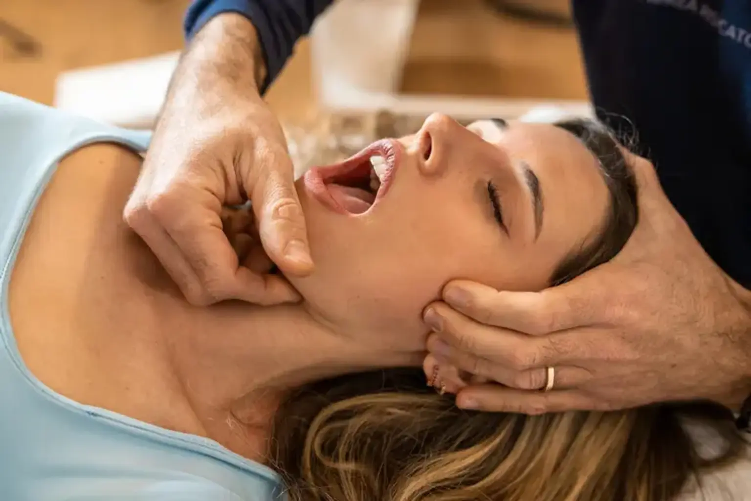
Craniofacial surgery involves the correction of facial and skull deformities, using a range of procedures to restore both form and function. The aim is not only aesthetic improvement but also the enhancement of speech, breathing, and eating functions. It may involve bone reshaping, soft tissue reconstruction, or tissue expansion.
How Craniofacial Surgery Works
The specific techniques used depend on the condition being treated. For example, cleft lip and palate repairs often require soft tissue procedures, while craniosynostosis may require more complex bone reshaping. In some cases, advanced technologies like 3D imaging or virtual planning can be used to ensure precision.
The Role of Plastic Surgeons and Maxillofacial Surgeons
Craniofacial surgery is typically performed by highly specialized plastic or maxillofacial surgeons, trained in both reconstructive surgery and aesthetic procedures. These professionals work in teams to provide the best outcomes for patients, especially in cases involving complex anomalies.
The Importance of Early Diagnosis and Intervention
How Early Treatment Affects Outcomes
Early intervention, especially in children, is crucial. The earlier craniofacial anomalies are treated, the better the outcome in terms of both function and appearance. For instance, repairing a cleft lip or palate early can reduce speech and hearing issues later in life.
Pre-Surgical Assessments
Before surgery, a thorough medical assessment is required to evaluate the child’s health, the severity of the condition, and any underlying factors. Imaging tests, like CT scans or MRIs, may be used to get a clearer picture of the skull and facial bones. Psychological support is also important, especially in young patients, to prepare them for surgery and recovery.
The Procedure: What to Expect from Craniofacial Anomaly Correction
Preparation for Surgery
Before surgery, patients undergo a thorough evaluation, including physical exams, imaging tests (CT or MRI), and consultations with the surgical team. This helps determine the best approach and ensures safety during the procedure. Psychological support may also be provided to help manage expectations and ease any anxiety.
Surgical Process and Techniques
The surgery itself varies depending on the anomaly being corrected. For conditions like cleft lip or palate, surgeons repair the lip and palate tissues, restoring the normal function of the mouth. In cases like craniosynostosis, surgeons may need to reshape the skull to allow proper brain growth. Advanced techniques such as bone grafting and tissue expansion are often used to optimize results.
Anesthesia and Pain Management
Patients are typically given general anesthesia, ensuring they remain unconscious and pain-free during the surgery. Post-surgery, pain management strategies include medications and local anesthesia to minimize discomfort. Recovery times vary but are closely monitored by the surgical team to ensure proper healing.
Recovery and Post-Operative Care
Initial Recovery
After surgery, patients may spend a few days in the hospital for observation. This allows doctors to monitor any complications like bleeding or infection. For children, there may be a need for extra comfort and care during their stay. During this time, a nutritional plan is also implemented to ensure proper healing.
Post-Surgical Care and Follow-Up
Once discharged, patients need to follow a strict post-operative care routine, which includes wound care, scar management, and avoiding any activities that might stress the healing area. Regular follow-up appointments are essential to track progress and ensure the surgical site is healing as expected.
Emotional and Psychological Support
Recovery isn't just physical. Craniofacial anomaly correction can significantly impact a person’s mental and emotional well-being. Emotional support, counseling, and peer groups help patients, especially children, adjust to their new appearance and manage any psychological stress during the healing process.
Risks and Complications of Craniofacial Anomaly Correction
Possible Risks
As with any surgery, craniofacial correction comes with risks such as infections, bleeding, or anesthesia complications. Nerve damage and scarring are also potential concerns, though skilled surgeons work to minimize these. There may also be issues with bone healing, especially in complex cases.
Long-Term Effects
While the surgery is highly effective, some patients may require additional procedures later on to address any growth-related changes or residual deformities. These revisions may involve small adjustments to optimize the final appearance and function.
Managing Complications
In rare cases where complications occur, prompt medical attention ensures that problems like infections or incorrect bone healing are managed. Surgeons may recommend further treatments, such as corrective surgeries or therapies, to address these issues and achieve the best possible outcome.
Advances in Craniofacial Anomaly Correction
Technological Advancements
In recent years, craniofacial surgery has seen major advancements, particularly with the integration of 3D imaging and virtual planning tools. These technologies allow surgeons to plan and simulate the surgery before performing it, improving precision and reducing risks. Robotic-assisted surgery is also becoming more common, offering greater control during delicate procedures.
Improvement in Surgical Materials and Techniques
Innovations in surgical materials, such as biocompatible implants and advanced bone grafting techniques, have improved healing times and the overall aesthetic results of surgery. Tissue regeneration methods are also evolving, allowing surgeons to grow and utilize natural tissue for more effective reconstructions.
Global Access to Advanced Craniofacial Surgery
Access to craniofacial surgery has improved globally, thanks to outreach programs and telemedicine, making high-quality care available to patients in underserved regions. Patients from different parts of the world now have access to world-class treatments, ensuring that craniofacial anomaly correction is accessible to all.
The Impact of Craniofacial Anomaly Correction on Quality of Life
Physical and Functional Benefits
Craniofacial anomaly correction doesn’t just improve appearance; it significantly enhances quality of life. Patients often experience better breathing, eating, and speaking capabilities after surgery. For example, repairing a cleft palate can improve speech clarity, and reshaping the skull in craniosynostosis can allow for proper brain development and facial symmetry.
Psychosocial Benefits
The emotional impact of craniofacial surgery is profound. Many patients, especially children, experience a significant boost in self-esteem after surgery, feeling more confident in their appearance. This, in turn, reduces social stigma and improves mental health, helping individuals feel more integrated into their communities.
Case Studies or Real-Life Examples
Take the case of a child with a cleft lip, who, after surgery, can eat and speak more easily and enjoys greater social acceptance. Such real-life success stories highlight how transformative craniofacial surgery can be, not just physically but also in improving overall life satisfaction.
Frequently Asked Questions (FAQs)
How long does recovery take after craniofacial surgery?
Recovery time varies based on the complexity of the surgery, but most patients can expect a few weeks of initial healing, followed by months of gradual recovery. Full recovery may take a year or more, especially for bone-related surgeries.
What are the costs involved in craniofacial anomaly correction?
Costs can vary widely depending on the country, hospital, and complexity of the procedure. In general, craniofacial surgeries are expensive, but many healthcare systems offer financial assistance, and insurance may cover part of the costs.
Can craniofacial anomalies be corrected in adulthood?
Yes, many craniofacial anomalies can be corrected at any age, though the procedures may be more complex in adults. Early correction is ideal, but adult patients can still benefit from surgery, improving both appearance and function.
Are there non-surgical treatments for craniofacial anomalies?
While surgery is the most effective treatment for most craniofacial anomalies, non-surgical treatments like orthodontics, speech therapy, and prosthetics may be used as supplementary options for improving function and appearance.
What is the success rate of craniofacial surgery?
Craniofacial surgeries generally have high success rates, especially with early intervention and skilled surgeons. While some patients may require additional procedures for refinement, most experience significant improvements in both appearance and quality of life.
Call to Action
If you or a loved one is considering craniofacial anomaly correction, consulting with a qualified surgeon is the first step. Early diagnosis and a tailored treatment plan can make a significant difference in outcomes. Don’t hesitate to reach out to specialists who can guide you through the process and provide the best possible care.
The Role of Multidisciplinary Teams in Craniofacial Anomaly Correction
Collaboration Among Specialists
Craniofacial surgery often requires a multidisciplinary team of experts. Surgeons, orthodontists, speech therapists, psychologists, and pediatricians all work together to provide comprehensive care. This collaboration ensures that both the physical and emotional needs of the patient are met throughout the treatment process.
Team Approach to Comprehensive Care
For example, while a surgeon focuses on the surgical correction, a speech therapist may help with speech development after a cleft palate repair, and a psychologist can provide emotional support. This holistic approach helps achieve the best outcomes, addressing all aspects of the patient’s health and well-being.
Patient-Centered Care
The team works closely with the patient and family members to ensure that every step of the treatment process, from pre-surgery assessments to recovery, is well-understood and managed. This ensures that patients feel supported and informed throughout their treatment journey.
Long-Term Monitoring and Adjustments
Ongoing Follow-Up Care
Even after the initial recovery period, craniofacial patients require long-term monitoring to ensure proper growth and development. Regular check-ups help detect any issues early, such as bone growth problems or the need for additional surgeries.
Growth Considerations for Children
For children, ongoing evaluation is crucial as they continue to grow. For instance, facial bones may shift over time, requiring minor corrections. Parents and caregivers are guided on what to expect during these growth phases, ensuring that the child receives timely interventions.
Late-Stage Interventions
In some cases, patients may require revisions or additional surgeries later in life as their bodies change. These interventions are often less invasive but can be necessary to maintain the desired aesthetic and functional outcomes.
The Psychological Impact of Craniofacial Anomaly Correction
Emotional Challenges Before and After Surgery
Patients with craniofacial anomalies often face emotional and social challenges. These challenges can include self-esteem issues, bullying, or social isolation. Surgery can have a profound positive impact, helping individuals feel more comfortable in their own skin.
Psychological Benefits of Surgical Correction
After surgery, many patients report feeling more confident and socially integrated. For children, this may mean less teasing at school and better interactions with peers. For adults, it can lead to improved job prospects and personal relationships.
Support Systems for Mental Health
To help with the psychological adjustment, support groups and counseling are often recommended. Peer support groups, where patients share their experiences, can provide comfort and reassurance, making the process smoother for both children and adults.
Global Trends in Craniofacial Anomaly Correction
Increased Access to Advanced Treatments Worldwide
Around the world, access to craniofacial surgery has expanded. Medical tourism has grown, with patients traveling to countries offering high-quality care at more affordable prices. Countries with advanced healthcare systems are increasingly offering these treatments to underserved regions, ensuring broader access.
Rising Popularity of Minimally Invasive Techniques
Minimally invasive procedures are becoming more common, reducing recovery times and risks. Advances in techniques like endoscopic surgery or robotic-assisted surgery have improved the precision of craniofacial procedures, leading to faster recovery and better results.
Emerging Global Standards in Craniofacial Care
As the field evolves, global standards for craniofacial care are being established, ensuring that patients everywhere receive the highest quality treatment. International collaboration among surgeons and researchers continues to improve techniques and patient outcomes.
The Role of Genetics in Craniofacial Anomalies
Genetic Factors in Craniofacial Disorders
Genetics plays a significant role in many craniofacial anomalies. Conditions like cleft lip and palate, craniosynostosis, and various syndromes (e.g., Crouzon syndrome, Apert syndrome) are often inherited. Mutations in specific genes disrupt the normal development of the skull and face during fetal development.
Genetic Counseling for Families
For families affected by craniofacial anomalies, genetic counseling is an essential part of the process. Genetic counselors can help assess the likelihood of recurrence in future pregnancies and provide information on the causes of the disorder. They also offer emotional support to families coping with genetic conditions.
Advancements in Genetic Research
Recent advances in genetics are improving our understanding of craniofacial anomalies. Researchers are working to identify the precise genetic causes of these conditions, which may lead to better prevention, earlier diagnosis, and more tailored treatments in the future.
Ethical Considerations in Craniofacial Anomaly Correction
Ethics of Cosmetic Surgery
Craniofacial surgery often raises ethical questions, especially when performed on children. Parents must decide when the right time is for surgery, balancing the child’s health, appearance, and psychological well-being. It’s important to consider the long-term effects and ensure that the surgery is performed for the right reasons—improving function and quality of life, rather than purely for cosmetic purposes.
Informed Consent and Patient Autonomy
Informed consent is critical in craniofacial surgeries, particularly for minors. Parents or guardians must fully understand the risks, benefits, and alternatives. As patients mature, it's important to involve them in the decision-making process to respect their autonomy and wishes.
Equity in Access to Care
Equitable access to craniofacial anomaly correction is a growing concern. Ensuring that all patients, regardless of socioeconomic background or location, have access to high-quality care is an ongoing challenge. Many initiatives aim to provide surgery to underserved populations, but there is still work to be done.
Cost and Insurance Considerations
The Cost of Craniofacial Surgery
Craniofacial anomaly correction can be expensive, with costs varying widely depending on the complexity of the procedure, geographic location, and healthcare system. These costs typically include surgery, hospital stays, follow-up care, and rehabilitation.
Insurance Coverage and Financial Assistance
Insurance companies often cover craniofacial surgeries, particularly those that are medically necessary, such as correcting birth defects. However, cosmetic procedures or revisions may not always be fully covered. Many hospitals offer financial assistance or sliding-scale fees to help make these surgeries more accessible to patients from lower-income families.
Medical Tourism and Affordability
Medical tourism is becoming more popular for patients seeking affordable craniofacial surgery. Countries with high-quality healthcare systems, such as Thailand, India, and Mexico, offer competitive pricing without compromising on the quality of care.
The Future of Craniofacial Anomaly Correction
Advances in Technology and Techniques
The future of craniofacial anomaly correction is promising, with continuous advancements in surgical techniques, imaging technology, and robotic surgery. Minimally invasive procedures are likely to become more common, leading to faster recovery and better outcomes for patients.
Personalized Medicine
As genetic research advances, personalized treatments tailored to an individual’s unique genetic profile may become a reality. This approach could improve surgical outcomes and minimize complications by addressing the specific causes of craniofacial anomalies at a genetic level.
Global Collaboration for Better Outcomes
The growing network of international medical professionals and researchers dedicated to craniofacial care is advancing knowledge and treatment. Collaboration across borders ensures that best practices are shared, and more patients around the world can benefit from improved care.
Conclusion
Craniofacial anomaly correction plays a transformative role in the lives of individuals affected by facial and skull deformities. The advances in surgical techniques, multidisciplinary care, and long-term monitoring have greatly improved both the functional and emotional outcomes for patients. From children born with clefts to adults with complex cranial deformities, the impact of surgery extends beyond just aesthetics—it restores confidence, improves quality of life, and enables individuals to live with fewer limitations.