Congenital tracheal stenosis

Last updated date: 16-Jun-2023

Originally Written in English

Congenital tracheal stenosis

Overview

Congenital tracheal stenosis (CTS) occurs in roughly 1 in 64,500 instances and manifests as stridor or respiratory insufficiency in the neonatal period or infancy. The prognosis and progress of this lesion set are determined by the degree of the stenosis and concomitant diagnoses. Some children require intubation and mechanical breathing for rescue, although extracorporeal support is rarely necessary.

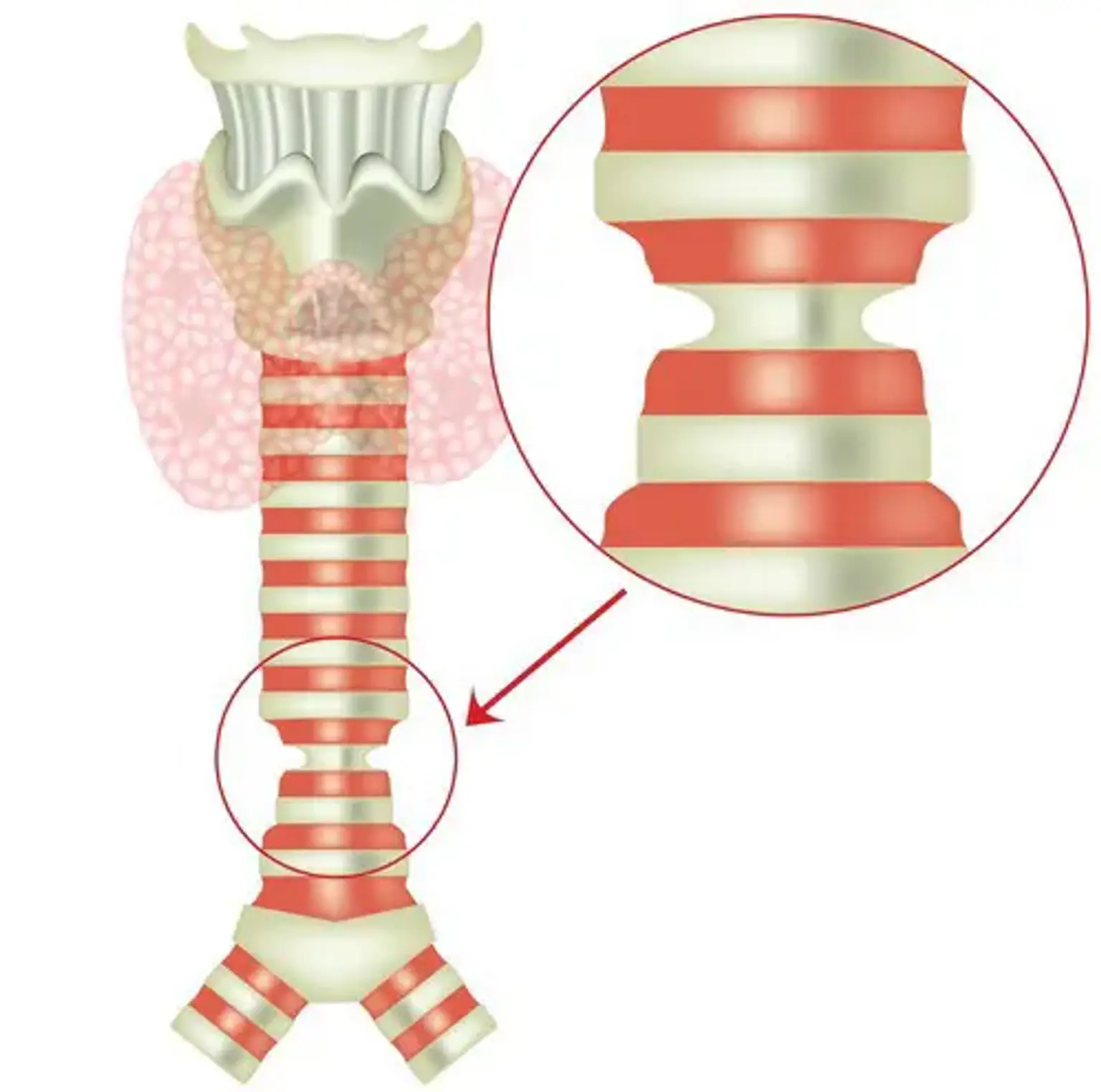
CTS is frequently caused by entire tracheal rings, which occur when the posterior membranous trachea is missing and replaced by complete circular cartilaginous rings. Tracheal involvement with such rings might be of either short or extended duration. Because this lesion is mechanical in character, medical treatment is frequently ineffective and is linked with a poor long-term prognosis. Surgical correction is still the basis of treatment. 


What is Tracheal Stenosis?

Tracheal Stenosis

Tracheal stenosis is characterized by a narrowing of the airway anywhere along the trachea, or windpipe. The trachea originates at the bottom of the voice box and extends down to the lungs via airways called bronchi. Tracheal stenosis can also spread into the bronchi, resulting in bronchial stenosis/narrowing.

This constriction might be present from birth (congenital tracheal stenosis), or it can be induced by a past damage to the airway, most commonly through a breathing tube or surgery. Acquired tracheal stenosis (narrowing caused by injury) occurs as a result of persistent irritation or damage. Chronic irritation from a breathing tube, reaction to tissue injury owing to pressure from a breathing tube cuff, or reaction to harm from external sources such as inhalational injury from fire are all possible causes.

  • Congenital tracheal stenosis

When a kid is born with tracheal stenosis, it is frequently caused by a condition known as full tracheal rings. Normally, the tracheal cartilage is horseshoe shaped and placed in front of the airway. This prevents the airway from collapsing during respiration. The airway's rear wall (in the open area of the horseshoe) is soft, allowing it to expand when breathing.

The airway does not enlarge when a youngster has full tracheal rings. Instead, the flexible section of the airway is gone, and the airway is constrained by the small, round cartilage. The stenosis typically narrows as it progresses down the trachea. Even in very young neonates, this can cause severe and hazardous respiratory distress. Typically, the issue is discovered soon after birth or early in infancy. It is occasionally discovered during adolescence or even maturity.


Who is at risk of tracheal stenosis?

Certain children are more likely to develop tracheal stenosis. It is linked to certain cardiac and lung abnormalities, as well as certain genetic diseases or syndromes, such as Down syndrome. Tracheal stenosis is more frequent in children with a blood vessel anomaly known as a left pulmonary artery sling (LPAS), a cardiac disorder in which the misdirected left pulmonary artery travels between the trachea and the esophagus. Tracheal stenosis affects more than half of all children with LPAS.


What are the symptoms of tracheal stenosis?

tracheal stenosis symptoms

Tracheal stenosis is characterized by two major symptoms. A child's breathing is frequently loud (also called stridor). Another indication is sudden and severe respiratory issues, which can cause a youngster to stop breathing and become blue. If the youngster does not receive urgent first assistance, this can be fatal.

Sometimes the issue may not become obvious until the kid develops a chest infection and experiences significant breathing difficulties that are out of proportion to the condition. This may need the use of a ventilator (breathing machine(

Tracheal stenosis can be related with other issues, such as a heart abnormality, therefore the physicians will extensively evaluate the youngster to see whether this is the case.


How is tracheal stenosis identified?

detecting tracheal stenosis

Microlaryngoscopy and bronchoscopy are the gold-standard methods for detecting tracheal stenosis. This is a minimally invasive treatment through the mouth that uses a camera to view through the voice box into the windpipe; it is conducted under anesthesia in the ICU or operating room. This allows our staff to assess the airway constriction and determine whether therapy is required.

Tracheal stenosis may be suspected in the following circumstances:

  • When a baby has trouble breathing and/or makes a lot of noise right after birth.
  • If an airway abnormality in the baby is discovered during pregnancy using ultrasonography and fetal magnetic resonance imaging (MRI).
  • When a prenatal ultrasound reveals vascular (blood vessel) abnormalities that are strongly linked to full tracheal rings (e.g., pulmonary artery sling).
  • When imaging examinations (such as a computed tomography [CT] scan) reveal a windpipe constriction.
  • When a youngster gets asthma-like respiratory symptoms that do not respond to standard asthma therapy.


What is the treatment for tracheal stenosis?

tracheal stenosis treatment

Tracheal stenosis treatment is determined on the kind and severity of the problem. There are several therapy methods available, including:

  • Observation 
    Milder types of tracheal stenosis that do not have a substantial impact on the patient can be closely managed by close monitoring and regular doctor visits. The problem may improve over time as the youngster and the airway mature.

  • Balloon dilation
    Scar tissue may be separated using a sickle knife and subsequently dilated with an angioplasty-like balloon during an endoscopic, non-surgical technique in less severe cases of tracheal stenosis. The creation of new scarring can be minimized by injecting a steroid into the tissue during balloon dilation.

  • Tracheal resection and primary anastomosis
    In more severe situations, open surgical surgery with reconstruction may be required. Tracheal resection and anastomosis may be performed for severe constriction involving a few tracheal rings. This entails surgically removing the faulty tracheal segment and then suturing the remaining ends back together to restore the airway. Surgery for entire tracheal rings typically includes more than a few faulty tracheal rings and, as a result, cannot be treated with a tracheal resection.

  • Slide tracheoplasty
    A sliding tracheoplasty is necessary for long-segment tracheal stenosis. The thin section of the trachea is sliced horizontally during this procedure. Then a vertical incision is made at the rear of one tracheal segment and the front of the other. Finally, the two parts are slid together and sutured together to form a larger tracheal airway.


What is a slide tracheoplasty?

slide tracheoplasty

To treat tracheal stenosis, a sliding tracheoplasty is the preferred procedure. The benefit of a'slide tracheoplasty' over other procedures is that it corrects tracheal stenosis using the body's own tissues, therefore there is no chance of rejection. The operation's goal is to expand the airway and thereby minimize breathing issues.

  • What happens before the operation?

The physicians will go through the procedure in further detail, address any concerns parents may have, and ask them to sign a consent form giving their permission for the procedure. Another doctor will come to the ward to explain the anesthesia to the family. Before the procedure, the physicians will request blood samples to ensure that the youngster is healthy. Please inform the physicians if the kid has any medical issues, such as allergies.

  • What does the operation involve?

The procedure takes roughly four hours and is performed while the youngster is under general anesthesia. During the procedure, he or she will be attached to a heart-lung bypass machine so that the surgeon may safely operate on the child's trachea.

The surgeon will create an incision (cut) in the child's chest and divide the trachea into top and bottom halves. The upper section will be cut at the rear of the trachea and the bottom section will be cut down the front. The surgeon will then slip the two pieces together, one on top of the other. He or she will then sew the two pieces together, making the trachea shorter but stronger and broader. You may obtain a decent notion of the operation's premise by sliding your index fingers over each other.

Three chest drains will be put into the incision's bottom to remove any fluid that gathers there. A drip into a vein in the child's neck, an arterial line in the wrist, another drip in his or her hand, and a urinary catheter to drain pee from the bladder will also be placed.

  • Are there any risks?

As with every procedure, there are risks and problems. We are not implying that these will occur, but we feel it is critical that people are aware of what may occur. The surgeon will go through the specific dangers of the child's procedure.

Bleeding shortly after surgery is a possibility. Chest drains allow blood to flow away from wounds, and bleeding normally ceases within a few hours. If the bleeding continues and treatments do not stop it, the kid may need to return to the operating room for further evaluation. There is always enough blood to replenish what has been lost. Chest drains also enable doctors check whether there is any air leak from the trachea repair, which is extremely unusual.

Infection is a possibility with every procedure, therefore the youngster will be given antibiotics to lessen the likelihood of infection. For the first two days, the nurses will wash out the surgical site with an antiseptic liquid through the chest drains.

Another concern, which affects around one-third of children, is that the trachea stays too narrow following surgery. Some children's tracheas develop floppy as a result, causing breathing issues. If the child's trachea is too small, it can be expanded with a balloon during another general anesthesia surgery. If the trachea is excessively floppy, a cage called a stent can be inserted to support it. This operation is also done while under general anesthesia.

There is a very tiny risk that the surgeon will be unable to operate due to the severity of the child's condition. Previous tracheal surgery can also make operations more difficult, especially if scar tissue has grown. It is also conceivable that the surgeon will uncover an undiagnosed cardiac problem, which might jeopardize the child's rehabilitation.

  • What happens afterwards?

The youngster will be admitted to the Cardiac Intensive Care Unit for treatment. To assist him or her breathe, a tube will be placed into the nose or mouth and attached to a ventilator. This helps the trachea to repair while resting. Even though the child is in critical care, parents can still participate in his or her care if they like.

The youngster will require daily chest physiotherapy during the first week to eliminate any secretions or mucus. This will be repeated until the youngster is able to cough. The youngster will undergo another bronchoscopy at the bedside two days following the procedure. They will have a bronchoscopy and bronchogram study one week later (without optical coherence tomography this time) to assess how the trachea is healing. The time required to 'wean' a youngster off the ventilator varies.

The strategy and the child's daily development will be explained by the team. When the children have been weaned from the ventilator, they will be transferred to Bear Ward to continue their recovery. The youngster will continue to receive frequent bronchoscopies as clinically necessary to monitor the repair of the trachea. The child's breathing may sound loud after each bronchoscopy, but this is normal.

It is also typical for youngsters to develop a fever when recuperating from illness. Parents can give their child conventional children's pain relievers like paracetamol or ibuprofen as directed on the label. Using a balloon to extend the tracheal diameter from the inside during this healing phase is occasionally essential. This is performed under anesthesia and may need to be repeated a few times.

  • Going home

During the child's time on Bear Ward, the team will begin to plan when he or she will be able to return home. There will be some instructional sessions to ensure that parents are confident in their ability to care for their kid following the procedure. To keep the secretions loose, the youngster may require a nebuliser or antibiotics to take home. The team will also describe the child's follow-up treatment, which includes frequent bronchoscopy and bronchogram examinations, though these may eventually be performed at a hospital closer to home.

We will notify the parent and the child's family doctor about the surgery (GP). Every year, the youngster should receive the flu vaccine as well as antibiotics if he or she has a cough or cold. After the procedure, the youngster should avoid hard play for a period, but can gradually resume normal activities.


What is the outlook for children with tracheal stenosis?

tracheal stenosis in children

In our experience, the first year following surgery can be the most difficult for parents. Parents will be concerned about their child's recovery after major surgery.

The child's breathing issues may persist, although they are likely to be less severe than before the procedure. The follow-up program might consume a significant amount of time, since appointments may be scheduled weekly at first, but will gradually decrease to once a month or so.

Some youngsters (about one-third) require further treatments to enlarge or strengthen the trachea if it becomes floppy (malacic). These operations are performed under general anesthesia and will necessitate a hospital stay.

Children appear to relax better and have less respiratory issues in the second year after surgery. A bronchoscopy will be required every six months or so, with yearly follow-up sessions. Although feeding may take some time to establish, the youngster should be able to have a relatively normal life, attending school or childcare. They may appear smaller than other children their age for the first several years, but with special feeding, the majority catch up. If you are concerned, please consult with the dietician.


Conclusion 

Congenital tracheal stenosis

Congenital tracheal stenosis is an uncommon but underdiagnosed abnormality that can cause life-threatening respiratory failure in newborns and infants. Initial airway control is required. Surgical repair is the mainstay of therapy and has a low fatality rate. The kind and amount of repair are mostly determined by the length of the stenosis.

Cardiac abnormalities are commonly connected with tracheal surgery and may be treated. Despite initially positive findings, post-operative morbidity from chronic granulation tissue is significant. These challenging individuals are best cared for by a multidisciplinary approach and continuous monitoring of the restored airway. The long-term quality of life is yet unknown.