Coronary Angiography
Overview
The anatomical and structural characteristics of the circulatory system, including the heart chambers, are identified by angiography. Angiography was originally utilized for structural diagnosis of intravascular diseases, but it has now been broadened to include functional evaluation and therapy choices.
Coronary angiography and cardiac catheterization are vital diagnostics for detecting and quantifying coronary artery disease, identifying valvular and other structural anomalies, and measuring hemodynamic parameters. The risks and consequences connected with these treatments are related to the patient's coexisting diseases as well as the operator's expertise and discretion.
Coronary angiography anatomy
In the last 50 years, medical imaging has benefited from a surge in innovation, allowing for fast advancement in the field of interventional radiology. Angiography is a technique for imaging anatomical and structural aspects of the vascular system by detecting contrast injected into a blood artery and projecting it on a series of x-rays to define the inner vessel wall and show flow through the lumen. Angiography, which began as a diagnostic tool, experienced a technical change during the previous century and evolved into a foundation for interventional therapy as well.
Angiography has progressed from a static two-dimensional record of the vasculature on screen films to a real-time two-dimensional presentation of the vasculature on television screens and three-dimensional reconstruction from computed tomographic scans. Angiography has several advantages since it provides real-time, dynamic imaging utilizing a typical imaging technology such as x-rays or computed tomography (CT), and it also provides treatment choices at the time of first diagnosis.
Angiography is an invasive procedure. In addition to treatment alternatives, invasive angiography is still the gold standard for identifying most intravascular diseases. In the last three decades, advances in imaging technology have broadened the scope of angiography to include non-invasive methods utilizing CT and magnetic resonance imaging technologies.
Intravenous contrast is frequently supplied via a peripheral vein during CT angiography (CTA), and triple-phase CT is usually collected. CTA is based on the gathering of high-resolution CT data, followed by angiographic two- or three-dimensional reconstruction. The advancements and modifications of CTA principles have proved very beneficial to individuals suffering from cardiovascular illnesses.
While this is a great first-line probe and provides information about the surrounding soft tissues, the tiny diameter makes it less effective for studying blood flow to the extremities. Interventional angiography gives greater resolution imaging and is capable of detecting changes in tiny arteries.
Magnetic resonance angiography (MRA): Clinical applications of MRA have grown in importance for both patients and clinicians, particularly in this era of fast developments in imaging technology. Magnetic resonance imaging (MRI) makes use of the inherent magnetic properties of bodily tissues in the presence of an external magnetic field.
Anatomy and Physiology
The aorta begins with the aortic root, which can measure up to 4.3 cm in healthy people. At the level of the sinus of Valsalva, the aorta branches into the left and right major coronary arteries. It then becomes the ascending aorta before becoming the aortic arch. The arch gives rise to three significant branches:
- The brachiocephalic trunk divides to form the right common carotid and right subclavian arteries.
- The internal and external carotid arteries are formed by the division of the left common carotid artery. The circle of Wilis is formed by the internal carotid artery and the vertebral artery, while the external carotid artery sends out the superior thyroid artery, ascending pharyngeal artery, and lingual artery. The facial artery, occipital artery, posterior auricular artery, maxillary artery, and superficial temporal artery are all branches of the external carotid artery.
- The left subclavian artery
The scalenus anterior muscle divides the subclavian artery into three parts. The first section feeds off the vertebral artery, which delivers blood to the vertebrobasilar system and Willis circle. The internal mammary artery and the thyrocervical trunk are two more arteries that branch out from the first section.
The costocervical trunk comes from the second section of the subclavian artery, while the dorsal scapular artery arises from the third. After crossing over the first rib, the subclavian artery changes its nomenclature to the axillary artery and serves the upper extremities.
The abdominal aorta contains three unpaired anterior branches to the viscera: the coeliac axis (which gives off the hepatic, splenic, and left gastric arteries); the superior mesenteric artery; and the inferior mesenteric artery. The abdominal aorta also includes three major lateral paired branches reaching the viscera: the suprarenal, renal, and testicular or ovarian arteries.
The abdominal aorta divides into common iliac arteries, which then divide into internal and external iliac arteries. The internal iliac artery separates into anterior and posterior divisions to serve the pelvic and perineal organs, whereas the external iliac artery joins the femoral artery below the inguinal ligament. The femoral artery provides blood supply to the lower extremities. Angiography catheters are commonly inserted into the femoral and radial arteries.
An intrinsic disadvantage of angiography is the inability to acquire pictures that are a lumenogram as compared to intravascular imaging modalities such as intravascular ultrasound, particularly in terms of identifying arterial calcification.
Coronary Angiography definition
Coronary angiography is a test that determines the degree of constriction in your arteries. Discover what to anticipate and when you might require a stent. Every year, about a million cardiac angiographic procedures are performed in the United States. They are relatively safe, with little problems. The risk of a major complication such as stroke, heart attack, or death during cardiac catheterization and angiography is one in 1,000. Fewer than one in ten thousand persons who receive these treatments die, and the majority of those who die already have a serious cardiac condition or another disorder. Older adults are at a higher risk of complications and mortality.
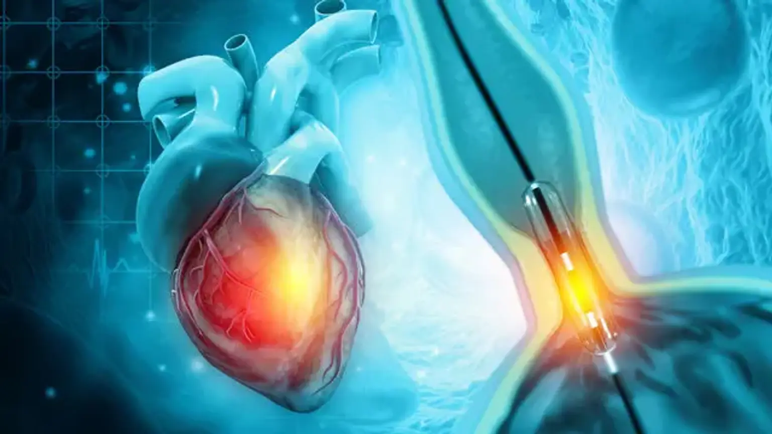
A coronary angiography is an X-ray of the heart's arteries. This demonstrates the amount and severity of any cardiac disease and can assist you in determining how effectively your heart is functioning. Your doctor will inject a liquid dye through a tiny, flexible tube called a catheter to generate the X-ray pictures. The catheter is threaded into the desired artery by the doctor from an access site. The access site is often in the arm, although it can also be in the groin.
The dye causes the blood flowing inside the blood vessels to be seen on an X-ray and highlights any constricted or blocked areas in the blood vessel. The dye is then excreted from your body via your kidneys and urine.
Why do I need coronary angiography?
Coronary angiography can be used to diagnose heart diseases, plan future therapies, and perform specific operations. It might be used in the following ways, for example:
- After a heart attack – where the heart's blood supply is blocked
- To help diagnose angina – where pain in the chest is caused by restricted blood supply to the heart
- To plan interventional or surgical procedures – such as a coronary angioplasty, where narrowed or blocked blood vessels are widened
Coronary angiography is also seen to be the best means of identifying coronary heart disease, which occurs when a buildup of fatty substances in the coronary arteries disrupts the blood flow to the heart.
Diseases diagnosed by coronary angiogram
Angiograms can identify a variety of heart disorders, including aneurysms (abnormal ballooning of the heart wall), cardiac arrhythmias (irregular heart beat), and birth abnormalities, such as a hole in the heart, in addition to damaged coronary arteries.
Medical issues to consider before having an angiogram
Before the procedure, you need to discuss a range of issues with your doctor including:
- Your medical history, including whether or not you have asthma, allergies, or renal disease, whether you have had adverse reactions to any medications, and any current medications you are taking
- You may need to stop taking certain medications before the test, such as blood thinners
- You must fast four to six hours before your test other tests –
- You may be subjected to a variety of tests prior to the angiogram, including blood tests, an electrocardiogram, a chest x-ray, and a cardiac CT (computed tomography).
Coronary angiography procedure
The majority of diagnostic coronary angio procedures are performed as outpatient procedures. That implies you'll be in and out of the hospital in a single day. A nurse will take your medical history before the operation, and you will change into a hospital gown. The nurse will prepare you for the treatment by inserting an IV cannula and, if required, shaving both sides of your groin and wrist.
During the process, a catheter, which is a long, thin, flexible tube, is placed into a blood artery in your groin or arm. The catheter tip is guided up to the heart and coronary arteries using X-ray pictures as a guide.
Contrast media, a specific type of dye, is injected through the catheter, and X-ray pictures (angiograms) are acquired. The contrast medium is visible on angiograms, revealing the blood arteries through which the fluid passes. This clearly shows any constricted or obstructed blood arteries.
The treatment is often performed under local anaesthesia, so you will be awake during the process, but the region where the catheter is put will be numbed.
When you arrive at the cath lab, you will lie down on a special table. During the exam, a cardiac monitor will record your heartbeat. An antiseptic wash is used to clean the skin on your wrist and both sides of your groin, and you are draped in sterile drapes.
A tiny dose of local anaesthetic is injected around the access point (wrist or groin) to numb the region before inserting a small catheter through the skin into the blood artery. The doctor monitors the catheter's progress using dynamic x-rays sent to a television display.
Because there aren't enough nerves in the blood arteries, you can't feel the catheter passing through the heart. A little quantity of contrast (x-ray sensitive dye) is administered into the catheter after it is in position. As the contrast passes through the blood vessels, more dynamic x-rays are acquired. As the contrast is administered, you may experience a warm flush or tingling. The angiography takes around 40 minutes.
Immediately after the coronary angiogram
After the angiogram, you can expect the following:
- Your blood pressure, pulse, respiration, and wound site are all examined and documented on a regular basis.
- You may be given intravenous fluids for a brief period of time, but you will be encouraged to eat and drink as quickly as possible.
- After four hours, you may be permitted to sit up.
- You may be released to go home up to six hours following your recovery.
- If you are not currently on a cholesterol-lowering diet, you will be advised to start one.
- The cardiologist who conducted the operation will provide you with preliminary results. You may need to schedule a follow-up consultation with a cardiologist to discuss your therapy.
Follow up after coronary angiography
After a period of rest and monitoring, you should be able to leave the hospital on the same day as your coronary angiography. Most individuals feel good a day or two following the treatment, however you may feel a little weary and the incision site may be sensitive for up to a week. Any bruising that occurs may remain for several weeks.
Certain activities, including as showering, driving, and moving heavy things, are normally discouraged for a day or two after the surgery. While you're healing, it's critical to keep an eye out for any signals of trouble.
If the swelling at the location of your cut worsens, or if you feel severe bleeding or circulation difficulties in your limbs, you should seek emergency medical assistance.
Following the surgery, you may be required to stay in the hospital for several hours or overnight. You may be instructed to take drinks to avoid dehydration and to flush the dye from your kidneys. Before you leave, a nurse will show you how to monitor the location for bleeding and what to do if it occurs. If the groin was utilized, you may be instructed to refrain from hard lifting and straining for a week to avoid bleeding.
Angioplasty, a treatment used to treat narrowed coronary arteries, may be performed during the angiography. To remove the obstruction, a special catheter is put through the blood channels and into the coronary arteries. A bypass procedure is another surgical option for severely constricted coronary arteries. This procedure entails transferring veins and arteries from other regions of your body to your heart.
Complications
As with any medical treatment, having a coronary angiography and angioplasty has both dangers and advantages.
Discuss the risks and benefits with your doctor, nurse, or other health professional, as well as any concerns you may have. Your healthcare team can provide you with further information about your specific situation and level of risk.
Minor complications may include:
- Bleeding under the skin at the incision site - this should go away within a few days, but if you are worried, please see your doctor.
- Bruising — It is usual to have a catheter-related bruise for a few weeks.
- Allergy to the contrast dye used in the operation, resulting in symptoms such as a rash–you should discuss any allergies you have with your cardiologist before undergoing the surgery.
More serious complications are uncommon, but may include:
- Damage to the artery in the arm or groin from the catheter, possibly affecting blood supply to the limb
- Heart attack
- Stroke
- Damage to the kidneys caused by the contrast dye
- Tissue damage caused by X-ray radiation if the procedure is prolonged
- Serious bleeding
- Death.
You are more likely to develop complications based on:
- Your age — the older you are, the more dangerous you are.
- If the surgery was planned or if it is an emergency treatment — emergency therapy is usually riskier since there is less time to arrange it and the patient is already ill.
- If you have renal problems, the dye used during an angioplasty might possibly cause further kidney damage.
- If you have one or more blocked coronary arteries, you should consult a doctor.
- If you have a family history of severe heart disease.
If the images from your angiogram indicate a narrowing or blockage that may be repaired right away, your cardiologist may opt to proceed with the surgery and place a stent at the same time (angioplasty).
Contraindications
- Absolute Contraindications
In North America, certain typical angiography tables have a listed weight restriction of 350 pounds. Patients who are morbidly obese and weigh more than this limit may be denied angiography due to technical safety concerns.
- Relative Contraindications
Pregnancy, unless there is a danger of maternal mortality due to persistent bleeding. Severe iodinated contrast medium allergy manifested as bronchospasm, laryngospasm, angioedema, and cardiovascular collapse in the past. When the technology is available, carbon dioxide angiography could be performed on these patients.
Steroids and antihistamines might be given to patients who have a history of minor allergic responses. Patients with preexisting renal impairment or dehydration are at a higher risk of developing renal function following contrast medium exposure. For this subset of patients, ultra-low contrast or zero contrast procedures have been proposed.
Patients with coagulopathy, INR more than 2, and platelet count greater than 50,000/microliter are at an increased risk of bleeding. However, with the introduction of vascular closure devices, the danger of bleeding has been reduced significantly.
Patients with diabetes who are taking metformin are at risk of having their renal function deteriorate and developing lactic acidosis, especially if they have underlying renal impairment. Patients who are extremely anxious or unable to lie still may require conscious sedation.
Conclusion
Cardiac catheterization and coronary angiography are non-surgical procedures used to examine the heart and the blood veins that supply the coronary arteries.
When noninvasive tests do not offer enough information, when noninvasive tests suggest a heart or blood vessel problem, or when a person shows symptoms that indicate a heart or coronary artery disease is quite likely, these tests are commonly conducted. One advantage of these tests is that doctors may treat a variety of illnesses during the process, including coronary artery disease.