Cranial base disease

Last updated date: 25-Aug-2023

Originally Written in English

Cranial Base Disease

Cranial Base Disease

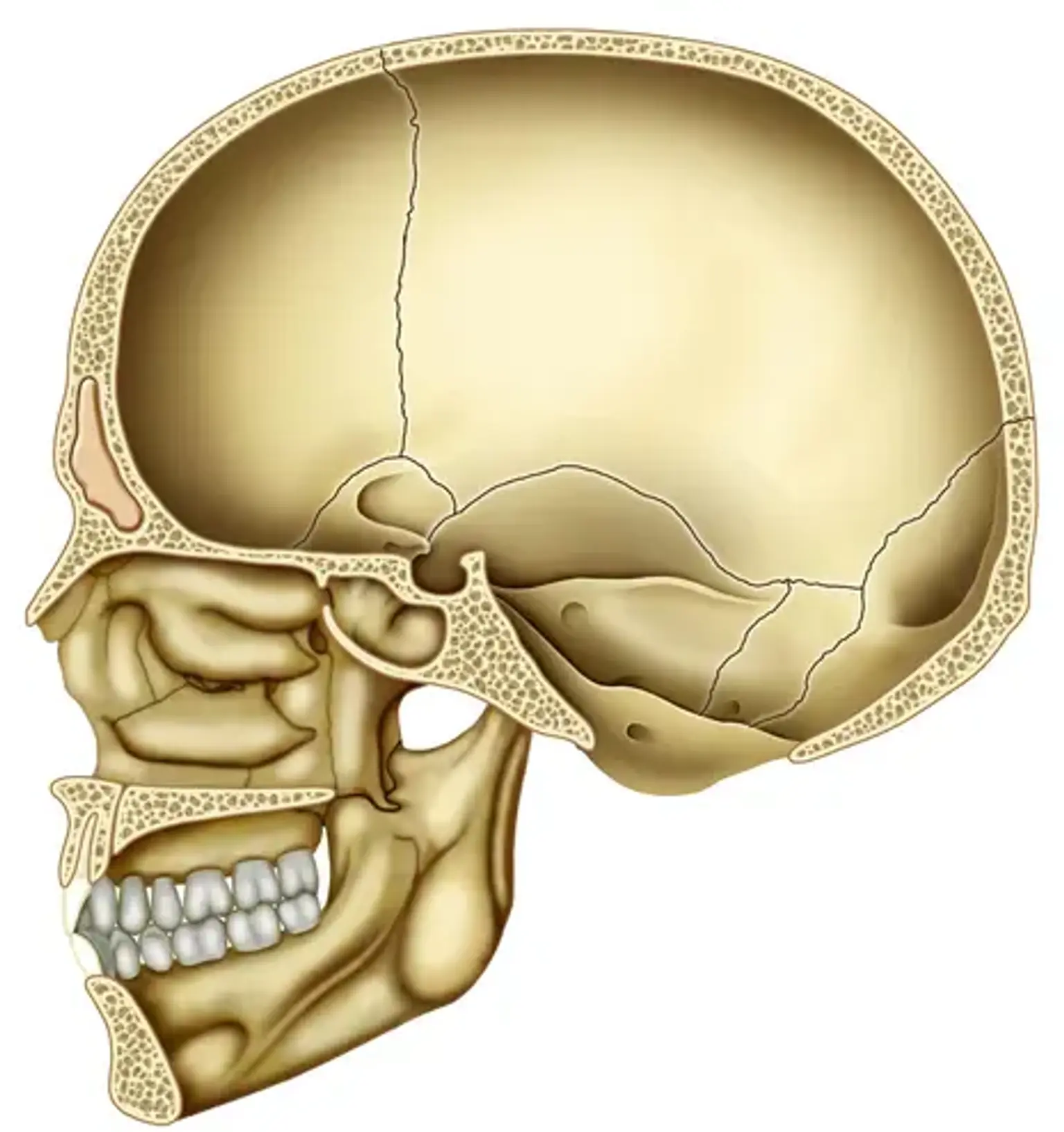
Every nerve that connects the brain to the rest of the body must pass via the cranial base, a tiny but incredibly complex region at the base of the skull. You may encounter neurological issues like headaches, facial pain, blurred vision, dizziness, or seizures if you have a cranial base disease. When the tumor has gotten big enough to press against the brain or the nerves, these symptoms frequently appear.


What is Cranial Base Disease?

Cranial Base Disease Definition

The term cranial base disease refers to a broad range of conditions that affect the head and neck, impacting areas from the collarbone to the top of the head. It is optimal for a collaborative team of subspecialists from several fields, including otolaryngology, neurosurgery, ophthalmology, plastic surgery, craniofacial surgery, cancer, radiation oncology, and rehabilitative medicine, to diagnose and treat cranial base disease. Skull base surgery can be carried out using a variety of surgical methods, including open surgery, minimally invasive approaches, and stereotactic radiotherapy. To get access to the surgical area during open surgery, small amounts of bone are removed through incisions created in the face and head (cranium). Various surgical methods can be employed to access lesions in different locations of the skull base region. Endoscopic surgery, for example, enables us to access the operating area through a smaller incision, reducing pain, infection and complication rates, and length of hospital stay. In stereotactic radiotherapy, cancers and vascular abnormalities are treated by precisely delivering x-rays to the targeted location.


Cranial Base Disease Types

Cranial Base Disease Types

The following are examples of skull base diseases:

  • Arachnoid cysts are a form of brain tumor that can obstruct fluid flow.
  • A branchial cleft cyst often referred to as a cleft sinus, appears in the neck or just below the collarbone. Branchial cleft cysts occur as the embryo develops. They form as a result of abnormal tissue development in the neck and collarbone region (branchial cleft).
  • A carotid body tumor is a growth on the side of the neck, close to where the carotid artery divides into smaller blood arteries that provide blood to your brain. It is also known as a chemodectoma or paraganglioma.
  • Cavernoma is an aberrant collection of blood vessels typically located in the brain and spinal cord. Sometimes they are referred to as cerebral cavernous malformations (CCM), cavernous hemangiomas, or cavernous angiomas. A typical cavernoma resembles a raspberry.
  • A cerebral aneurysm (also referred to as a brain aneurysm) is a weak or thin area on a brain artery that inflate or bulges out and fills with blood. The nerves or brain tissue may be compressed by the bulging aneurysm. Additionally, it could burst or rupture, leaking blood into the nearby tissue (called a hemorrhage).
  • A craniopharyngioma is an uncommon form of benign (noncancerous) brain tumor. The pituitary gland, which secretes hormones that regulate numerous bodily functions, is where the craniopharyngioma first appears. A craniopharyngioma's slow growth may affect the pituitary gland's and other surrounding brain structures' ability to function.
  • Encephaloceles are rare neural tube anomalies and characterized by the brain and the membranes that protect it protruding like sacs through holes in the skull. These defects result from the neural tube's inability to fully close during fetal development.
  • Glomus jugulare tumor is a tumor in the area of the temporal bone in the skull that affects the middle and inner ear tissues. The ear, upper neck, base of the skull, and nearby blood vessels and nerves can all be compromised by this tumor.
  • Meningocele is a congenital abnormality characterized by a sac that protrudes from the spinal column. Although there is spinal fluid in the sac, there is no neural tissue. It might have meninges (the membranes that cover the central nervous system) or skin on top of it. Frequently, the sac can be seen from the outside of the back.
  • Meningioma is a tumor that develops from the meninges, the membranes that cover the brain and spinal cord. It is included in this group even if it isn't strictly a brain tumor since it might compress or pressure the nearby brain, nerves, or blood vessels.
  • Neurofibroma is a type of nerve tumor that manifests as soft lumps on or beneath the skin. Anywhere in the body, a neurofibroma can form within a primary or minor nerve. Common benign nerve tumors of this kind typically develop more centrally within the nerve.
  • Rathke cleft cyst (RCC) is a cranial base disease and fluid-filled benign (non-cancerous) growths that form between the pituitary gland's lobes. They are congenital malformations, which means that they appear as the fetus develops inside the mother's womb. A portion of the fetus' growing Rathke pouch, which eventually develops into the pituitary gland, gives rise to an RCC.
  • A schwannoma is a rare form of tumor that develops in the nervous system. Schwann cells are the source of schwannoma formation. The nerve cells of the nervous system are supported and shielded by Schwan cells. The majority of schwannoma tumors are benign, meaning they are not cancer. However, they can occasionally evolve into malignancy.
  • Tumors of the pituitary gland are abnormal growths that form there. Some pituitary tumors cause the hormones that control vital body processes to be produced in excess. Your pituitary gland may produce fewer hormones as a result of some pituitary tumors.


Cranial Base Disease Symptoms

Cranial Base Disease Symptoms

The region behind the eyes and nasal cavities, which comprises the floor of the skull, is where tumors frequently form or spread. These tumors, which may be benign or cancerous, have the potential to cause complications with the spinal cord, blood vessels, and nerves that travel through the base of the skull.

Nearly all cranial base diseases, whether benign or malignant, need to be treated to eradicate the tumor and avoid consequences as it grows or the tumor spreads. Surgery, radiotherapy, chemotherapy, or a combination of these treatments may be used to treat skull base tumors. Here are some signs you should speak with a skull base specialist about:

  • A growth or wound that does not go away
  • Persistent throat infection
  • Persistently swollen neck glands
  • Abnormal growth in the neck
  • Having trouble swallowing
  • Having trouble moving your mouth or jaw
  • Voice change or hoarseness
  • Gums, the tongue, or the mouth's lining may develop white or red spots.
  • Alterations in denture fit
  • Undiagnosed face pain or ongoing ear pain
  • loss of smell
  • Bloody nasal flow or bloody cough
  • Chronic and unrelieved nasal congestion
  • Upper body obesity
  • Increase in neck fat
  • Extreme headache and vomiting or nausea
  • Double vision
  • Gait disorder
  • Loss of hearing
  • Seizure
  • Sensational numbness or diminution.
  • Alteration in mental state.


Cranial Base Disease Diagnosis

Cranial Base Disease Diagnosis

Your doctor will check your medical history, conduct a physical examination, and prescribe diagnostic testing to identify the source of your symptoms. Depending on your symptoms and signs, different tests and exams may be performed.

  • A small mirror, light, or specialist examination scope may be used by your doctor to check the neck, throat, tongue, nasal and oral cavities, and other areas of the body. Additionally, your doctor might feel for lumps in the cheeks, lips, gums, and neck.
  • It is possible to order laboratory tests that check samples of blood, urine, or other body fluid. Additionally, you could require one or more of the imaging tests listed below: X-rays, a CT (or CAT) scan, an MRI, or a PET scan are examples of imaging techniques.
  • A biopsy may also be requested by your doctor. A biopsy entails the removal of cells or tissues for microscopic analysis.

Your doctor will want to know the stage (or severity) of the disease if cancer has been diagnosed. It is carefully determined through staging if and where cancer has spread throughout the body. The doctor can better plan your care and give you details about your prognosis if they are aware of the disease's stage.


Cranial Base Disease Treatment

Skull Base Surgery

 Skull Base Surgery

The tumor is removed during surgery. It's possible that oral surgery to remove a minor tumor won't have any long-term effects. To offer a margin of safety during surgery to remove a larger tumor, cancer may also be removed along with some nearby healthy tissue and structures. Talking, chewing, and swallowing may be impacted by this operation. The neck's lymph nodes will also be removed if the surgeon feels that cancer has spread there. A neck dissection is what is done in this situation. To solve cosmetic and functional problems, the surgeon will frequently need to reconstruct specific parts and will cooperate with other experts.

Questions to consider before skull base surgery:

  • Which kind of surgery would you advise?
  • Will my lymph nodes need to be removed? Why?
  • What will I feel like after surgery? How long will I be hospitalized? 
  • What are the surgical risks?
  • Will it be difficult for me to swallow, speak, or eat?
  • What will the scars look like? How will they appear?
  • Will my appearance change?
  • Will I require plastic or reconstructive surgery? When will that be possible?
  • Do I have any teeth left? Can they be replaced?
  • Will I require the assistance of a specialist to improve my speech?
  • When can I return to my regular activities?

Your face and neck may swell after surgery, and you might appear different. Within a few weeks, swelling in the face and neck normally goes away. The removal of lymph nodes, or neck dissection, can restrict the flow of lymph, which can cause lymph to solidify in the tissues and prolong swelling. After a neck dissection, your shoulders and neck could also feel weak and stiff. These issues might be improved by physiotherapy, including the proper exercises and treatments.


Radiation Therapy

Radiation Therapy

High-energy sources are used in radiation therapy, often known as radiotherapy, to kill cancer cells. Radiation can come from a device outside the body (external radiation therapy) or radioactive substances (radioisotopes) injected into or put close to the site of the cancer cells (internal radiation therapy, or brachytherapy).

Both radiation treatment and chemotherapy have a wide range of choices. Not all radiation treatments, nor all options that can be used to treat diseases of the skull base, are accessible to all medical facilities.

Radiation to the head and neck region can damage normal cells, including those in the mouth and oropharynx, in addition to cancer cells. Patients who receive radiation therapy to the head and neck region run the risk of experiencing a variety of issues, including dry mouth, malnutrition, and dental issues. Other conditions, such as thyroid dysfunction and tissue hardening (fibrosis), can affect the head and neck. However, because every person is different, every patient's response to treatment may vary. There may be changes in the form and severity of treatment-related adverse effects for you.

In radiation therapy, a process known as fractionation divides the total radiation dose into several smaller, equal doses that are administered over several days or weeks. The radiation oncologist can select from a variety of radiation schedules, some of which are described below:

  • Five days a week, for a predetermined amount of time, outpatient standard radiation therapy is often administered. Radiation therapy has the benefit of avoiding surgical risk, potentially preserving organs and thereby maintaining organ function, and being able to treat massive tumors or potential tumor involvement that would otherwise be challenging to reach with surgery and result in more post-operative dysfunction and defect.
  • Accelerated fractionation is a form of therapy that involves administering a total dose in several daily fractions that are roughly the same amount and spaced out over a shorter period.
  • Hyperfractionated radiotherapy is the use of numerous radiation fractions (doses) per day that are smaller than usual but result in a larger total dose, with little to no change in overall treatment time. The justification for this sort of therapy is that it gives tumor cells less time between treatments to recuperate from radiation damage. However, this type of therapy carries a higher risk of acute radiation-related side effects.


Chemotherapy

Chemotherapy

Chemotherapy is a routine treatment used to eradicate cancer cells. Squamous cell carcinoma of the head and neck (SSCHN) has traditionally been treated with surgery and/or radiation, especially when the cancer was localized and a cure was possible. Chemotherapy does not treat these cancers on its own. However, chemotherapy and radiotherapy work better together to treat locally advanced head and neck tumors and oral tumors, and they also lessen the likelihood that cancer will spread to other body areas. In some cases, it helps patients avoid surgery and preserves organs and organ function when combined with radiation.

Neoadjuvant (induction chemotherapy), concurrent chemotherapy, or post-treatment chemotherapy (adjuvant chemotherapy) are all possible forms of chemotherapy. Even if specific tumors have shown local control rates of 75-85%, there is still a big proportion of toxicity; a feeding tube may be needed, and severe mucositis, skin reactions, anemia, malnutrition, and dehydration are frequent side effects.

Questions to consider before chemotherapy:

  • Will I also require radiotherapy and another chemotherapy?
  • Why do I require chemotherapy?
  • Which medication will I be taking?
  • What will the chemotherapy administration look like? How do the treatments work?
  • How will the radiation treatment be carried out?
  • Do I need to visit a dentist before starting my treatment?
  • How long must I rest from dental work before starting chemotherapy?
  • What are the dangers and adverse reactions?
  • Are there any long-term consequences?
  • When do the treatments start and end?
  • How many sessions will I require?
  • Do I have to remain in the hospital? How many days?
  • What effects will chemotherapy have on my daily activities?


Targeted Therapy

The goal of targeted therapy is to stop the growth and spread of cancer by using medications to locate and target specific proteins on cancer cells. These chemicals disrupt particular molecules necessary for carcinogenesis, the conversion of healthy cells into cancer cells, and tumor growth.

The number of targeted drugs being tried in clinical trials has dramatically increased in recent years. Tumor hypoxia (low oxygen), angiogenesis (formation of new blood vessels), and EGFR are promising targets. The use of targeted cancer therapies alone, in conjunction with other targeted cancer therapies, and conjunction with other cancer treatments including radiotherapy and chemotherapy are all being studied. Targeted therapy might be a part of your treatment plan, depending on your cancer form or whether you've previously undergone cancer treatment.


Cranial Base Fracture

Cranial Base Fracture

A basilar skull fracture involves at least one of the bones that make up the base of the skull and is typically brought on by severe blunt force trauma. Basilar skull fractures typically affect the temporal bones, but they can also affect the frontal bone's orbital plate, occipital, sphenoid, and ethmoid.  Hemotympanum, cerebrospinal fluid (CSF) otorrhea or rhinorrhea, Battle sign (retroauricular or mastoid ecchymosis), and raccoon eyes (periorbital ecchymosis) are a few clinical exam findings that are highly predictive of basilar skull fractures. Facial fractures, cervical spine injury, cerebral bleeding, cranial nerve injury, vascular injury, and meningitis are frequently linked to basilar skull fractures. Due to their propensity to engage in high-risk activities, younger persons are more likely to suffer basilar skull fractures. Conservative treatment is used to treat the majority of basilar skull fractures.


Conclusion

The cranial base disease affects the upper neck, nasal cavities, ear canals, and bony shelf that divides the brain from the eyes. Because important nerves, blood vessels, and other systems pass through this region, treating disorders of the skull base is challenging. Skull base disease provides unique challenges for surgeons. Many doctors have extensive experience in the most recent minimally invasive techniques that have completely changed the field of skull base surgery. They combine a multidisciplinary approach with the most cutting-edge technology and techniques.