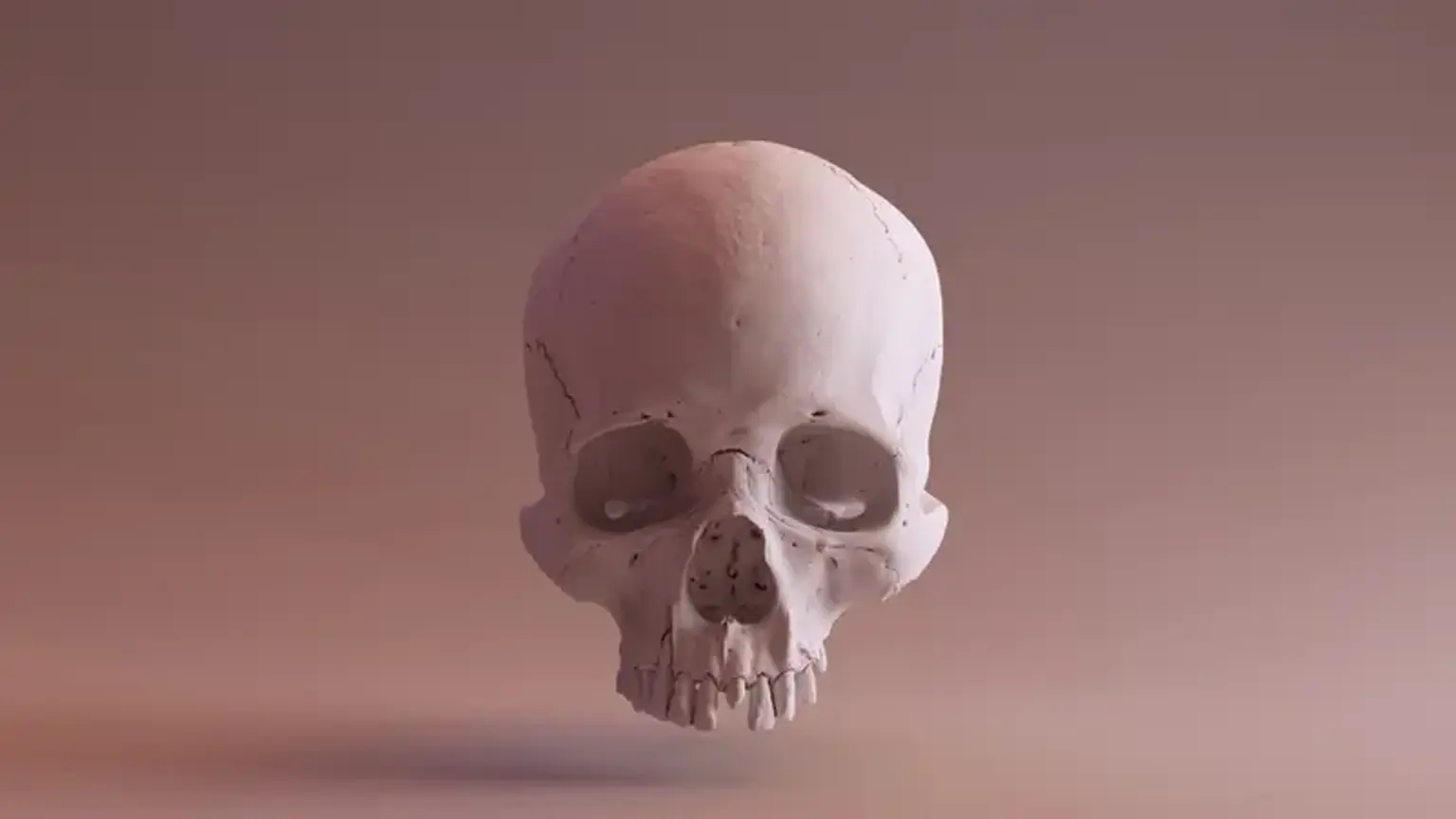
Craniostenosis
Craniostenosis is becoming more well-known in clinical practice, particularly in pediatric and neurosurgical settings. Patients are seeking medical help earlier, perhaps as early as the neonatal age, for surgical repair of deformities. Although it may appear that the incidence has grown (following evaluation) it is possible that this is merely a relative increase and not an absolute increase. This is due to heightened awareness among all medical professionals, particularly pediatricians and general practitioners. The days of blindness, delayed development, massive proptosis, and advanced neurologic impairment in these patients are long gone. The pendulum has swung completely in the other direction, and early detection and referral appear to be the norm. Several centers in India deal with such instances.
What is Craniostenosis?
In its broadest sense, the term craniostenosis, or contraction of the skull, refers to any situation in which the size of the skull and its contents are out of proportion. This can be seen in malignancies of the brain or meninges, as well as hydrocephalus.
However, in a more limited sense, the term craniostenosis refers only to abnormalities that cause a reduction in the size of the skull cavity.
Craniostenosis Causes
Premature closure of the skull sutures is the most frequent cause of such a contraction. Because the obliteration of these sutures does not usually occur until later in life, any closure of these sutures in the earlier decades must be regarded as premature. Suture closure, on the other hand, is only useful when it occurs during intrauterine life or the first year of post-fetal life. Because of the suture gaps, the development of the skull is potential during this time. Later, as in the basal synchondroses, the skull develops solely through periosteal apposition and resorption.
Premature synostosis of the sutures is a congenital deformity that can be passed down through the generations. The bigger the number of sutures implicated in the synostosis and the earlier it occurs, the greater the degree of craniostenosis. The skull remains so small after all the sutures are closed that it appears as a microcephalic skull. Because the sutures that remain open perform a compensating function, closing simply single sutures may result in no craniostenosis at all. The uneven development of the skull in different directions is a result of this compensation.
The several forms of craniostenosis are named after the characteristics of the malformation in the skull. This is defined by the relative significance of the contraction and compensation components. As a result, there are terms like tower skull (oxycephaly), boat skull (scaphocephaly), flat skull (plagiocephaly), pseudo-microcephaly, and so on.
Craniosynostosis
Craniosynostosis is a congenital condition in which the bones of a baby's skull fuse together prematurely. This occurs before the baby's brain has completed its maturation. The skull might become increasingly deformed as the baby's brain develops. Sutures are flexible materials that are used to fill the crevices between the bones of a baby's skull. These sutures allow the skull to expand in conjunction with the baby's brain. Because the sutures become bone, a child's skull bones start to fuse together at the age of two. The suture is termed as close when this happens. One or more sutures fuse prematurely in a baby with craniosynostosis. The infant's brain growth may be restricted or slowed as a result of this.
The baby's head will discontinue growing in only that area of the skull if a suture closes and the skull bones meet together too early. The baby's head will continue to expand in the portions of the skull where the sutures have not fused together. The brain inside the skull will have matured to its normal size, but the skull will have an abnormal shape. However, there are cases when more than one suture closes early. In certain cases, the brain may not have enough space to grow to its normal size. This can cause pressure to increase within the skull.
Craniosynostosis Causes
In most newborns, the cause of craniosynostosis is unknown. Because of genetic mutations, some babies are born with craniosynostosis. In some situations, craniosynostosis is caused by a single gene mutation, which can lead to a genetic syndrome. In most cases, however, craniosynostosis is believed to be caused by a combination of genes and other circumstances, such as what the mother comes into touch with in her surroundings, what she eats or drinks, or specific drugs she takes while pregnant.
Many other families with children who have birth abnormalities want to know what causes these problems. Understanding the factors that are more likely in babies born with a birth defect will aid in our understanding of the reasons. The Centers for Birth Disorders Research and Prevention are funded by the CDC and work together on large studies like the National Birth Defects Prevention Study to learn more about the causes and dangers of birth defects like craniosynostosis.
The Centers for Disease Control and Prevention (CDC) recently released key findings from research studies on various factors that raise the risk of having a baby with craniosynostosis:
- Maternal thyroid disease. Women who have thyroid issues or are managed for thyroid disease while pregnant have a higher risk of having a craniosynostosis-affected child than women who do not have thyroid issues.
- Medications in particular. When compared to women who did not take clomiphene citrate (a fertility medication) right before or early in pregnancy, they are more prone to having a baby with craniosynostosis.
The CDC is still researching birth disorders like craniosynostosis and ways to prevent them. If you're pregnant or planning to get pregnant, talk to your doctor about how you may improve your chances of having a healthy baby.
Craniosynostosis Types
Craniosynostosis comes in a variety of forms. They have a distinct appearance depending on which suture, or sutures, are affected, and are named for both the head shape (names ending in -cephaly) and the abnormally fused suture.
Craniosynostosis Coronal
It is still sometimes known as plagiocephaly (crooked head), but the word is more widely used to describe the more common disorder of flattening of the back of the head in infants (deformational or positional plagiocephaly), which is unrelated to suture problems. Coronal synostosis occurs when the right, left, or both sides of the suture that extends from ear to ear over the top of the head fuse together. When it is one-sided, this condition causes abnormal growth of the forehead, flattening the forehead and brow on the affected side and overgrowth, or bossing (bulging forward) on the opposing side. The impacted eye may also appear to be of a different shape. The forehead may look wide and towering when both sides are engaged, although the skull is short from front to back (referred to as brachycephaly). The syndromic types are more likely to have this issue. In contrast to positional plagiocephaly, the top of the head seems to be more of a trapezoid than a parallelogram. Unilateral lambdoid suture synostosis, which is an uncommon kind of synostosis that might be confused with positional plagiocephaly, can also produce plagiocephaly. The difference is a conspicuous bony ridge or spherical shape on the flattened side of the back of the skull, which is evident after the lambdoid suture fuses.
Craniosynostosis Metopic
The suture that extends from the top of the head down the middle of the forehead, toward the nose, fuses in metopic synostosis. It's the only one that's supposed to close before the brain stops growing, but if it does so too soon, it can cause a pronounced ridge to form over the forehead. In severe cases, the forehead resembles the front of a ship, and when seen from above, it resembles a triangle (thus the term trigonocephaly to express this shape), with eyes set close together (hypotelorism).
Craniosynostosis Sagittal
Early fusion of the suture that spans front to back, down the middle of the top of the head is referred to as sagittal synostosis. The result of this union is a long, narrow cranium. Scaphocephaly or dolichocephaly are words used to characterize this morphology. Because the skull cannot efficiently expand sideways, it overgrows front to back, resulting in a small occiput (back of the skull) and an abnormally wide forehead (also known as frontal bossing).
Craniosynostosis Lambdoid
Along the back of the head (occiput), the lambdoid suture runs. If this suture is closed too soon, the back of the baby's skull may flatten (posterior plagiocephaly). One of the most uncommon forms of craniosynostosis.
Craniosynostosis Symptoms
Changes in the shape of the head and face are common in baby craniosynostosis. When compared to the other side of the child's face, one side of the child's face may not look the same. The following are examples of clinical findings:
- A soft spot that no longer exists (anterior fontanelle)
- Sutures correspond to bony ridging.
- When measured by circumference, there appears to be an increasing pace of head growth.
- Asymmetric eye sockets, broad or narrowly separated eye sockets
The pressure inside the skull may be increased in some situations due to the fact that the skull bones cannot expand as quickly as the brain. This only happens when more than one suture fuses prematurely. Additional symptoms that may emerge as a result of this include:
- Irritability spikes, especially when lying down
- Feeding problems
- Delays in development
The signs and symptoms of craniosynostosis can be confused with those of other diseases or medical disorders. Always seek a diagnosis from your pediatrician (children’s doctor).
Craniosynostosis Diagnosis
Craniosynostosis can be congenital (existing at birth) or discovered later in life, most commonly during a physical examination in the first year of life.
A thorough physical examination and diagnostic testing are used to make the diagnosis. Your child's doctor will begin by inquiring about any family history of craniosynostosis or other head or face anomalies, as well as a detailed pregnancy and birth history.
Because craniosynostosis is linked to other neuromuscular problems, the doctor may inquire about developmental milestones. Developmental delays may necessitate additional medical attention to address underlying issues.
The doctor will measure the size of your child's head during the checkup to determine normal and abnormal ranges. The physical examination can detect craniosynostosis. Your neurosurgeon may suggest imaging studies if necessary.
Craniosynostosis Treatment
There are several surgical methods for this condition, and not every case of synostosis is addressed surgically. Your child's surgeon will provide specific treatment suggestions for craniosynostosis determined by a several criteria, including:
- The patient's age
- The suture involved.
- The severity of the illness (e.g., complete versus partial fusion)
- Expectations about what will happen if you don't get treatment
- Other medical issues that could exist
Treatment is not required for the mildest cases of craniosynostosis. Mild ridging without considerable deformity defines these cases. The majority of cases, however, necessitate surgical intervention. The treatment goals are to provide enough cranial volume for the brain to avoid the negative effects of decreased volume and increased intracranial pressure, and improve the physical appearance to avoid psychosocial concerns as the children grow older.
Craniosynostosis Surgery
Surgical options vary depending on the patient's age and the severity of their craniosynostosis. Minimally invasive surgery with postoperative helmet therapy falls into one of two subgroups: traditional open surgery and minimally invasive surgery with postoperative helmet therapy.
- Endoscopic surgery with post-operative helmet therapy
The endoscopically assisted strip craniectomy, or minimally invasive approach, is a simple procedure that involves removing a tiny strip of bone that includes the fused suture. This can be accomplished with one or two minor incisions, resulting in little blood loss and a rapid recovery time. Despite these benefits, there are also drawbacks to this method. In order to reliably produce a good cosmetic effect, this treatment must be performed at a young age (preferably less than 3.5 months of age). Patients must also wear a cranial-molding helmet for the first 7-9 months following surgery. Helmet therapy is likely as crucial as surgery in getting the intended results with this method. Because the first year of life is marked by rapid head growth, this strategy necessitates more frequent helmet changes and replacements. The brain's growth, combined with a series of molding helmets worn for several months, gradually reshapes the head in the minimally invasive approach.
- Traditional open surgery
Traditional open surgery refers to a number of procedures that involve a more direct correction of the cranial problem. These procedures necessitate larger incisions, more time in the operating room, more blood loss (and frequently blood transfusion), and longer hospital stays. These drawbacks are compensated by a slew of benefits. Children as young as 10-11 months can usually have open surgery without affecting the cosmetic result. There is an instant increase in cranial volume and shape without the need for a postoperative helmet. After surgery, the baby has a different head shape that is immediately noticeable.
There is no one-size-fits-all solution. Although open surgical approaches have a longer track record, both minimally invasive and open surgery are thought to have equal results in terms of normalizing brain volume, intracranial pressure, cosmesis, and risks. Major problems, such as stroke or death, are uncommon in craniosynostosis surgery. Minor complications, such as infection, hematoma, and healing issues that necessitate repeated surgery, are more common.
On the day of the operation, patients are taken to the ward. It is typical for a child to wear a turban-like wrap around his or her head after surgery. After this type of procedure, the face and eyelids may swell. When patients are comfortable with only oral medication, have sufficient oral intake, and show baseline behavior, they are ready to go home. Patients who have minimally invasive surgery are frequently able to return home on the same day. Patients who have open surgery usually stay in the hospital for 3 to 4 days. Patients are closely monitored following surgery and then less regularly as time passes. Typically, children are observed until they reach the age of six.
Conclusion
Craniostenosis is a congenital condition that primarily affects males and causes a deformation in the skull due to the early fusion of cranial vault sutures. It's especially significant when it happens during a period of rapid brain development. Despite the fact that the condition has been known for a long time, no review of a random sample of cases has been published.