Craniosynostosis
Overview
The premature fusion of the cranial sutures, known as cranial synostosis, poses a number of classification and therapeutic issues. Single gene mutations or chromosome abnormalities are responsible for at least 20% of instances.
Craniosynostosis Definition
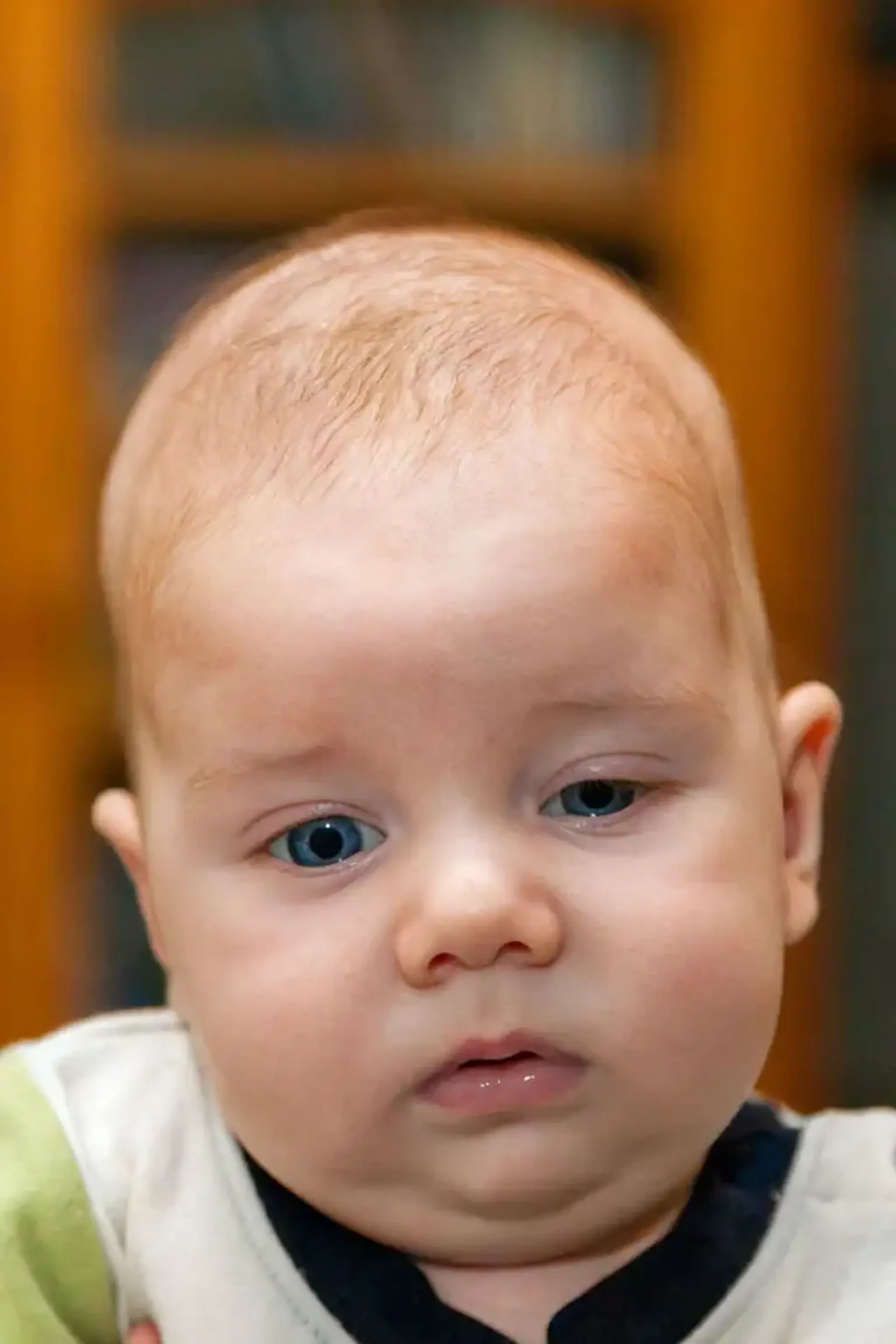
The early fusing of cranial sutures causes cranial synostosis. These sutures exist to aid the baby's passage through the birth canal and, later, to allow the brain to expand and thrive. When one or more sutures close prematurely, the structure of the skull changes, growing along the path of least resistance (perpendicular to the closed suture) and resulting in an atypically shaped skull, increasing intracranial pressure (ICP) and affecting the respiratory and neurologic systems, as well as the child's development.
The ensuing development pattern may give enough space for the growing brain, but it can also result in an aberrant head shape and facial traits. When the compensation fails to give enough space for the growing brain, craniosynostosis causes an increase in intracranial pressure, which can cause vision impairment, sleeping issues, eating difficulties, or mental development impairment with a considerable loss in IQ.
Epidemiology
Craniosynostosis affects one out of every 2000 to 2500 live births. It has risen through time, with predisposing variables being either environmental (maternal smoking, in utero exposure to teratogens, intrauterine constraint, fetal positioning) or genetic (maternal smoking, in utero exposure to teratogens, intrauterine constraint, fetal placement) (mutations). Almost 20% of all craniosynostoses are caused by genetic factors, the majority of which are inherited in an autosomal dominant pattern, however new mutations occur in 50% of instances. Non-syndromic craniosynostosis accounts for 75% of instances, while syndromic craniosynostosis accounts for 25%.
Sagittal sutures are impacted in 55 percent to 60 percent of cases, coronal (20 percent to 25 percent), metopic (roughly 15%), and lambdoid sutures are affected in approximately 15% of cases (3 percent to 5 percent ). Usually within the first year of life, a clinical diagnosis is made.
Craniosynostosis causes
When a single suture is involved, cranial synostosis is classified as simple, and when many sutures are involved, it is classified as complicated. Another frequent approach to categorize them is as syndromic (Apert, Crouzon, Pfeiffer) or non-syndromic (Apert, Crouzon, Pfeiffer) with an isolated finding.
Pathophysiology
The skull vault (calvaria) increases during childhood to accommodate the expanding brain. This growth occurs mostly in the cranial sutures, which are narrow seams of undifferentiated mesenchyme that run between separate bones. The metopic and sagittal sutures divide the paired frontal and parietal bones in the midline; coronal sutures split the frontal and parietal bones; and lambdoid sutures separate the parietal bones from the single occipital bone.
Craniosynostosis is a condition in which one or more of the cranial sutures fuse prematurely, resulting in secondary skull distortion due to a combination of lack of growth perpendicular to the fused suture and compensatory regrowth at the non-fused sutures. The prevalence of craniosynostosis is estimated to be between 1 in 2100 and 1 in 2500 births worldwide.
It's critical to recognize and treat craniosynostosis because it's linked to a slew of problems that compromise sensory, respiratory, and neurological function.
Craniosynostosis types
A comprehensive history and physical examination, as with any other disease, are essential in determining the diagnosis.
It's critical to know if there's a family history of atypical head shapes, in utero exposure to teratogenic medicines, intrauterine restraints, or an abnormal fetal position, as well as any pregnancy difficulties and missed milestones.
The physical examination allows the doctor to determine whether a suture fusion is present and whether any concomitant symptoms, such as congenital defects or dysmorphic traits, would constitute the craniosynostosis part of a syndrome.
The importance of examining the cranium cannot be overstated. To determine the cephalic index, look at it from all angles and measure the head circumference (maximum breadth of the skull multiplied by 100 divided by the maximum length of the skull). The examiner should also inspect and measure the fontanelles, as well as observe and touch the scalp for any sutural ridging and conspicuous vessels over the head.
Apart from these, depending on the severity of the disorder, the patient may experience other clinical manifestations as a result of the craniosynostosis, which should be noted and followed. If any indicators of intracranial pressure (ICP) are present, an ophthalmologic examination should be performed, which will reveal papilledema. Examine any potential airway blockages or feeding issues.
The different non-syndromic craniosynostoses are described below:
Scaphocephaly
Scaphocephaly is an exemplary example of this disorder, with the term hinting at the deformity of the skull. The Greek-derived word'scaphocephaly' literally means 'boat-head.' 'Dolichocephaly' is a similar term
Premature sagittal suture closure limits growth in a perpendicular plane, causing the skull to remain narrow and not grow sideways. Standing above the infant and looking down at the top of the head, this is best seen. Compensatory development occurs forward at the coronal suture and backward at the lambdoid suture, resulting in a pronounced frontal bossing and a prominent back section of the head, respectively, known as frontal bossing and coning.
Trigonocephaly
The premature closure of the metopic suture causes trigonocephaly. Using Virchow's law to forecast the deformity, this fusion will result in a narrow forehead, which is highlighted even more by the suture being removed. At both the coronal sutures, compensatory development occurs, pushing the forehead forward. The final shape can best be judged from a top view, which reveals a roughly triangular shape to the skull.
Trigonocephaly is another Greek-derived term that means "triangular-shaped head." Hypotelorism is a facial trait of metopic synostosis; in the frontal view, the width between the eyes is narrower than typical.
Plagiocephaly
Anterior plagiocephaly
Unilateral coronal synostosis is referred to as anterior plagiocephaly. Children born with unilateral coronal synostosis acquire a skew head, known as plagiocephaly, as a result of compensatory processes.
The sagittal suture 'divides' the coronal suture into two half; unilateral means that the sagittal suture is fused on either the right or left side. This fact brings up an essential point right away. In unilateral coronal synostosis, right and left are not the same as when the sagittal or metopic sutures are closed. This asymmetry is visible in the deformities of the skull, as well as the facial deformity and problems.
Using Virchow's law, the skull malformation can only be anticipated in part this time. The forehead is flattened and growth is stopped in the plane perpendicular to the fused suture, but only on the ipsilateral side of the head. The same side of the head where the suture is closed is referred to be ipsilateral.
Compensatory growth can occur in both a parallel and perpendicular plane. The parallel plane is accounted for by increased growth at the metopic and sagittal sutures, which results in temporal fossa bulging. On the side of the skull with the patent coronal suture, the contralateral side, compensatory growth in the perpendicular plane occurs. The front half of the brow will protrude forward.
Asymmetry of the frontal bones, greater width of the skull, and forward displacement of the ear on the ipsilateral side of the head are all visible from a top view of the skull. A frontal view of the skull will reveal asymmetrical aspects of the face, such as a shift in the chin point of the jaw and a deviation of the tip of the nose.
Due to the ipsilateral forward displacement of the temporomandibular joint and the ear, the chin point is more to the contralateral side of the head. The tip of the nose will also point in the opposite direction. Malocclusion of the jaw occurs in up to 90% of cases; a subtle form of strabismus is generated by the uneven positioning of the orbits; and refractive error, particularly astigmatism, is caused by the asymmetrical development of the orbits.
Posterior plagiocephaly
Unilateral lambdoid synostosis is also known as posterior plagiocephaly, signifying that it causes a'skew head,' similar to unilateral coronal synostosis. The difference this time is that the abnormality is primarily seen at the occiput.
According to Virchow's law, growth restriction occurs on the ipsilateral side of the skull, whereas compensatory growth occurs on the contralateral side. This growth pattern has an effect on the kid's base of the skull, which is not visible when viewed from behind the child, as well as the cervical spine, which has a curve. Furthermore, a protrusion of the mastoid can be visible from behind the kid. There are usually only minor asymmetries in the forehead.
Brachycephaly
The closure of both coronal sutures results in brachycephaly, or a'short head.' According to Virchow's law, this will result in a child's head being restricted in both forward and backward growth, with recessed frontal bones and a flattened occiput. Because of the sagittal and lambdoid sutures, compensatory growth will occur sideways and upwards.
Oxycephaly
Oxycephaly is a form of cephalic condition that is also known as turricephaly and high-head syndrome. This word refers to the premature closure of the coronal suture as well as any other suture, such as the lambdoid suture.
The different syndrome presenting with craniosynostoses are described below:
- Apert: Coronal suture is affected. Midface hypoplasia, hypertelorism (bulging and wide-set eyes), beaked nose, underdeveloped jaw (leading to crowded teeth), syndactyly of hands and feet, and hearing loss are all symptoms of this condition. They may have intellectual deficits ranging from mild to moderate.
- Crouzon: Midface hypoplasia, beaked nose, exophthalmos, hypertelorism, cervical vertebral fusion, and hearing loss are all symptoms of this condition, which affects coronal, sagittal, and/or lambdoid sutures. Some people have cleft lips and/or palates. They are usually intelligent people.
- Pfeiffer: features brachycephaly (bi-coronal sutures), hypertelorism, maxillary hypoplasia, broad thumbs, great toe, syndactyly, brachydactyly, hearing loss.
- Muenke: affecting coronal suture (uni or bilateral); features midface hypoplasia, hypertelorism, macrocephaly, and hearing loss.
Diagnosis
The most precise way is a CT scan with 3D reconstruction, which allows all sutures to be assessed, however this option must be carefully considered due to the radiation risk. Plain x-rays are inexpensive. As a result, they're effective for assessing infants at low risk of craniosynostosis, but they're not precise enough. When compared to CT, MRI is less accurate, but it is still a useful tool. It is often reserved for children whose CT indicated any brain irregularities.
Ultrasound is a low-cost (but technician-dependent) modality that can only be utilized with open fontanelles but is extremely effective for seeing and monitoring sutures at each inspection. GRASE (gradient-and-spin-echo), a kind of MRI that emphasizes bone-soft tissue boundaries and shows the cranial sutures as hyperintense, is one of the newer modalities in use.
When two or more sutures are afflicted by syndromic craniosynostosis, genetic testing is the standard follow-up (recommended especially when two or more sutures are affected), testing for FGF receptor genes, which have been the most commonly linked genes to these syndromes. FGFR2 and FGFR3, as well as transcription factors, are put to the test (TWIST, MSX2). The amount of study being done to understand and pinpoint the genetic origins of these diseases is increasing. To date, 57 genes have been identified as having a link to craniosynostosis and being the underlying cause, the most prevalent of which are the ones stated above.
Prenatal diagnosis
When a family member has a molecular anomaly, the challenges surrounding prenatal or preimplantation diagnosis are similar to those encountered with other genetic illnesses. The topic of whether craniosynostosis can be diagnosed prenatally using ultrasound or other imaging modalities is more difficult.
Because cranial sutures form late (16 weeks gestation) compared to most other embryonic structures, there is insufficient time for growing deformation of the skull to have happened by the time most conventional ultrasound diagnosis is performed (20 weeks), unless in the most severe cases.
Although a few reports of craniosynostosis diagnosis by ultrasound have surfaced around this time, the majority of even syndromic craniosynostosis is not diagnosed during pregnancy. When it comes to checking for recurrence of craniosynostosis in an older sibling, evidence of Apert syndrome can be found by looking for syndactyly in the limbs.
In most other cases, a late ultrasound to check for cephalopelvic disproportion is recommended, but caution should be exercised in offering any confident conclusion in the prenatal period that craniosynostosis has been excluded, unless in the most severe cases.
Treatment for Craniosynostosis
Acute management
Maintenance of the airway, support of feeding, eye protection, and treatment of elevated ICP are all priorities in the care of neonates and babies with severe multisuture synostosis.
Depending on the anatomical cause, respiratory difficulty may necessitate urgent sleep study evaluation by specialist paediatric respiratory physicians and ENT surgeons, necessitating either continuous positive airway pressure support, nasal stenting, tonsillectomy/adenoidectomy, choanal dilatation, early midface advancement, or tracheostomy.
If the eyelids are unable to seal, exposure keratitis develops, and an immediate ophthalmology opinion should be sought, with tarsorraphies or early midface advancement as options for eye protection.
There are various causes of elevated ICP in craniosynostosis, each of which necessitates a distinct treatment. A shunt is required for hydrocephalus, and obstructive sleep apnea is treated by improving the airway . Calvarial enlargement is required for craniocerebral disproportion (a result of craniostenosis). If there is tonsillar herniation, foramen magnum decompression may be required. When operating on the rear of the skull in multisuture synostosis, irregular venous drainage is a considerable risk.
Elective management
The treatment of these patients is determined on the type of craniosynostosis they have. When compared to some syndromic forms that require urgent surgical intervention due to involvement of the airway, ophthalmologic, and neurological systems, the simple and non-syndromic varieties can be addressed surgically but electively. In cases where unilateral craniosynostosis is not too severe, a conservative approach with remodeling helmets could be tried first.
The extent, as well as the type of surgery, depends on the age and presentation of the patient. Two modalities are in use:
- Endoscopic suturectomy: done in a patient less than six months of age because the bone is more flexible and manageable by an endoscope. In comparison to an open craniotomy, the postoperative recovery is faster, there is less blood loss, and the surgery is shorter. The sole disadvantage is that in most cases, the surgery must be combined with a 4- to 6-month postoperative use of a remodeling helmet.
- Open craniotomy: done in patients older than six months because the bones are more rigid and cannot be manipulated as well with an endoscope. This technique provides for improved skull remodeling and reduces the necessity for postoperative helmet wear.
The primary purpose of surgery is to allow enough space in the cranial vault for the brain to grow and develop normally, as well as to give the child a more attractive appearance.
When there are no evidence of increased ICP or airway compromise, the optimal time to correct is between 6 and 12 months of age, which coincides with the period when the infant's brain and skull expand the greatest. Additional intervention may be required at any time, but it is more likely in syndromic instances.
Differential Diagnosis
Differentiating positional plagiocephaly from craniosynostosis is necessary. There are no sutures that have fused prematurely. A parallelogram of the head, ipsilateral anterior displacement of the ear and head, and ipsilateral occipital flattening with contralateral occipital bossing are all symptoms of this condition.
Due to the "return to sleep campaign" to lower the incidence of sudden infant death syndrome, the prevalence has risen over time. Still, the only issue is one of appearance, which can be alleviated by alternating the baby's sleeping side of the head. Helmets can be remodeled as well. This issue does not necessitate surgery and has no impact on neurologic development. Parents should be comforted and told to keep their kid sleeping on his back.
Prognosis
If left untreated, craniosynostosis can impede a child's development by restricting brain growth and causing damage to brain tissue owing to increasing intracranial pressure. The type of craniosynostosis determines the degree of developmental delay. Learning difficulties are less common in children with sagittal synostosis than in those with metopic, uni-coronal, or lambdoid synostosis. A bad academic and cognitive outcome for these children could be mitigated, if not averted, with early diagnosis of developmental gaps and placement in assistance programs.
Cases with immediate surgical surgery had an excellent result, with reasonably normal growth and development. Follow-up for additional suture fusion and head growth is critical to determining whether these individuals require re-intervention, with a special focus on syndromic craniosynostosis.
Complications
Postoperative hyperthermia is the most prevalent consequence, followed by infections (meningitis), seizures, subgaleal hematoma, subcutaneous hematoma, and cerebrospinal fluid leaking. Reintervention, as well as open craniotomy, increase the risk of complications . Death and morbidity rates can exceed 50% in cases of significant blood loss.
Conclusion
In the treatment of patients with craniosynostosis, especially those with syndromic craniosynostosis, an interdisciplinary approach is critical. Pediatricians, neurosurgeons, plastic surgeons, maxillofacial surgeons, ophthalmologists, geneticists, nurses, respiratory sleep physicians, orthopedics, and, subsequently, a developmental expert should all be on the team.
Referral to a developmental pediatric nurse for early intervention and developmental monitoring is critical in the implementation of a special education plan to address the patient's potential shortcomings, especially if they are at risk of developmental delay. Early detection and referral for treatment are critical to providing these people the best chance possible.
Physical therapy, occupational therapy, speech therapy, and possibly a hearing or vision aid will be required, as well as placement in an appropriate school setting.
Each of these disciplines must collaborate with the other members of the multidisciplinary healthcare team. They must share their findings and ensure that other clinicians are aware of any changes or therapy outcomes that have interprofessional repercussions. Only then can optimal outcomes be steered.
Because physical and mental problems may emerge as the infant grows, the follow-up will be extended. A bad academic and cognitive outcome for these children is mitigated, if not prevented, with early diagnosis of developmental gaps and placement in assistance programs.
Most children's outcomes are uncertain and dependent on the sort of genetic condition they have. However, some children's lives can be improved with proper nursing care.