Crystal Arthropathies

Last updated date: 27-Aug-2023

Originally Written in English

Crystal Arthropathies

Overview

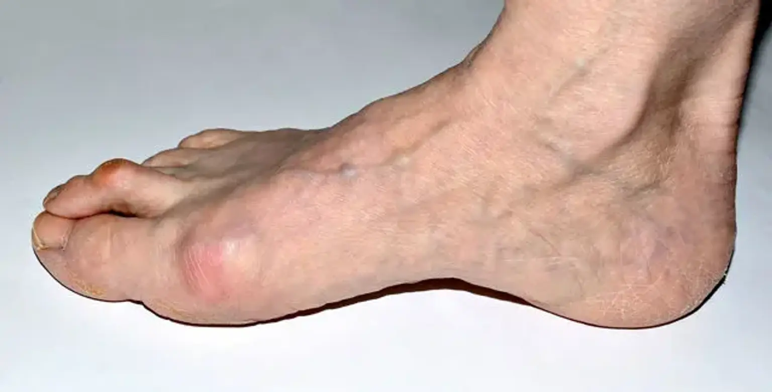
Crystal arthropathies are a set of joint illnesses characterized by crystal deposits in the joints and surrounding soft tissues. Gout and calcium pyrophosphate deposition are the two most prevalent kinds (CPPD). Crystalline arthropathies can cause joint degeneration and kidney illness in the long run.


Crystalline arthropathies definition

Crystal arthropathy is a type of arthropathy (joint disorder) marked by the formation of small crystals in one or more joints. The identification of diverse microcrystals such as monosodium urate, calcium pyrophosphate dihydrate, calcium hydroxyapatite, and calcium oxalate has improved thanks to polarizing microscopy and other crystallographic techniques.

The most common types are gout and calcium pyrophosphate deposition (CPPD).


Gout

Gout

Gout is one of the most common causes of chronic inflammatory arthritis in the United States, and it is marked by the deposition of monosodium urate (MSU) monohydrate crystals in the tissues. Even before the common era, gout was acknowledged. As a result, it is possibly the most well-understood and managed of all the rheumatic disorders.


Causes of Gout

Causes of Gout

Risk Factors

The most common cause of gout is hyperuricemia. Higher serum urate levels not only increase the risk of gout flare-ups, but they also lead to more frequent flare-ups over time. In a study of more than 2000 older persons with gout, those with levels greater than 9 mg/dl were three times more likely than those with levels less than 6 mg/dl to experience a flare in the next 12 months.

Gout is caused by a combination of factors, including hyperuricemia. In fact, only a small percentage of these patients develop gout. Older age, male sex, obesity, a purine diet, alcohol, drugs, concomitant conditions, and heredity have all been linked to gout and/or hyperuricemia. Diuretics, low-dose aspirin, ethambutol, pyrazinamide, and cyclosporine are among the offending drugs.

Consumption of animal foods such as shellfish (e.g., shrimp, lobster), organs (e.g., liver and kidney), and red meat can all lead to hyperuricemia and gout (pork, beef). Alcohol, sugary beverages, sodas, and high-fructose corn syrup are all possible causes of this condition.


Triggers

A flare-up can be triggered by any disease that induces changes in extracellular urate concentration. Stress (surgery, recent trauma, or famine), dietary considerations (e.g., fatty foods, beer, wine, and spirits), and medicines are all examples of these circumstances (e.g., aspirin, diuretics, or even allopurinol).


Epidemiology of Gout

Gout prevalence varies by age, gender, and country of origin. Gout affects 1 to 4% of people in the United States. Globally, older age and male sex are two common risk factors. Gout is 2 to 6 times more common in men (3 to 6%) than it is in women in Western countries (1 to 2 %).

The prevalence rises with age, but reaches a halt after 70 years. In the years 2007-2008, around 3.9 percent of adults in the United States were diagnosed with gout. Individuals with chronic conditions such as hypertension, chronic renal disease, diabetes, obesity, congestive heart failure, and myocardial infarction are more likely to develop gout.


Comorbidities:

Gout is frequently linked to hypertension, diabetes, hyperlipidemia, and metabolic syndrome. Psoriasis patients produce more urate and are more likely to develop gout. Patients with renal insufficiency, on the other hand, have lower urate excretion, which can lead to gout attacks. 


Pathophysiology of Gout

Pathophysiology of Gout

Hyperuricemia

Hyperuricemia is a major element in the development of gout because it reduces urate solubility, which promotes monosodium urate crystal nucleation and growth. Both exogenous and endogenous purine breakdown produce uric acid in the blood, which is then eliminated by the kidneys. The foundation for the elevation in serum uric acid levels is overproduction and/or underexcretion of uric acid.


Inflammatory Response

When macrophages phagocytize monosodium urate crystals, cytosolic protein complexes develop and become activated, causing inflammation (NLRP2 inflammasome). After that, these complexes attract caspase-1, which converts pro-IL-1beta to IL-1beta. The inflammatory response of gout is mostly mediated by IL-1beta. It causes vasodilation, monocyte recruitment, and initiates and intensifies the inflammatory cascade. Increased IL-1beta secretion can lead to the breakdown of bone and cartilage. Other cytokines involved in the inflammatory response include TNF-1, IL-6, CXCL8, and COX-2.


Symptoms of Gout

Symptoms of Gout

The symptoms of a gout flare-up are frequently visible in a patient. The first metatarsophalangeal joint is the most usually affected joint. In some cases, the talar, subtalar, ankle, and knee are also involved. Although the above-mentioned joints are frequently affected by gout, the physician should pay special attention to other joints, particularly those with underlying osteoarthritis. Other periarticular structures, such as tendons and bursa, may be damaged in addition to joints.

The majority of patients present with sudden onset joint discomfort. The pain may be sudden, rousing the patient from sleep, or it may have developed gradually over a few hours prior to the presentation, reaching its peak severity at 24 hours. The pain is frequently acute and resistant to common home cures; even touching the joint can be terrible. Local inflammation caused by gout flare-ups commonly manifests as erythematous, swollen, and heated joints. Fever, general malaise, and weariness are some of the systemic symptoms of joint inflammation.

The physical exam findings correspond to the patient's medical history. Typically, the injured joint is red, swollen, heated, and tender. The flare-up in chronic gout sufferers might affect many joints. It can induce a systemic inflammatory response syndrome, which can be misdiagnosed as sepsis, when multiple joints are involved.

Patients with prolonged hyperuricemia may develop tophi, which are nodules formed by subcutaneous urate depositions. Joints, ears, finger pads, tendons, and bursae are all common places for tophi to appear.


Diagnosis of Gout

Synovial Fluid Analysis

Monosodium urate crystal identification is the gold standard for gout diagnosis. During a gout flare-up, synovial fluid is often yellow in color and cloudier in appearance. It comprises crystals and white blood cells. The synovial fluid in patients with septic arthritis will be more opaque, with a yellow-green color. Septic arthritis synovial fluid has a higher white blood cell count (above 50000/ml) and a positive gram stain when examined under a microscope. Additionally, bacteria will be present in the cultures, whereas crystals will be absent.

Under polarized microscopy, needle-shaped, negatively birefringent crystals can be seen in synovial fluid or tophus aspiration. Arthrocentesis is also required to confirm the diagnosis and rule out other causes of septic arthritis, such as Lyme disease or pseudogout (calcium pyrophosphate).


Laboratory Study

During a gout flare-up, the white blood cell count, erythrocyte sedimentation rate (ESR), and C-reactive protein (CRP) levels are typically elevated, however these features are non-specific and do not support the diagnosis.

Serum urate levels may also be normal during an acute gout flare-up. After the flare-up has passed, the doctor should check the serum urate level in patients with an unclear gout diagnosis. In symptomatic individuals, hyperuricemia aids in the clinical diagnosis of gout, but it does not definitively confirm the diagnosis. In the general population, asymptomatic hyperuricemia is not uncommon. Gout is less likely to be diagnosed if serum urate values are consistently low.

Urinary fractional excretion of uric acid can be assessed, especially in young people who have hyperuricemia for no apparent reason. It can assist distinguish between uric acid overproduction and underexcretion and serve as a therapeutic guidance.


Imaging

Ultrasonography and dual-energy CT (DECT) can help diagnose gout, albeit they aren't commonly utilized. A hyperechoic enhancement over the cartilage, often known as a twofold contour sign, indicates monosodium urate deposition on ultrasonography. Due to beam attenuation following exposure to two different X-ray spectra, DECT can detect urate.


Management of Gout

The therapy of gout is determined by the treatment goals. The main goal during acute flares is to minimize inflammation and discomfort. The long-term goal is to lower serum urate levels in order to prevent flare-ups and tophi regression.

Acute Flares

The goal of acute flare management is to reduce inflammation and the discomfort that comes with it. To lessen the intensity and duration of the flare-up, the physician should begin medication within the first 24 hours of beginning.

Non-pharmacological therapy, such as rest with cold packs applied topically, can be used with anti-inflammatory drugs. NSAIDs, colchicine, or systemic glucocorticoids are the first-line treatments for gout flares. To avoid a return flare-up, the therapies should last at least 7 to 10 days.

There is no evidence to suggest that one NSAID is better than the other. Naproxen or diclofenac are examples of high-dose, fast-acting NSAIDs. Due to its toxicity profile, indomethacin is not recommended.

When compared to a placebo, colchicine has been demonstrated to lower pain by over 50% in a randomized control experiment after 24 hours. The FDA recommends starting with 1.2 mg and then switching to 0.6 mg one hour later. Twelve hours following the last treatment, a preventive dose (0.6 mg once or twice daily) should be given.

Doses must be reduced in older populations, as well as those with chronic kidney disease (CKD) and/or hepatic impairment. Due to an increased risk of colchicine toxicity, patients taking other medicines that affect cytochrome P450 (CYP) 3A4 and P-glycoprotein may need to cease or change their prescription regimen. 

When NSAIDs and/or colchicine are contraindicated in people with gout, glucocorticoids are recommended. Patients with renal insufficiency benefit from these medications as well. For monoarticular gout flare-ups, glucocorticoids can be given intra-articularly, and for polyarticular flare-ups, they can be given orally. Anakinra, canakinumab, and rilonacept are IL-1 antagonists that are effective for acute gout flares, although they are not approved for this usage in the United States.


Non-acute Flares

Pharmacologic

The clinician should not start urate-lowering therapy (ULT) in patients with asymptomatic hyperuricemia or gout with rare attacks (1 flare/year). The American College of Rheumatology 2012, Guidelines for starting ULT include the following:

  1. Frequent flares (greater than or equal to two per year)
  2. CKD stage 2 or more
  3. Tophus diagnosis on physical examination or imaging
  4. Past urolithiasis.

To evaluate adverse effects and therapeutic response, urate-lowering medication is started at a modest dose. In individuals with tophi, the dose is titrated every 2 to 6 weeks to attain serum urate levels of less than 6 mg/dl or 5 mg/dl.

To lower the risk of gout flare-ups at the commencement of ULT, colchicine prophylaxis is recommended for 3 months in patients without tophi or 6 months in patients with tophi after achieving serum urate target.

ULT can categorize into three classes (based on the mechanisms).

  • Xanthine oxidase inhibitors (XOI) - XOI works by preventing uric acid production. Allopurinol and febuxostat are members of this class. In the case of gout, allopurinol is the first-line pharmacologic ULT. The liver enzymes, renal function, and blood count should all be checked on a regular basis by the doctor. Skin rashes to life-threatening severe allopurinol hypersensitivity (particularly in HLA-B*5801 positive patients) are all possible side effects of allopurinol.
  • Uricosuric 

The uricosuric drugs function by enhancing urate clearance in the kidneys. These drugs are ineffective as monotherapy in patients with low creatinine clearance (less than 30 ml/minute) and are contraindicated in those who have had nephrolithiasis in the past. Probenecid and lesinurad are examples of drugs in this class. Probenecid is the only drug that has been licensed for use alone. Uricase is exclusively prescribed to people who have refractory gout.

Uricase converts uric acid to allantoin, which is soluble in water. The authorized uricase drugs for treating hyperuricemia include Pegloticase and Rasburicase. Patients must stop taking other ULT medications before commencing this therapy because they may develop antibodies against uricase, which can only be discovered by monitoring the serum urate level, which is unaffected by other treatments. This drug class is also contraindicated in persons who have a deficit in the enzyme glucose-6-phosphate dehydrogenase (G6PD); thus, possible treatment should be checked for this enzyme before starting treatment.

  • Interleukin-1 (IL-1) inhibitor - Inflammation in gout is largely mediated by interleukin-1 (IL-1). Blocking agents like anakinra and canakinumab, which block this interleukin, are crucial, especially during gout flare-ups. This class is still undergoing research. As a result, these agents have not yet been approved in the United States for gout flare-ups.


Non-pharmacologic

Gout patients are encouraged to change their habits to avoid future attacks.

Reducing alcohol consumption, restricting purine-rich meals (meat, shellfish, high fructose corn syrup, and sweetened soft drinks), and substituting low-fat or non-fat dairy products for higher fat content dairy products are among the diet recommendations. Weight loss and proper hydration can also assist to lessen the frequency of gout flare-ups.


Differential Diagnosis

  • CPPD deposition
  • Septic arthritis
  • Osteoarthritis
  • Rheumatoid arthritis
  • Psoriatic arthritis
  • Cellulitis


Prognosis of Gout 

The prognosis of gout is determined on the individual's comorbidities. Individuals with cardiovascular comorbidities have a higher mortality rate. When gout is treated properly, most individuals can live a normal life with only minor side effects. Patients with symptoms that appear at a younger age frequently have a more serious condition when they show. Recurrent flare-ups are common in persons who do not change their lifestyle.


Complications of Gout

  • Tophi, Joint deformity, osteoarthritis, bone loss
  • Urate nephropathy and nephrolithiasis.
  • Ocular complications, such as conjunctivitis, uveitis, or scleritis from the urate crystal precipitation.


Calcium Pyrophosphate Deposition Disease

Calcium Pyrophosphate Deposition Disease

The crystal deposition arthropathy calcium pyrophosphate deposition disease (CPPD) affects the synovial and periarticular tissues. It might have asymptomatic, acute, or chronic inflammatory arthritis as a clinical manifestation.

Chronic CPP deposition arthritis, also known as pseudo-rheumatoid arthritis, is a kind of rheumatoid arthritis that affects the wrists and metacarpophalangeal (MCP) joints and has a waxing and waning clinical history that can last several months.

Calcium pyrophosphate deposition illness is caused by calcium pyrophosphate dihydrate crystals that often affect bigger and weight-bearing joints, such as the hips, knees, and shoulders.


Causes of CPPD

An imbalance between the generation of pyrophosphate and the levels of pyrophosphatases in sick cartilage is thought to be the cause of calcium pyrophosphate deposition disease. Pyrophosphate reacts with calcium in the synovium and surrounding tissues to generate CPP.

CPPD has been linked to a number of comorbidities. Hyperparathyroidism was found to have the strongest favorable correlation with CPPD in several investigations, followed by gout, osteoarthritis, rheumatoid arthritis, and hemochromatosis. Osteoporosis, hypomagnesemia, chronic renal disease, and calcium supplementation are some of the other comorbidities.

Calcium pyrophosphate deposition is thought to activate the immune system, resulting in inflammation and additional soft tissue injury.


Symptoms of CPPD

Acute calcium pyrophosphate arthritis has symptoms that are comparable to acute urate arthropathy, such as joint edema, erythema, and discomfort. A low-grade fever may be present in up to half of these patients. The knee is the most usually affected joint, but other weight-bearing joints, such as the hips and shoulders, can also be impacted.

Chronic CPP arthritis affects a subset of patients, with waxing and waning episodes of non-synchronous, inflammatory arthritis affecting several non-weight-bearing joints such wrists and MCP joints, similar to rheumatoid arthritis.

Crystal deposition disease should be suspected in elderly patients who present with acute degenerative arthritis in weight-bearing joints. The elderly are more likely to take a gentler approach. Following catastrophic injuries, some individuals will experience acute flares.


Diagnosis of CPP arthritis

Patients with calcium pyrophosphate deposition disease should have arthrocentesis for synovial fluid analysis, as well as radiography of the affected joints, after a thorough physical examination.

The presence of rhomboid crystals in the synovial fluid aspirate observed under polarized microscopy could be used to confirm the diagnosis. Positive birefringence is common in these crystals.


While the presence of chondrocalcinosis on imaging supports the diagnosis of CPPD, its absence does not rule it out. Ultrasound (US) may reveal early indications such as cartilage irregularities. Chondrocalcinosis, or calcification of joint cartilage, can be seen on radiographic imaging.

Previous research has shown that magnetic resonance imaging (MRI), particularly gradient-echo sequences, can be used to assess the burden of calcium pyrophosphate crystal formation in joint cartilage.


Management of calcium pyrophosphate deposition disease

Reduced inflammation and stabilization of any underlying metabolic condition that may predispose to calcium pyrophosphate crystal deposition are the goals of treatment for patients with calcium pyrophosphate deposition disorder. Joint suction and intra-articular glucocorticoid injection are usually the treatment of choice in individuals with acute flares involving one or two joints, unless contraindicated.

When a patient has acute inflammation involving three or more joints, the treatment is usually systemic, with nonsteroidal anti-inflammatory medications being used often (NSAIDs). Colchicine or systemic glucocorticoids may be used to treat patients who are allergic to NSAIDs.

After septic arthritis has been ruled out or deemed improbable, joint aspiration should be performed. Deferring glucocorticoid therapy until synovial fluid cultures are negative is recommended for individuals with signs and symptoms indicative of septic arthritis.

To reduce additional inflammation, further methods include the use of cold packs and joint rest with weight-bearing restrictions. Low-dose colchicine may be given daily to patients with repeated bouts of acute CPP arthritis.

While various drugs reduce serum urate levels and prevent the production of urate crystals, there is currently no treatment that specifically targets CPP crystal deposition. As a result, treating underlying metabolic disorders, soft tissue inflammation, and symptomatology is crucial in the therapy of CPPD.


Prognosis of CPPD

Acute calcium pyrophosphate arthritis is usually self-limited, and the inflammation goes away after a few days or weeks of treatment.

Patients with chronic CPP inflammatory arthritis may experience symptoms that are similar to those seen in rheumatoid arthritis, such as morning stiffness, localized edema, and limited range of motion. Tenosynovitis can also be associated with carpal or cubital tunnel syndrome in some cases. Multiple joints are frequently implicated, and inflammatory episodes may occur in a nonsynchronous, waxing and waning clinical course that lasts several months.

Acute CPP arthritis is more likely in patients with underlying joint comorbidities, such as osteoarthritis. CPP crystals trigger the immune system when they are deposited, causing inflammation and fibrocartilage damage.


Complications of CPPD

Calcium pyrophosphate's molecular structure has the ability to cause inflammatory responses. The occurrence of chondrocalcinosis is linked to the breakdown of menisci and synovial tissue.

After many episodes of acute CPP crystal arthritis, individuals may develop palpable nodules or lumps that resemble gout tophi. These nodules form in the periarticular tissue and are caused by CPP crystal accumulations in the synovium and nearby soft tissue, which can lead to additional joint deterioration.

A small percentage of patients will develop spinal involvement, which can manifest as spine stiffness and bony ankylosis similar to ankylosing spondylitis. Some people may develop symptoms that resemble diffuse idiopathic skeletal hyperostosis (DISH), such as calcification of the posterior longitudinal ligament and spinal cord compression.


Conclusion

Crystal Arthropathies

Gout is an arthritic condition that causes severe discomfort. Sharp crystals can form in the big toe or other joints when your body has too much uric acid, causing gout attacks, which are episodes of swelling and discomfort. Gout can be treated with drugs as well as dietary and lifestyle changes.

Patients with chondrocalcinosis or previous bouts of acute calcium pyrophosphate arthritis will benefit from knowing that specific circumstances, such as surgical procedures or trauma, are linked to arthritic flares. People with CPPD who present at a younger age may be screened for underlying metabolic disorders including hyperparathyroidism or hemochromatosis. It is also vital to obtain a complete family history.