CT(chest/abdomen/pelvis)

Last updated date: 03-Mar-2023

Originally Written in English

CT(chest/abdomen/pelvis)

Overview

CT is a diagnostic imaging technique that produces comprehensive pictures of internal organs, bones, soft tissue, and blood arteries. The cross-sectional pictures produced by a CT scan can be reformatted in different planes and even produce three-dimensional images that can be seen on a computer display, printed on film, or transmitted to electronic devices.

CT scanning is frequently the most effective way for diagnosing many different types of cancer since the pictures allow your doctor to confirm the presence of a tumor as well as its size and location. CT scanning is quick, painless, noninvasive, and precise. It can disclose internal injuries and bleeding fast enough in an emergency situation to help save lives.
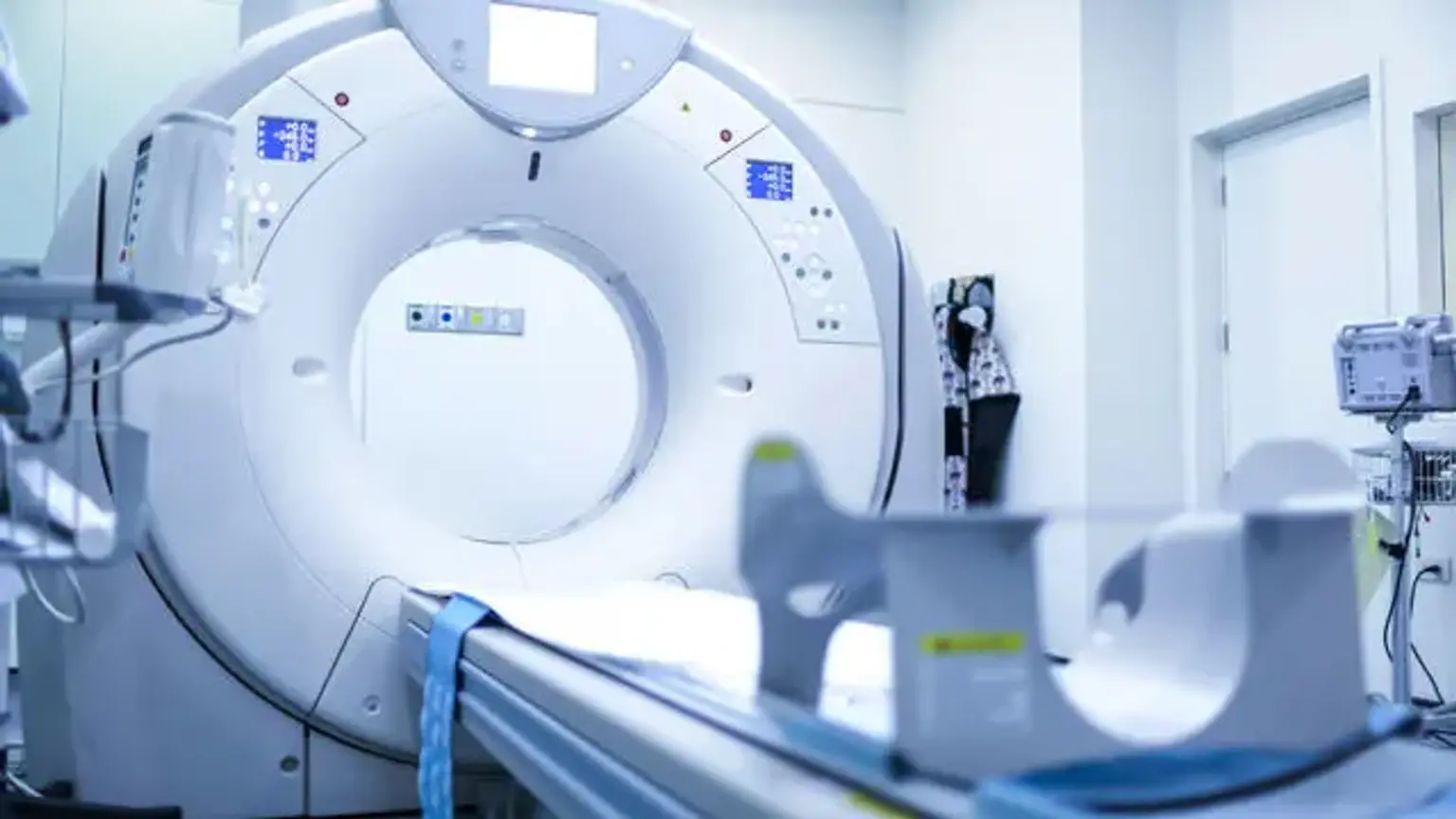
 

What is Computed Tomography (CT)?

Computed Tomography

A computed tomography (CT) scan, sometimes known as a CT scan, is a type of radiological imaging investigation. Allan MacLeod Cormack, a physicist, and Godfrey Hounsfield, an electrical engineer, created the machine. In 1979, they were given the Nobel Prize in Physiology or Medicine for their achievements. In 1974, the first scanners were deployed. Since then, developments in technology and mathematics have enabled single pictures to be computed into two-dimensional instructive images.

The CT scan is essentially an X-ray examination in which a succession of beams are spun around a specific body component, producing computer-generated cross-sectional pictures. When compared to ordinary X-rays, the benefit of tomographic pictures is that they contain comprehensive information about a particular area in cross-section, avoiding image superimposition, which gives a significant advantage over plain films. For a suspected disease, CT scans reveal excellent clinicopathological correlation.

The use of CT scans improves a physician's ability to effectively diagnose a patient's ailment. Low-dose CT scans are being used in cancer screening and prevention. The research was first referred to as a CAT scan, which stood for computer axial tomography, because the table moved after each axial image was collected.

The table travels constantly throughout a spiral or helical scan as the x-ray source and detectors spin. This considerably shortens the period of the investigation, allowing for faster findings in emergency situations. It quickly replaced cerebral angiography for diagnosing head trauma injuries and brain masses in a quick and highly reliable manner. A qualified radiologist interprets and reports on CT scans obtained by a radiologic technician. 


CT Advantages

CT Advantages

Compared to standard two-dimensional medical radiography, CT scanning provides significant advantages. For starters, CT removes the superimposition of pictures of structures beyond the region of interest. Second, CT scans feature higher picture quality, allowing for more detailed investigation. CT can discriminate between tissues with radiographic density differences of 1% or less. Third, CT scanning allows for multiplanar reformatted imaging: depending on the diagnostic goal, scan data can be displayed in the transverse (or axial), coronal, or sagittal plane.

CT's enhanced resolution has enabled the creation of novel studies. CT angiography, for example, avoids the intrusive insertion of a catheter. CT scanning can perform a virtual colonoscopy with improved accuracy and less patient discomfort than a regular colonoscopy. Virtual colonography detects cancers significantly more accurately than a barium enema and utilizes a lower radiation dosage.

CT is a diagnostic technology that uses moderate to high levels of radiation. The radiation dosage for a specific examination is determined by several criteria, including the volume scanned, the patient's build, the number and kind of scan sequences, and the required resolution and picture quality. Two helical CT scanning parameters, tube current and pitch, are easily adjustable and have a significant impact on radiation. In examining anterior interbody fusion, CT scanning is more accurate than two-dimensional radiographs, however they may still overestimate the amount of fusion.


CT Types

CT Types

  • Spiral CT

Spinning tube, also known as spiral CT or helical CT, is an imaging method that involves spinning an entire X-ray tube around the center axis of the region being scanned. These are the most common types of scanners on the market since they have been in production for a longer period of time and have a cheaper cost of production and buying.

The fundamental restriction of this form of CT is the equipment's bulk and inertia (X-ray tube assembly and detector array on the opposite side of the circle), which restricts the speed at which it can spin. To increase temporal resolution, some systems employ two X-ray sources with detector arrays offset by an angle.

  • Electron beam tomography

Electron beam tomography (EBT) is a kind of CT in which a big enough X-ray tube is built so that only the electrons' route between the cathode and anode of the X-ray tube is twisted using deflection coils. This kind offered a significant benefit since sweep rates could be significantly quicker, allowing for less fuzzy imaging of moving structures like the heart and arteries. When compared to spinning tube types, fewer scanners of this form have been built, owing to the higher expense of developing a considerably bigger X-ray tube and detector array, as well as restricted anatomical coverage.

  • Dual source CT

In contrast to standard single tube systems, dual source CT is a sophisticated scanner with two X-ray tube detection systems. These two detector systems are positioned at 90° in the same plane on a single gantry.

  • CT perfusion imaging

CT perfusion imaging is a kind of CT that assesses blood artery flow while injecting a contrast agent. Blood flow, transit time, and organ blood volume may all be estimated with a high degree of sensitivity and specificity. Although this sort of CT may be utilized on the heart, its sensitivity and specificity for identifying abnormalities are still lower than those of other types of CT. This technique may also be utilized on the brain, as CT perfusion imaging can typically detect low brain perfusion much before a standard spiral CT scan. This form of CT scan is superior than others in terms of stroke diagnosis.


CT Indications

CT Indications

CT scans give information that is as near to real-time as possible, allowing for optimal illness treatment. Patients with stomach discomfort, fever, and an increased white blood cell count would have been brought to the operating room for an exploratory laparotomy before the CT scan.

The discovery of CT has provided physicians and surgeons with knowledge to avoid unneeded laparotomies, saving millions of dollars in healthcare expenditures. Finally, the patient benefit outweighs the risk of radiation, and it has remained a mainstay in clinical disease diagnosis. CT scan technological developments have considerably improved patient treatment by making it more efficient and cost-effective.

Until the development of MRI and PET scans, the CT scan was the primary tool for detecting and monitoring malignancies. Tumors respond positively to iodine contrast. Cancer patients can be detected and staged with a combined PET CT scan.

Depending on the organs to be evaluated, a CT scan is utilized for a variety of clinical purposes. CT scans are useful in both inpatient and outpatient therapeutic settings. It can rule out severe sickness in an emergency situation. The purpose of obtaining a CT scan is to assist the physician in diagnosing, narrowing the differential diagnosis, and confirming the doctor's concerns. It is also useful for cancer screening, staging, and monitoring. Its use aids in the proper performance of biopsies as well as during surgical procedures.

  • Brain: The CT scan is the primary examination for patients with hydrocephalus to measure ventricular size and compare the size in situations of shunt dysfunction. The CT scan is typically normal for pediatric patients who have headaches, loss of consciousness, or seizures; consequently, conservative use is advised to prevent radiation exposure. For a patient involved in a traumatic event, a CT scan can quickly reveal skull fractures, traumatic hematomas (epidural, subdural, and intracerebral), and edema, allowing for prompt therapy.
  • Abdomen: primary tumors, metastases, abscess, ascites, cholecystitis, appendicitis, renal calculi, pancreatitis, obstruction, lymphadenopathy, foreign body
  • Spine: fractures, degenerative changes, stability, and osteomyelitis A cervical CT scan can be used to evaluate the complete cervical spine in the emergency room. The whole spine can be adequately viewed with acute trauma.
  • Screening: colon and lung cancer
    • CT colonography/colonoscopy is used to diagnose colon disease and early-stage cancer with good sensitivity and specificity. 
    • Low-emission CT can be used to diagnose lung cancer in smokers and former smokers with a high smoking history aged between 55 and 80 years old using a low radiation dose. 
  • CT Angiography: brain, heart, lung, kidney, extremities
  • Intraoperative CT scans have been utilized in neurosurgery, breast cancer, and lung cancer to provide real-time pictures during or soon after an operation. Its usage during spine surgery allows for accurate screw placement. In the case of cerebral pathology, the intraoperative CT scan aids in the appropriate insertion of a ventricular catheter or cyst drainage. For intraoperative remaining tumors that can still be removed, MRI clearly outperforms CT scanning. The CT, on the other hand, can assist correct the amount of resection, which is occasionally overestimated by the surgeon.


  • Neck: used for tumors, benign masses, thyroid nodules, and lymphadenopathy. For head and neck pathology, the CT scan is the first-line imaging examination. If an adult patient presents with a neck mass, the study localizes and characterizes it and shows if adenopathy is present.
  • Lung: to detect pulmonary embolisms, hemothorax, pneumothorax, excess fluid, emphysema, fibrosis, and pneumonia that may be missed on traditional X-ray. With the growth of Covid-19 cases, the utilization of chest CT scans for diagnostic reasons has grown. In Covid-19 cases, an abdominal CT scan seldom contributes new information.
  • Bone: for the detection of complicated bone fractures, degraded joints, knees, malignancies, and osteomyelitis. The CT scan is more sensitive than X-ray imaging in detecting elbow fractures, particularly those affecting the growth plate. Intraoperative CT is utilized for difficult procedures such excision of a talocalcaneal coalition.
  • Gyn: to identify cysts, fibromas, and tumors. Ultrasound is the primary diagnostic tool in gynecological pathology; however, CT plays an essential role in those cases where the sonogram is inconclusive. 
  • Biopsy: CT guided biopsy to different organs for tumor diagnosis and pathogen identification. 
  • Abscess: CT guided aspiration of deeper abscesses that would previously require surgical exploration and removal.


A CT scan can be used to diagnose subarachnoid hemorrhage, hematomas, and stroke. The presence of calcifications may indicate the existence of cerebral vascular diseases such as arteriovenous malformations and aneurysms. CT-perfusion uses color-coded maps to assess cerebral blood flow to particular brain regions. CT angiography determines whether or not blood flow is adequate inside certain organs such as the brain, heart, lung, kidney, or extremities.

CT-myelography can be used to examine the spinal cord for thecal sac compression and leakage in individuals who cannot get an MRI. Tumors, ascites, effusion, cholecystitis, and blockage are all treated in the abdomen. In most cases, a CT scan can pinpoint the source of abdominal discomfort; however, its utility in oncologic pain is relatively restricted.


The Procedure

CT scanner

The CT scanner equipment moves the X-ray tube around the patient's body through the gantry, which is a circular frame. Computerized data is collected every time the machine spins. The subject is gently moved up and down the table, resulting in various cross-section photographs. A 2D picture slice is created with each rotation.

The thickness of each consecutive imaging slice is determined by the operator and the physician/radiologist, although it typically varies from 1 to 10 millimeters. To accommodate the best cross-sectional image, the gantry may be shifted at the required angle. When the necessary number of slices is acquired, a scan is copied into the computer picture, which may then be simply replicated and saved.

The picture is constructed using pixels based on their radiosensitivity and shown with Hounsfield scale units, which are compared to known tissue density. Water has a value of zero, whereas air has a value of 1000 and bone has a value ranging from 400 to 2000. To demarcate blood arteries, malignancies, and infectious processes, intravenous iodine can be administered into the circulation.

To see the digestive tract, intravenous iodine-based or oral barium-based contrast is employed. The photos may be digitally stitched together to create a 3D depiction of the region of interest. CT scans are taken in the cranial direction, that is, from the feet to the head. It is vital to remember that contemporary CT machines display the picture on the other side of the patient since the image is generated as seen from the patient's foot. As a result, the image's right side corresponds to the patient's left side.


Interfering Factors

If artifacts obscure the pictures, CT scans may be declared inconclusive. Interfering variables such as dental implants, shrapnel, gunshot fragments, surgical clips, pacemakers, and body piercings generate a "flare" known as streak artifacts. These picture distortions conceal underlying structures, making appropriate visualization and evaluation of current disease difficult.

To compensate for the flaring photos, metal artifact reduction methods and normalized metal artifact reduction improve the photographs and lower the chance of inaccuracy. The Gaussian diffusion sinogram can be used to eliminate streak artifacts from dental implants in some circumstances where earlier photos are available; however, this is a restricted research because previous images may not be accessible.

When utilizing intravenous contrast for comprehensive CT scans, the iodine-radio-labeled dye may interfere with laboratory testing of certain biological markers and chemical substances like troponin, angiotensin-converting enzyme, or electrolytes like zinc or iodine. The CT scan has a severe drawback in that it does not effectively reveal tendons, ligaments, the spinal cord, or intervertebral discs. Magnetic resonance imaging (MRI) is the preferred test in such instances.


Complications

Ionizing radiation is used in CT scans, which has the potential to damage biological tissue. CT scans can have a radiation exposure that is 50 to 1,000 times greater than traditional X-rays. After natural/environmental sources, they account for the majority of radiation to the population. CT scans account for around half of all medical radiation.

It is predicted that every 1.0 mSv of exposure increases the probability of acquiring deadly cancer by 5%. As a result, a radiation exposure of 100 mSv carries a 0.5 percent chance of cancer. For every 1000 CT scans conducted on a juvenile patient, approximately one deadly malignancy develops. Using the A-bomb statistics, the lifetime risk of leukemia from a single juvenile head CT scan is around one in 10,000, and the risk of brain cancer is about one in 2,000 to 10,000.

This radiation exposure is especially dangerous in pediatric patients due to the fragility of developing organs when conducted under 10 years of age and the cumulative lifetime exposure. Multiple tests should be avoided. They should be carried out only if the benefit substantially outweighs the danger.

Contrast agents can produce allergic responses, which are generally minor and involve an itchy rash; nevertheless, serious events such as bronchospasm and anaphylactic shock can occur. A deadly response has a one-in-a-million chance of occurring. If the patient is allergic to iodine, steroids must be administered to mitigate any potential adverse effects if contrast is used.

Renal failure owing to iodine contrast material can occur in 2 to 7% of patients, with those with previous kidney disease being at a higher risk. When contrast-induced nephropathy is severe, dialysis may be required to remove the dye. Adequate hydration prior to the post-contrast injection will eradicate contrast from the body in non-serious situations.


What is a CT contrast agent?

As with all x-rays, solid structures such as bone are easily seen, but soft tissues vary in their capacity to block x-rays and hence may be dim or difficult to view. As a result, intravenous (IV) contrast agents that are highly visible in an x-ray or CT scan and are safe to employ in patients have been created. Contrast agents comprise chemicals that are more effective at halting x-rays and hence appear more prominently on an x-ray picture.

To evaluate the circulatory system, for example, an iodine-based contrast agent is injected into the circulation to assist brighten blood vessels. This sort of test is used to examine for probable blood channel blockages, particularly those in the heart. For imaging the digestive system, including as the esophagus, stomach, and GI tract, oral contrast agents, such as barium-based compounds, are utilized. 


Conclusion 

CT, or computed tomography, is a computerized x-ray imaging method in which a narrow beam of x-rays is targeted at a patient and swiftly rotated around the body, creating signals that are processed by the machine's computer to create cross-sectional pictures or "slices" of the body. These slices are referred to as tomographic pictures, because they include more comprehensive information than traditional x-rays.

After the machine's computer collects a number of successive slices, they may be digitally "stacked" together to generate a three-dimensional picture of the patient, allowing for simpler identification and localization of fundamental structures as well as suspected tumors or anomalies.