Cytogenetic analysis of chorionic villi

Last updated date: 08-Jan-2023

Originally Written in English

Cytogenetic analysis of chorionic villi

Overview

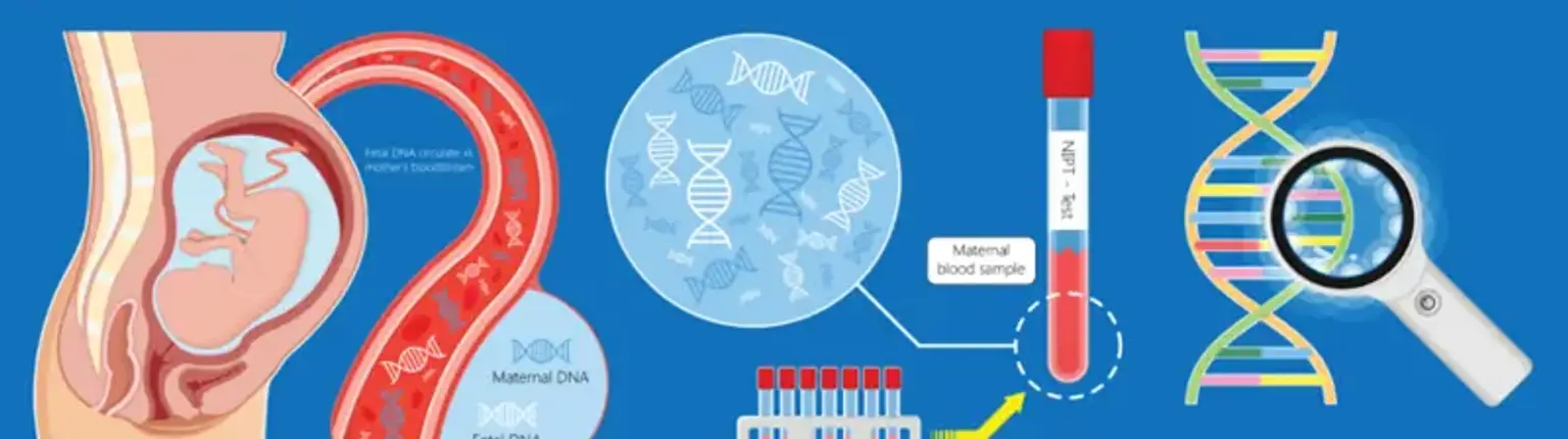
Prenatal diagnostics makes use of chorionic villus sample to discover fetal genetic disorders. Its benefits include the ability to do the operation during the first trimester of pregnancy, a reasonably quick outcome, and a risk of miscarriage equivalent to that of amniocentesis.


What is Chorionic Villus Sampling?

Chorionic Villus Sampling

Chorionic villus sampling is a prenatal genetic testing process that involves biopsying placental tissue between 10 and 13 weeks gestation. The fundamental benefit of chorionic villus collection is that genetic findings are available earlier in pregnancy. If the results are abnormal, individuals can seek counseling for obstetric treatment and suggestions, early referral to pediatric subspecialists, or earlier and safer means of pregnancy termination.

The chorionic villi are small placental tissue projections that resemble fingers. They are genetically identical to the infant. Other genetic abnormalities and illnesses can be tested for. This will be determined by the family history as well as the sorts of lab testing available at the time of the surgery.

During CVS, your doctor removes a little bit of tissue from the placenta. The sample will be used to assess your baby's health. CVS can occur between 10 and 13 weeks of pregnancy. Because there is a slight danger of miscarriage following the test, CVS is not offered to all pregnant women. CVS differs from another prenatal test known as amniocentesis (also called amnio). Amniocentesis is performed later in pregnancy. Consult your clinician about having CVS, amnio, or other prenatal testing performed.

There are 2 types of CVS procedures:

  • Transcervical. A catheter is put through the cervix into the placenta to get the tissue sample.
  • Transabdominal. A needle is put through the belly and uterus into the placenta to get the tissue sample.


Anatomy of the baby in the uterus

Baby uterus

  • Amniotic sac. During pregnancy, this is a thin-walled sac that surrounds the fetus. The sac contains amniotic fluid. This is the baby's fluids. The amnion is likewise housed in the sac. This is the membrane that covers the placenta's fetal side. This protects the infant from harm and aids in temperature regulation.
  • Anus. This is the opening at the end of the anal canal.
  • Cervix. This is the lowest portion of the uterus that enters the vagina. It is largely formed of fibrous tissue and muscle. It is round in form.
  • Fetus. This term refers to an unborn infant from the eighth week following conception till delivery.
  • Placenta. This is a flat organ that develops solely during pregnancy. It transfers nutrients and other elements from the mother to the child. The placenta permits the fetus to absorb oxygen, food, and other nutrients. It also allows the youngster to expel carbon dioxide and other waste.
  • Umbilical cord. This is a rope-like cord between the infant and the placenta. The umbilical cord has two arteries and one vein. They provide oxygen and nutrition to the infant while transporting waste items away from the newborn.
  • Uterine wall. This is the wall of the uterus.
  • Uterus (womb). This is a pear-shaped hollow organ in a woman's lower belly. It is located between the rectum and the bladder. During menstruation, it removes its lining. A baby develops when a fertilized egg (ovum) is inserted in the lining.
  • Vagina. This is a canal located behind the bladder and ahead of the rectum. It connects the uterus to the vulva.


Indications for Chorionic Villi Sampling

Chorionic Villi Sampling

Women of all ages, regardless of maternal age or other risk factors, should be provided prenatal examination for aneuploidy by screening or diagnostic testing, according to the American College of Obstetrics and Gynecology. Because prenatal genetic testing cannot detect every abnormality, testing should be tailored to the patient's risk, reproductive objectives, and preferences. Genetic testing should ideally be considered during the first obstetric appointment.

Indications for chorionic villus sampling include. 

  • Abnormal early genetic screening on a non-invasive prenatal screening (NIPS), first trimester combined screening or abnormalities on ultrasound
  • A prior child with a structural birth defect 
  • A prior child with autosomal trisomy or sex chromosome aneuploidy 
  • Advanced maternal or paternal age 
  • Parental carrier of a chromosomal rearrangement 
  • Parental aneuploidy or aneuploidy mosaicism 
  • Parental carrier of a genetic disorder, such as Tay Sachs, Sickle Cell Disease, or Neurofibromatosis


Contraindications

Patients on anticoagulation should exercise caution. Because the surgery may result in more severe fetal hemolytic illness, maternal alloimmunization is a relative contraindication. Furthermore, patients with bloodborne infectious disorders such as HIV and hepatitis should be informed of the possibility of vertical transmission through prenatal diagnostic testing.


How do I get ready for chorionic villus sampling?

Ready for chorionic villus sampling

  • The technique will be explained to you by the healthcare practitioner. Inquire about any concerns you have regarding the surgery.
  • You will be asked to sign a consent document, which authorizes your healthcare professional to perform the treatment. Before signing, please read the form. If anything is unclear, ask questions.
  • In general, there are no specific dietary or physical restrictions before CVS.
  • Inform your doctor if you are sensitive to or allergic to any medications, latex, iodine, tape, or anesthetic drugs (local and general).
  • Inform your doctor about all of the medications you are taking. Prescription and over-the-counter medications, vitamins, herbs, and other supplements are all included.
  • Inform your doctor if you have a history of bleeding problems or if you are on any blood-thinning (anticoagulant) medications, aspirin, or other medications that may interfere with blood clotting. You may need to discontinue these medications prior to the operation.
  • If you are Rh negative, inform your doctor. Blood cells from the mother and baby may mingle during the CVS procedure. This can result in Rh sensitization and the breakdown of the baby's red blood cells. In most circumstances, prenatal blood testing will have shown whether or not you are Rh negative. Before the procedure, you may be requested to give these lab results.
  • It is possible that you will be requested to have a full bladder just before the surgery. A full or empty bladder may assist shift the uterus into a better position for the surgery, depending on the location of the uterus and placenta.
  • Your healthcare physician may suggest further specialized preparation based on your medical condition.


Technique

Technique

The path taken to do the procedure is determined by the practitioner, however placental location may impact the decision. The optimum spot exposing the placenta's longest axis is determined during the transabdominal approach. The patient is positioned supine, and his or her abdomen is cleansed with a chlorhexidine or iodine solution. To establish a sterile field, sterile drapes are used. A local anesthetic may be administered. Under continuous ultrasound monitoring, an 18 or 20 gauge spinal needle is introduced into the placenta.

Once the stylet is removed, a 20 cc syringe containing collection media is attached to the needle's tip. The needle is moved up and down through the placenta, collecting tissue under negative pressure. After the sample is taken, it is examined to confirm that enough chorionic villi were aspirated.

The transcervical method involves placing the patient in a lithotomy posture and inserting a sterile speculum into the vagina. An iodine solution is used to clean the cervix. To facilitate catheter passage, a single tooth tenaculum may be applied to the anterior lip of the cervix. A transcervical CVS catheter is introduced into the placenta under continuous ultrasonography supervision.

The catheter has a pliable guidewire with an echogenic tip that may be seen on ultrasound. Once in place, the stylet is withdrawn, and a 20 cc syringe containing media is placed to the end of the catheter to produce negative pressure. Prior to concluding the operation, the sample is examined for adequacy. Small biopsy forceps can also be used to do transcervical CVS.

Tissue samples are submitted to the lab for culturing and further testing, such as conventional karyotyping, fluorescence in situ hybridization, and chromosomal microarray. Rapid evaluation findings are available in 2 to 4 days, whereas cultured samples are available in 1 to 2 weeks. Families should meet with their provider to discuss next steps. At 16 weeks, further ultrasounds may be conducted, and MSAFP may be taken to check for open neural tube abnormalities.


Complications of Chorionic Villus Sampling

The hazards of chorionic villus sampling are comparable to those of amniocentesis and include pregnancy loss, hemorrhage, infection, membrane rupture, and inconclusive findings.

The pregnancy loss rate has reduced as ultrasound guidance and ability and technique have improved; nonetheless, chorionic villus sampling has a steep learning curve. A comprehensive analysis of the consequences of chorionic villus sampling found that utilizing a transabdominal technique, total fetal loss was 0.7 percent within 14 days, 1.3 percent within 30 days, and 2% for loss at any point throughout pregnancy. The overall rate of fetal loss within 14 days in the amniocentesis group was 0.6 %.

The number of times the needle or catheter is inserted, the operator's expertise and ability, pregnancies following assisted reproductive procedures, and the use of transcervical cannula instead of biopsy forceps are all predictors of higher fetal loss.

Contained placental mosaicism, which occurs in 1-2% of instances, can be detected by chorionic villus sampling. This happens when there is a chromosomal difference between the placenta and the fetus. In 10% of cases, the fetus is really mosaic; nonetheless, mosaicism is associated with an increased risk of poor placental function and postnatal problems such as fetal growth restriction and maternal hypertension. In the second trimester, mosaicism should be followed with amniocentesis.

Furthermore, limb reduction abnormalities and oromandibular hypogenesis have been mentioned as hazards of chorionic villus sampling in the literature. Early CVS (before 10 weeks of pregnancy) has been established as a risk factor for several problems. The World Health Organization estimates that the risk of limb deformities is around 6 per 10,000, which is the same as the overall population risk. Although the danger is modest, patients should be counseled about the evidence if CVS is considered before 10 weeks.

Up to 32% of women have suffered vaginal spotting, which is more likely following transcervical CVS. The reported rate of culture failure, amniotic fluid leaking, or infection following CVS is less than 0.5%.


How will I feel after the procedure?

After the procedure

During and after CVS, most women suffer relatively moderate discomfort, cramping, and period-like pain. The process itself is only a few minutes long, however you will most likely be required to sit and relax in a waiting room for around 30 minutes following the surgery before being allowed to go home.

If you have a negative blood type, you will be given an Anti-D injection following the surgery. If you suffer light period-like pain the first night following the surgery, it is fine to take ordinary paracetamol.

If you experience vaginal bleeding, severe stomach discomfort, a high temperature, or unusual discharge from your vagina in the first few days or weeks following the treatment, go to the nearest hospital emergency room.


Cytogenetic results of chorionic villus sampling

Cytogenetic results

Seven medical centers in the United States have published cytogenetic findings of first-trimester chorionic villus sample. With the direct approach, long-term culture, or both, the rate of achieving a cytogenetic diagnosis was 99.6% for 6033 patients who underwent a successful chorionic villus sampling operation. There were no mispredictions of sex and no diagnostic mistakes concerning trisomies 21, 18, or 13, sex chromosome aneuploidies, or structural abnormalities.

There were no reports of a normal cytogenetic diagnosis followed by the birth of a cytogenetically aberrant child. Three examples of uncommon aneuploidies discovered exclusively by the direct approach (tetraploidy, trisomy 16, and trisomy 22) were not confirmed by cytogenetic follow-up. Mosaic cytogenetic abnormalities were found in 0.83% of all instances when chorionic villus sample was performed, although only 7 of 30 cases (23.3%) were verified by amniocentesis or in fetal tissues.

Maternal cell contamination occurred in 1.9% of long-term cultures, although this presented no cytogenetic diagnostic challenges. Overall, either cytogenetic approach resulted in extremely high laboratory success and diagnostic accuracy, however the long-term culture method resulted in fewer predicted mistakes and none when both methods were utilized.


Chorionic villi and stem cells

Stem cells

Recent research has revealed that chorionic villi can be a rich source of fetal stem cells as well as multipotent mesenchymal stem cells.

One possible advantage of employing fetal stem cells over embryonic stem cells is that they avoid the ethical concerns of anti-abortion campaigners by getting pluripotent lines of undifferentiated cells without harming a fetus or destroying an embryo. These stem cells would also avoid the donor/recipient problem that has so far hindered all attempts to employ donor-derived stem cells in treatments if used to treat the same individual from whom they originated.

Fetal stem cells have been used to create artificial heart valves, functional tracheas, muscle, fat, bone, heart, neural, and liver cells.


How much does a chorionic villus sampling cost?

The price of a CVS treatment varies depending on where you receive it and whether or not you have insurance. Many insurance policies pay at least a portion of the cost of CVS, but check with your provider to make sure. You will have to pay for the operation yourself if you do not have insurance or if your plan does not cover it. Your doctor's office can provide you with further information about CVS prices and payment plans.


Other Types of Pregnancy Tests

Pregnancy Tests

Other pregnancy tests that look for fetal genetic disorders are:

  • A tiny needle is guided by ultrasonography into the cervix and into the side of the placenta during vaginal CVS. Complications from vaginal CVS are somewhat greater than those from transabdominal CVS.
  • Amniocentesis is the removal of a tiny volume of amniotic fluid using a thin needle introduced through the abdomen. The ultrasonography is used to guide the needle. The cells in the fluid sample are analyzed for chromosomal abnormalities. The chance of miscarriage after amniocentesis is around one in 250. If your CVS test findings were ambiguous, you may be offered amniocentesis.


Conclusion 

Chorionic villus sampling is a well-established and safe method of doing early diagnostic prenatal genetic testing. With a qualified practitioner and the improvement of ultrasonography, complications are kept to a minimum. The results of a chorionic villus sample can assist families in making educated pregnancy decisions.