Introduction
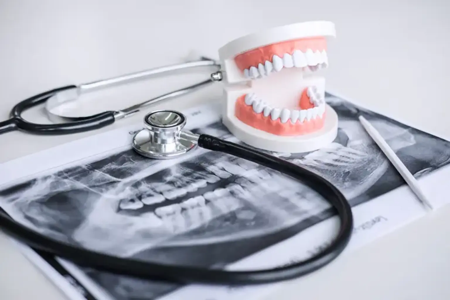
Dental imaging is a critical component of modern dentistry, helping professionals diagnose and treat oral conditions with precision. Oral and Maxillofacial Radiology (OMFR) is a specialty within dentistry focused on using imaging techniques to evaluate the mouth, jaw, face, and neck. This field has evolved significantly over the years, from traditional X-rays to advanced digital imaging systems, providing dentists with high-quality, detailed views of the oral and facial structures.
OMFR plays a pivotal role in diagnosing dental and maxillofacial conditions, such as tooth decay, infections, bone abnormalities, and tumors. Today, technology has enhanced the accuracy of these images, ensuring more effective treatment planning and better patient outcomes.
The Basics of Oral and Maxillofacial Radiology
Oral and Maxillofacial Radiology focuses on using advanced imaging techniques to diagnose conditions affecting the mouth, jaw, and facial regions. Unlike traditional dentistry, which focuses on teeth, OMFR specializes in the complex structures of the oral cavity, including bones, tissues, and joints.
Key areas of focus in OMFR include:
TMJ Disorders: Imaging is used to assess the temporomandibular joint (TMJ), helping to identify issues like arthritis or disc displacement.
Pathology: OMFR plays a key role in detecting tumors, cysts, and infections in the oral and facial regions.
Dental Implants and Surgery: Detailed imaging helps in planning dental implants and reconstructive surgeries.
This specialty requires deep knowledge of anatomy and pathology, as OMFR specialists interpret the images and provide comprehensive reports that guide treatment decisions.
Common Imaging Techniques in Dentistry
There are several types of imaging techniques used in dentistry, each with specific purposes and advantages:
Intraoral Radiography: This is the most common type of dental X-ray. It provides clear images of individual teeth, helping diagnose cavities, infections, and root issues. It’s a quick, effective method for detecting localized problems.
Panoramic Radiography: This technique provides a broad view of the entire mouth, including all teeth, jawbones, and surrounding structures. It’s typically used for assessing wisdom teeth, jaw alignment, and evaluating bone loss.
Cone Beam Computed Tomography (CBCT): CBCT offers 3D imaging, providing detailed, high-resolution views of the teeth, bone, and soft tissues. This technology is especially useful for dental implants, orthodontics, and complex maxillofacial surgeries. It helps plan procedures with remarkable precision.
Magnetic Resonance Imaging (MRI): While not as commonly used in routine dental imaging, MRI is valuable for evaluating soft tissues, such as muscles and joints. It is often used to diagnose TMJ disorders or soft tissue abnormalities in the face.
Each imaging method serves a specific purpose, and the choice of technique depends on the condition being assessed and the level of detail needed for diagnosis and treatment planning.
The Role of Cone Beam Computed Tomography (CBCT)
Cone Beam Computed Tomography (CBCT) has revolutionized dental imaging, offering a 3D view of the oral and maxillofacial structures. Unlike traditional X-rays, which provide flat, 2D images, CBCT captures detailed images from all angles, making it invaluable for precise diagnostic and treatment planning.
CBCT is particularly beneficial in the following areas:
Dental Implants: It allows for accurate measurement of bone volume and density, essential for successful implant placement.
Orthodontics: CBCT helps assess jaw alignment and detect structural abnormalities that may require surgical intervention.
Surgical Planning: Surgeons use CBCT to evaluate the anatomical structures and plan complex surgeries, such as reconstructive jaw surgery or removal of tumors.
This imaging method provides far more detailed information compared to traditional methods, improving the accuracy of diagnoses and outcomes for various dental and maxillofacial procedures.
Panoramic Radiography: A Broader View
Panoramic radiography provides a broad, comprehensive image of the entire mouth in a single shot. This technique captures the teeth, jawbones, and surrounding structures, offering a "bird’s-eye view" of the oral and maxillofacial area. Unlike traditional X-rays that focus on a small area, panoramic radiographs show the full picture, making them invaluable for certain diagnoses.
Panoramic radiographs are commonly used in:
Wisdom Teeth Evaluation: Panoramic X-rays can identify impacted wisdom teeth and their relationship to adjacent teeth and nerves.
Bone Assessment: They help detect bone loss due to periodontal disease, a crucial aspect of oral health.
Orthodontic Planning: These images are essential for assessing jaw alignment and planning orthodontic treatments.
While they do not offer the same level of detail as other techniques like CBCT or intraoral radiography, panoramic X-rays provide a quick, comprehensive overview of the mouth and are often used as a first diagnostic tool.
Intraoral Radiography: Precision for Individual Teeth
Intraoral radiography is a common and highly effective diagnostic tool in dentistry. It involves taking X-rays inside the mouth, typically focusing on individual teeth or specific regions. This technique is essential for examining detailed areas like tooth roots, the surrounding bone, and areas prone to cavities or infections.
There are three main types of intraoral X-rays:
Periapical X-rays: Focus on the entire tooth, from crown to root, and the surrounding bone. These are used to check for cavities, root infections, and bone loss.
Bitewing X-rays: Captures the crowns of the upper and lower teeth, typically used to detect cavities between teeth.
Occlusal X-rays: Shows the floor of the mouth or the roof of the mouth, often used to assess larger dental conditions or abnormalities.
Intraoral X-rays provide highly detailed images that allow for early detection of problems like cavities, abscesses, or bone infections, which can be treated before they progress into more serious conditions.
Magnetic Resonance Imaging (MRI) in Oral Health
Magnetic Resonance Imaging (MRI) is a non-invasive imaging technique that uses strong magnetic fields and radio waves to create detailed images of soft tissues. While it is more commonly used in medicine for brain and spinal imaging, MRI also has valuable applications in oral and maxillofacial radiology.
In dentistry, MRI is primarily used to evaluate soft tissues, such as muscles, ligaments, and the temporomandibular joint (TMJ). It is particularly helpful in:
TMJ Disorders: MRI can visualize joint structures, disc displacement, and signs of inflammation, aiding in the diagnosis of disorders like temporomandibular joint dysfunction (TMD).
Soft Tissue Pathologies: MRI helps in detecting tumors, cysts, and other abnormalities in the soft tissues of the face and mouth that may not be visible on X-rays.
Assessment of Facial Muscles: It can assess the condition of muscles involved in chewing and facial expressions, crucial for surgical planning and injury recovery.
Although not commonly used for routine dental exams, MRI is essential for diagnosing complex conditions related to the soft tissues of the mouth and face.
The Importance of Image Interpretation in Oral Radiology
The interpretation of dental images is as crucial as the imaging process itself. While advanced imaging techniques like CBCT and panoramic X-rays provide detailed views of the oral and maxillofacial regions, it is the expertise of the radiologist that ensures accurate diagnosis and effective treatment planning.
Oral and maxillofacial radiologists have specialized training in understanding complex anatomy, pathology, and developmental variations within the mouth, jaw, and face. Their role includes:
Identifying Pathologies: Radiologists can detect dental issues like cavities, infections, cysts, or tumors that may not be immediately visible during a physical exam.
Assessing Treatment Planning: For procedures like dental implants or jaw surgeries, radiologists ensure that the imaging data is used to guide precise, individualized treatment plans.
Evaluating Surgical Sites: Accurate interpretation is key when planning for surgeries, as it ensures that all relevant anatomy is taken into account, minimizing risks and improving recovery outcomes.
Without proper image interpretation, even the most advanced technology can miss critical findings. Radiologists provide a thorough review of images, ensuring that all relevant findings are noted and guiding healthcare providers to make informed decisions for patient care.
Advancements in Dental Imaging Technology
Dental imaging technology has come a long way in recent years, significantly improving the accuracy and efficiency of dental diagnoses. Some of the most notable advancements include:
Digital Radiography: Traditional X-rays are being replaced by digital systems that offer faster image capture and higher resolution. Digital radiographs expose patients to less radiation and provide instant results, making them more convenient for both patients and practitioners.
Cone Beam Computed Tomography (CBCT): CBCT technology provides 3D images that offer far more detail than conventional 2D X-rays. This has become a game-changer in planning dental implants, evaluating bone structure, and diagnosing complex dental conditions.
Artificial Intelligence (AI) in Imaging: AI algorithms are now being integrated into dental imaging systems. AI can help identify patterns in radiographs that may not be immediately apparent to human eyes, enhancing diagnostic accuracy and helping to detect issues like cavities, tumors, and periodontal disease earlier.
Portable Imaging Systems: New portable X-ray units allow for imaging in more flexible settings, such as dental offices without heavy imaging equipment or even in emergency situations.
These innovations have not only improved diagnostic capabilities but have also made dental imaging safer, faster, and more accessible.
Safety Protocols in Dental Imaging
While dental imaging is crucial for diagnosis and treatment planning, it’s essential to ensure that it is done safely. Radiation exposure, though minimal in modern dental imaging techniques, is still a concern. Safety protocols are in place to minimize risks, both for patients and dental staff.
Use of Digital X-rays: Digital radiographs expose patients to significantly less radiation compared to traditional film X-rays. This is particularly beneficial for patients who need frequent imaging, such as those undergoing orthodontic treatment.
Lead Aprons and Thyroid Collars: Patients are often provided with lead aprons and thyroid collars to protect sensitive areas of the body, such as the thyroid gland, from radiation exposure.
ALARA Principle (As Low As Reasonably Achievable): This principle is followed to ensure radiation is kept at the lowest possible level needed for effective imaging. For example, only necessary images are taken, and protective measures are used to shield other areas of the body.
Training and Certification for Technicians: Dental professionals and radiology technicians undergo rigorous training to ensure they operate imaging equipment correctly and follow safety protocols. This reduces the likelihood of overexposure to radiation.
With these safety measures in place, dental imaging can be a safe and effective tool for diagnosis and treatment planning, ensuring patients receive the best care with minimal risk.
The Impact of 3D Imaging on Treatment Planning
3D imaging, especially through technologies like CBCT, has significantly enhanced treatment planning in dentistry. The ability to visualize teeth, bones, and soft tissues in three dimensions provides unparalleled accuracy and precision.
Dental Implants: Before CBCT, implant planning was based on 2D X-rays, which offered limited information. Now, with 3D images, implant placement can be planned with greater precision, ensuring proper positioning and minimizing complications.
Orthodontics: 3D imaging allows orthodontists to evaluate jaw relationships and tooth positioning more effectively. This leads to more personalized treatment plans that consider the entire 3D structure of the mouth.
Surgical Planning: For complex oral surgeries, such as reconstructive jaw surgery or tumor removal, 3D imaging provides a detailed map of the anatomy, allowing surgeons to plan and execute procedures with greater accuracy and safety.
Accuracy and Efficiency: With 3D imaging, the chances of complications and mistakes during treatment are minimized. This improves the success rates of procedures and shortens recovery times for patients.
The ability to plan dental and maxillofacial procedures with 3D images enhances both the predictability and outcomes of treatment, benefiting both patients and dental professionals.
Applications of Dental Imaging in Orthodontics
Dental imaging plays a pivotal role in orthodontics, the branch of dentistry focused on diagnosing, preventing, and treating dental and facial irregularities. Accurate imaging is crucial for assessing tooth positioning, jaw alignment, and the development of treatment plans that will yield the best results.
Panoramic X-rays in Orthodontics: These X-rays provide a full view of the teeth, jawbones, and surrounding tissues, helping orthodontists evaluate overall alignment and detect issues like impaction or crowding.
CBCT in Orthodontics: Cone Beam CT is revolutionizing orthodontics by offering 3D views of the jaw and teeth. This allows orthodontists to more precisely plan the movement of teeth and evaluate bone structure. With CBCT, orthodontists can assess the severity of skeletal issues and decide on the most effective treatment approach, whether it’s braces, aligners, or surgical intervention.
Monitoring Growth and Development: For children and teens, dental imaging helps monitor the development of the teeth and jaws, allowing orthodontists to identify potential issues early and intervene when necessary. This early intervention can lead to more effective treatments with better long-term results.
Treatment Progression: Imaging is also used to track the progress of orthodontic treatment. Regular radiographs allow practitioners to assess the movement of teeth and ensure they are on track to achieve the desired alignment.
Overall, dental imaging has made orthodontic treatments more precise, efficient, and predictable, leading to better outcomes and shorter treatment times for patients.
Dental Imaging and Early Detection of Oral Diseases
One of the most significant advantages of modern dental imaging is its ability to detect oral diseases in their earliest stages. Early detection is crucial for effective treatment and improved patient outcomes. Imaging allows dental professionals to identify issues before they become severe, reducing the need for extensive treatments and minimizing the risks of complications.
Cavities and Tooth Decay: Intraoral X-rays are highly effective in identifying cavities, especially those located between teeth or beneath existing fillings. Detecting decay early can prevent more extensive tooth damage and the need for root canals or extractions.
Periodontal Disease: Bone loss caused by periodontal disease is often invisible to the naked eye. Panoramic X-rays and CBCT scans can detect bone density loss early, allowing dentists to intervene before the disease progresses to more advanced stages.
Oral Cancer: Dental imaging can help detect the early signs of oral cancer, such as tumors or lesions that might not yet be visible during a clinical exam. Early detection significantly improves the chances of successful treatment and recovery.
The ability to identify these issues early means that patients can undergo less invasive treatments, leading to better oral health outcomes and overall well-being.
The Role of Dental Imaging in Cosmetic Dentistry
In cosmetic dentistry, where aesthetic outcomes are of utmost importance, dental imaging plays an essential role in planning and executing treatments. Whether it's for teeth whitening, veneers, crowns, or smile makeovers, accurate imaging ensures that procedures align with a patient's goals and expectations.
Smile Design: Digital imaging allows cosmetic dentists to create a "smile design" based on the patient’s facial features, tooth proportions, and overall aesthetic goals. This ensures that the results will look natural and harmonious with the rest of the face.
Veneers and Crowns: Accurate imaging is critical for creating veneers and crowns that fit perfectly and look natural. CBCT or digital impressions can help craft precise restorations that mimic the shape, size, and color of the patient's natural teeth.
Predicting Outcomes: Imaging technology can also be used to predict the outcome of cosmetic procedures before they are performed. This allows patients to visualize their potential results, which can help reduce anxiety and ensure satisfaction with the final result.
By incorporating advanced imaging, cosmetic dentists can plan treatments with greater precision, creating results that improve both the appearance and function of a patient’s smile.
The Global Popularity of Dental Imaging
As dental technology continues to evolve, dental imaging has gained worldwide popularity due to its effectiveness and accessibility. From advanced imaging centers to local dental practices, this technology is transforming dental care across the globe.
Global Reach: Dental imaging technologies like CBCT, digital radiography, and intraoral cameras are now common in many countries. Whether in developed nations with state-of-the-art facilities or in emerging markets, dental imaging is becoming a standard practice.
Cost-Effective and Time-Saving: In countries with established healthcare systems, dental imaging offers cost-effective solutions for diagnosis and treatment planning, helping reduce the need for multiple appointments or unnecessary procedures. In remote or underserved areas, portable imaging units bring advanced diagnostic capabilities to locations where traditional imaging equipment may be unavailable.
Advancements in Access and Training: The growing accessibility of dental imaging has led to improvements in training and education for dental professionals worldwide. Continuing education programs and international conferences are helping dental practitioners stay updated on the latest imaging technologies and techniques.
The increasing popularity of dental imaging is reshaping the dental landscape, making precise and efficient care available to more people globally.
The Future of Dental Imaging
As technology continues to advance, the future of dental imaging looks even more promising. Innovations in AI, machine learning, and imaging software are poised to take dental diagnostics to new heights, improving both accuracy and patient care.
Artificial Intelligence and Automation: AI has the potential to revolutionize image interpretation. AI algorithms are already being developed to detect signs of dental conditions, such as cavities, tumors, and bone loss, with high accuracy. In the future, AI could automatically analyze images, reducing the workload of radiologists and ensuring faster, more consistent diagnoses.
Integration with Virtual Reality (VR): The integration of VR with dental imaging could allow for more immersive diagnostic experiences. Dental professionals might soon be able to "walk through" 3D images, enhancing their ability to plan complex procedures, such as reconstructive surgeries or implant placements.
Personalized Imaging: Advances in personalized medicine will likely lead to more customized imaging techniques. By tailoring imaging protocols to each patient's unique anatomical features, future systems may provide even more precise and relevant diagnostic data.
Improved Patient Experience: In the future, dental imaging may become even less invasive and more comfortable for patients. Faster, more efficient systems will minimize radiation exposure and shorten appointment times, while innovative tools might make imaging less intimidating for anxious patients.
With these advancements, dental imaging will continue to play a crucial role in improving diagnostic accuracy, treatment planning, and patient care in the years to come.
Patient Comfort and Dental Imaging
While dental imaging is essential for accurate diagnosis and treatment planning, patient comfort is always a priority. Innovations in dental imaging are focused on reducing discomfort and anxiety during procedures.
Quicker Imaging Times: Digital X-rays and CBCT scans are faster than traditional X-rays, which reduces the time a patient spends in the dental chair. This is especially helpful for patients who feel anxious during long procedures.
Reduced Radiation: Newer imaging techniques, such as digital radiography, use significantly lower radiation, making the process safer and more comfortable for patients.
Non-Invasive Procedures: Many imaging technologies, such as 3D scans, require no physical contact or discomfort, making them more patient-friendly.
Efforts to improve patient comfort help make dental imaging a less stressful experience, leading to better compliance and more thorough care.
Cost Considerations in Dental Imaging
While advanced dental imaging technologies offer numerous benefits, they also come with cost considerations that both patients and practitioners need to keep in mind.
Initial Costs: The initial investment for advanced imaging equipment like CBCT or digital radiography can be expensive for dental practices. However, this cost is often offset by the improved accuracy and efficiency these technologies provide, reducing the need for follow-up appointments and treatments.
Insurance and Coverage: Dental insurance coverage for imaging varies. Some plans may cover the costs of routine X-rays, while others may limit coverage for more advanced imaging techniques. It's important for patients to check with their insurance provider to understand what’s covered.
Long-Term Value: Despite the upfront costs, advanced imaging ultimately leads to more effective treatments, reducing the need for complex procedures and enhancing the longevity of dental work. For patients, this can mean fewer visits and lower overall treatment costs.
While dental imaging may seem expensive at first glance, its long-term value in enhancing care and treatment outcomes often justifies the investment.
Common Misconceptions About Dental Imaging
Although dental imaging is a vital part of modern dental care, there are still several misconceptions surrounding its use. Addressing these myths helps patients make informed decisions about their care.
"X-rays are harmful and should be avoided.": While it’s true that X-rays involve radiation, modern dental imaging techniques use extremely low doses, especially digital radiography. The benefits far outweigh the risks, particularly when it comes to early detection and preventative care.
"Dental imaging is only for major procedures.": Many people think imaging is only necessary for complex surgeries or treatments, but routine X-rays are an integral part of preventive care. Regular imaging helps catch issues early and guide simple treatments, preventing bigger problems later.
"3D imaging is only for specialists.": Though CBCT and other 3D imaging technologies are commonly used by specialists, general dentists also use them to plan procedures like implants, crowns, and orthodontic treatments, making them accessible for everyday dental care.
By understanding the facts, patients can approach dental imaging with confidence, knowing that these technologies are safe, effective, and crucial for maintaining good oral health.
Conclusion
Dental imaging has revolutionized the field of dentistry, enhancing both the diagnosis and treatment of various oral health issues. From advanced 3D imaging to digital X-rays, the technologies available today enable dentists to deliver precise and effective care, ensuring better outcomes for patients.
Early Detection: Imaging techniques allow for early detection of issues like cavities, periodontal disease, and oral cancer, which leads to more effective and less invasive treatments.
Improved Treatment Planning: Technologies such as CBCT offer a 3D view of the mouth, making it easier for dentists to plan complex procedures like implants or orthodontics with great accuracy.
Patient-Centered Care: Modern imaging systems focus on patient comfort, with faster processes, less radiation, and non-invasive procedures that make visits more pleasant and less intimidating.
As dental imaging continues to evolve, it will play an increasingly important role in improving the quality of care and the patient experience. By embracing these advancements, dental professionals can ensure that patients receive the most accurate diagnoses and effective treatments available.