What is Microscope-Assisted Dentistry?
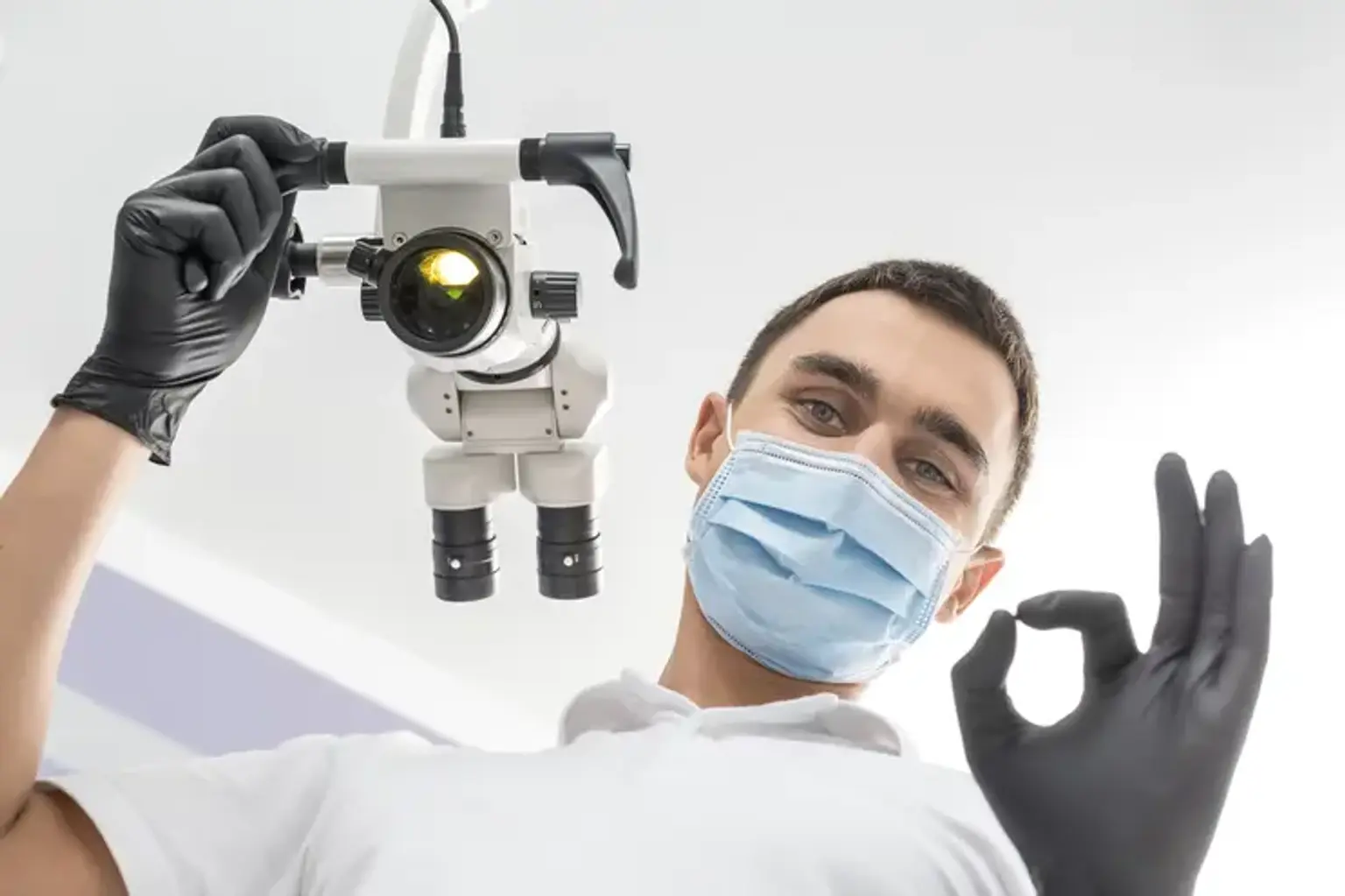
Microscope-assisted dentistry refers to the use of high-powered dental microscopes to enhance precision during dental procedures. These specialized tools provide a magnified, detailed view of the teeth, gums, and surrounding tissues, allowing dental professionals to perform treatments with greater accuracy. The technology has become a cornerstone in modern dentistry, especially for complex procedures like root canals, restorations, and surgeries.
Unlike traditional dental tools that offer limited visibility, dental microscopes enable a much higher magnification, often up to 20x. This allows dentists to detect even the smallest issues that may not be visible with the naked eye, improving treatment outcomes significantly. The introduction of this technology has reshaped how dentists approach dental procedures, ensuring higher success rates and fewer complications.
The Technology Behind Dental Microscopes
Dental microscopes are equipped with advanced optical technology that magnifies dental structures. These microscopes typically feature multiple lenses, built-in lighting, and adjustable focus settings. This gives dental professionals clear, bright views of the treatment area, even in the deepest parts of the tooth or gum. Many modern microscopes also include features like digital integration for capturing images and videos, which can be useful for patient education and record-keeping.
Traditional dental magnification tools, like loupes, offer limited magnification and do not provide the same level of detailed visualization. In contrast, a dental microscope’s high magnification allows dentists to see fine structures within the tooth, such as tiny cracks, early cavities, or complex root canal systems, which are often missed with the naked eye.
The ability to visualize these fine details helps in diagnosing dental issues more accurately and performing procedures with minimal invasiveness, reducing the risk of errors and complications.
How Dental Microscopes Improve Treatment Accuracy
Microscope-assisted dentistry offers unparalleled precision, which is crucial for certain procedures that demand high levels of accuracy, such as root canals, tooth restorations, and dental surgeries. The enhanced visualization allows dentists to work in tight spaces, such as the intricate canals of a tooth during a root canal treatment. With a microscope, they can see the entire root system and any underlying issues, ensuring that all infected or damaged tissue is removed.
This level of detail also aids in placing dental implants or performing restorations like crowns and bridges. The dentist can ensure that the work fits perfectly, with no gaps, and can assess the surrounding bone or gum structure to avoid damage.
For patients, this means fewer complications, quicker recovery times, and better overall results. With the ability to perform procedures more accurately, dental professionals can preserve more of the natural tooth structure, offering long-term benefits for the patient’s oral health.
Microscope-Assisted Endodontics: Root Canal Therapy
Root canal therapy is one of the most common procedures enhanced by dental microscopes. A root canal is needed when the tooth’s pulp becomes infected or inflamed, which can cause severe pain. In traditional root canal procedures, dentists often rely on their experience and limited visibility to treat the infection. However, microscopes allow for a much more detailed view of the tooth’s internal structure.
Using a dental microscope, the dentist can identify the exact location of the infection, ensuring that all infected tissue is removed thoroughly. The microscope’s magnification also helps in locating and cleaning the tiny, often curved, canals within the tooth that may be missed during a traditional procedure. This increases the chances of success, reduces the likelihood of infection recurrence, and improves the longevity of the tooth after treatment.
By using this technology, endodontists can perform root canal therapy more efficiently, with fewer post-treatment issues and better long-term outcomes.
Microscope Use in Dental Restorations and Implants
Microscope-assisted dentistry plays a crucial role in dental restorations and implant procedures. Dental restorations, such as crowns, bridges, and fillings, require precision to ensure they fit comfortably and function well within the patient's mouth. With the aid of a dental microscope, dentists can achieve a high level of accuracy when preparing the tooth, placing the restoration, and making adjustments for a perfect fit.
For dental implants, microscopes allow the dentist to assess the surrounding bone and soft tissues with precision, ensuring proper placement of the implant. This minimizes the risk of complications such as infection or improper alignment, which can lead to implant failure. The enhanced visualization also helps in avoiding vital structures, such as nerves or blood vessels, during surgery.
By using a microscope, dental professionals can provide more effective and long-lasting results, improving both the aesthetics and function of the patient's smile.
Microscope-Assisted Dental Surgery: A Surge in Precision
Microscope-assisted dental surgery has revolutionized the way oral surgeries are performed. Procedures such as tooth extractions, gum surgeries, and other complex oral surgeries benefit greatly from the magnified view provided by a dental microscope. The ability to see every detail clearly allows for more controlled and less invasive surgeries, reducing trauma to surrounding tissues and promoting faster healing.
For example, during a tooth extraction, a dental microscope helps the dentist to work with greater precision, ensuring that the surrounding bone and gum tissues are preserved as much as possible. This not only speeds up recovery but also lowers the risk of complications like infection or damage to neighboring teeth.
In addition, the microscope helps in delicate surgeries, such as soft tissue grafting or bone regeneration, where exact precision is crucial for successful outcomes.
Global Popularity of Microscope-Assisted Dental Procedures
Microscope-assisted dental treatments are gaining popularity worldwide due to their ability to provide greater precision and better outcomes. Countries such as the United States, Japan, and Germany have been early adopters of this technology, and the demand for it is growing globally.
In many developed countries, patients are becoming more informed about the benefits of microscope-assisted procedures and are actively seeking dentists who offer these advanced treatments. As the technology becomes more accessible and affordable, even dental practices in developing regions are starting to integrate microscopes into their services. This trend is helping elevate the standard of care across the globe.
The widespread use of dental microscopes reflects a larger movement toward minimally invasive treatments that prioritize patient safety, comfort, and long-term success. With its ability to improve treatment accuracy and outcomes, microscope-assisted dentistry is quickly becoming a preferred choice for both patients and dental professionals alike.
Benefits of Microscope-Assisted Dental Treatments
The benefits of microscope-assisted dental treatments are extensive, not only for the dentist but for the patient as well. Here are the key advantages:
Enhanced Accuracy: With up to 20x magnification, dental microscopes allow practitioners to identify and treat issues that would otherwise go unnoticed, leading to more accurate procedures.
Minimally Invasive: The precision offered by microscopes means less need for aggressive techniques, reducing trauma to the surrounding tissues and speeding up recovery times.
Reduced Pain and Discomfort: Because procedures are more precise, there is less need for extensive drilling or cutting, resulting in less pain and a more comfortable patient experience.
Higher Success Rates: With improved visualization, dentists can perform treatments with greater confidence, leading to better long-term outcomes and fewer complications.
Faster Recovery: Less invasive procedures and the precision provided by microscopes contribute to shorter recovery times and a quicker return to daily activities for patients.
These benefits make microscope-assisted dentistry an attractive option for patients seeking effective, efficient, and less painful dental treatments.
Common Dental Procedures That Benefit from Microscopes
Microscope-assisted dentistry enhances the precision of many common dental procedures. Here are a few examples:
Root Canal Therapy: The microscope allows dentists to see the tiny, often complex structures of the tooth's root system, ensuring that all infected tissue is removed. This minimizes the risk of infection recurrence and improves long-term success rates.
Dental Extractions: When removing a tooth, the microscope helps the dentist to work with precision, preserving surrounding bone and tissue. This minimizes the need for additional procedures, such as bone grafts, and speeds up healing.
Implants: For implant placement, microscopes provide clear visibility of the surrounding bone, ensuring that the implant is placed correctly and safely. This reduces the risk of complications and improves implant success rates.
Restorative Dentistry: Whether it's placing fillings, crowns, or bridges, microscopes ensure that restorations fit precisely, providing better aesthetics and functionality.
Gum Surgeries: In procedures like gum grafts or pocket reduction surgery, microscopes enable precise manipulation of delicate tissues, ensuring better outcomes and reduced recovery times.
Safety Protocols in Microscope-Assisted Dental Procedures
While dental microscopes significantly enhance precision, safety protocols are still crucial. Here are key safety measures in place for microscope-assisted treatments:
Sterilization and Hygiene: As with all dental procedures, ensuring that all tools and the treatment area are sterile is vital. The dental microscope is also regularly disinfected to prevent cross-contamination.
Accurate Diagnosis: The high magnification of a dental microscope allows dentists to spot potential issues early, even before symptoms appear, reducing the risk of misdiagnosis or overlooked problems.
Minimizing Invasive Techniques: Because of the precision afforded by the microscope, dentists can perform less invasive procedures. This reduces trauma to tissues, lowers the risk of complications, and leads to a safer, faster recovery.
Training and Expertise: Only trained professionals should operate dental microscopes, as they require a high level of skill to use effectively. Dental practices that offer microscope-assisted treatments ensure their practitioners are thoroughly trained.
Who Can Perform Microscope-Assisted Dental Treatments?
Microscope-assisted dental treatments require specialized training. While any dentist can learn to use a dental microscope, those who perform procedures with microscopes typically undergo additional training to master the technology.
Endodontists, periodontists, and oral surgeons are the specialists most likely to use microscopes regularly due to the nature of their work. However, general dentists can also incorporate microscope technology into their practices, particularly for complex restorative and surgical procedures.
When seeking a dentist who offers microscope-assisted treatments, it's essential to inquire about their experience with the technology. Ask whether they use dental microscopes for specific procedures and if they are trained to operate them effectively. Many practices also display certifications or endorsements from professional organizations that emphasize the importance of advanced technology in patient care.
Is Microscope Dentistry More Expensive?
One common question patients have is whether microscope-assisted dental treatments are more expensive than traditional methods. While it’s true that these procedures may cost more upfront due to the specialized equipment and expertise involved, the long-term benefits can make them a worthwhile investment.
Higher Success Rates: Microscope-assisted procedures often lead to fewer complications, fewer follow-up appointments, and longer-lasting results, which can ultimately save patients money over time.
Reduced Need for Follow-up Procedures: With the increased precision provided by dental microscopes, the need for repeat treatments or additional interventions is less likely.
Cost of Technology: The dental microscope itself is a significant investment for practices, and some of this cost is passed on to patients. However, as technology becomes more widely adopted, the cost of treatments is expected to become more competitive.
Insurance Coverage: In some cases, dental insurance may cover microscope-assisted procedures, particularly if they are deemed medically necessary. It’s worth checking with your insurance provider to determine what treatments are covered.
Ultimately, while microscope-assisted treatments may be more expensive in the short term, their higher success rates, reduced complication risks, and improved long-term outcomes make them a smart choice for many patients.
Patient Experiences and Testimonials
Many patients who have undergone microscope-assisted dental treatments report highly positive experiences. The added precision means less pain, faster recovery, and a higher likelihood of a successful outcome. Some patients have shared that their procedures felt less invasive due to the focused approach provided by the microscope, resulting in less post-treatment discomfort.
For example, a patient who underwent a root canal with a microscope-assisted approach shared that the procedure was quick, and they experienced minimal swelling afterward. Similarly, patients who received dental implants have noted a faster healing time and fewer complications compared to traditional methods.
Patients often appreciate the clearer communication with their dentist, as microscopes allow for real-time visualizations of the procedure. This helps reduce anxiety, as patients can better understand what is happening during their treatment.
Training and Education for Dental Professionals
Dental professionals must undergo specialized training to effectively use microscopes in their practice. This training typically includes not only the technical aspects of using the microscope but also the ability to interpret magnified images accurately and make precise adjustments during procedures.
Programs that offer microscope training are often part of postgraduate education for specialists, such as endodontists or periodontists, but even general dentists can benefit from this training. As more dental schools incorporate advanced technology into their curricula, the next generation of dentists will likely be better equipped to offer these high-precision treatments.
Advanced workshops and continuing education programs also help dentists stay up-to-date with the latest advancements in microscope technology and techniques. This ensures that patients receive the best care possible.
The Future of Microscope-Assisted Dentistry
The future of microscope-assisted dentistry looks bright. As technology continues to evolve, dental microscopes are becoming more affordable, portable, and integrated with digital tools. Innovations like 3D imaging and intraoral cameras are being combined with dental microscopes, providing even more detailed and dynamic views of the mouth.
The integration of Artificial Intelligence (AI) into microscope-assisted treatments is also on the horizon. AI could help identify dental issues automatically, further enhancing the dentist's ability to make accurate diagnoses. This may also speed up the treatment process and improve overall patient outcomes.
As more dental practices adopt these technologies, patients can expect greater accessibility to microscope-assisted procedures, and the overall quality of dental care will continue to improve.
Is Microscope-Assisted Dentistry Right for You?
Deciding whether microscope-assisted dentistry is right for you depends on the complexity of the procedure you need and your personal preferences. If you’re facing a more complicated treatment, such as a root canal, dental implant, or a restoration requiring high precision, opting for a dentist who uses a microscope can lead to better results and a more comfortable experience.
For patients with anxiety or concerns about the invasiveness of a procedure, the precision and less traumatic nature of microscope-assisted treatments may offer peace of mind. While these procedures may come at a higher cost, the long-term benefits of increased accuracy, faster recovery, and fewer complications often make it worthwhile.
If you're considering microscope-assisted dental treatments, it’s a good idea to consult with a dentist who is experienced in using this technology. Discuss your treatment options, ask about the benefits of using a microscope, and determine if this advanced approach aligns with your needs.
Combining Microscope-Assisted Treatments with Other Technologies
In modern dentistry, microscope-assisted treatments are often combined with other advanced technologies to enhance patient outcomes. For example, 3D imaging, digital radiographs, and cone beam CT scans allow for a more thorough evaluation of the patient’s dental and bone structure before treatment. These images can be integrated with the microscope's magnified view, helping the dentist to plan and execute procedures with even greater precision.
By combining these tools, dentists can identify issues that may not be visible through traditional methods and ensure that procedures are performed safely and effectively. This holistic approach to dentistry is especially beneficial for complex cases, such as implants, orthodontics, or reconstructive surgeries.
Accessibility of Microscope-Assisted Dentistry
While microscope-assisted dental treatments are incredibly effective, their accessibility can vary depending on the region. Larger cities and more advanced dental practices tend to have the equipment and expertise necessary to offer these treatments. However, as the technology becomes more widespread, even smaller practices are beginning to integrate microscopes into their services.
In some cases, dental insurance may cover certain microscope-assisted treatments, especially if they are deemed medically necessary. However, coverage can vary, so it’s important to check with your provider. As demand for these advanced procedures grows, costs may decrease, making them more accessible to a broader range of patients.
The Role of Patient Education in Microscope-Assisted Dentistry
Patient education is crucial in microscope-assisted dentistry. Many patients may not be aware of the advantages that microscopes offer for precision and safety. Dentists play an essential role in educating their patients about the technology, how it works, and how it can improve treatment outcomes.
This education can help alleviate concerns, especially for patients who are nervous about undergoing complex procedures. Dentists can use visual aids, such as before-and-after images, videos, and live demonstrations, to help patients understand how microscopes enhance their care.
Understanding the technology allows patients to make informed decisions about their treatments and feel more confident in their choice of dentist.
Conclusion
Microscope-assisted dentistry represents a significant leap forward in dental care, providing precision, efficiency, and improved patient outcomes. With its ability to magnify treatment areas, reduce invasiveness, and enhance accuracy, it’s clear that this technology is transforming the way dental professionals approach complex procedures.
As this technology becomes more common and affordable, more patients will be able to benefit from the improved results it offers. Whether you’re undergoing a routine dental procedure or need specialized treatment, microscope-assisted dentistry offers a level of care that was once unimaginable. By embracing this innovation, both dental professionals and patients can look forward to a brighter, more comfortable dental future.