Diffuse Toxic Goiter (Graves' Disease)
Overview
Patients with goiter frequently present to outpatient clinics with a wide range of problems. Goiter has a wide range of causes and morbidities, and a correct diagnosis is critical for determining the best treatment strategy. Goiter, whether simple or nodular, harmless or poisonous, may have a significant influence on a patient's quality of life and well-being, as well as long-term physical and aesthetic health impacts.
Diffuse toxic goiter is an autoimmune disorder characterized by a diffusely hyperplastic thyroid gland and excessive thyroid hormone production. Graves disease, the most prevalent cause of hyperthyroidism, is distinguished by symptoms such as diffuse toxic goiter, oculopathy, and pretibial myxedema/acropachy. Diffuse toxic goiter is also found in various autoimmune thyroid disorders that induce hypothyroidism, the most prevalent of which being Hashimoto thyroiditis.
Introduction
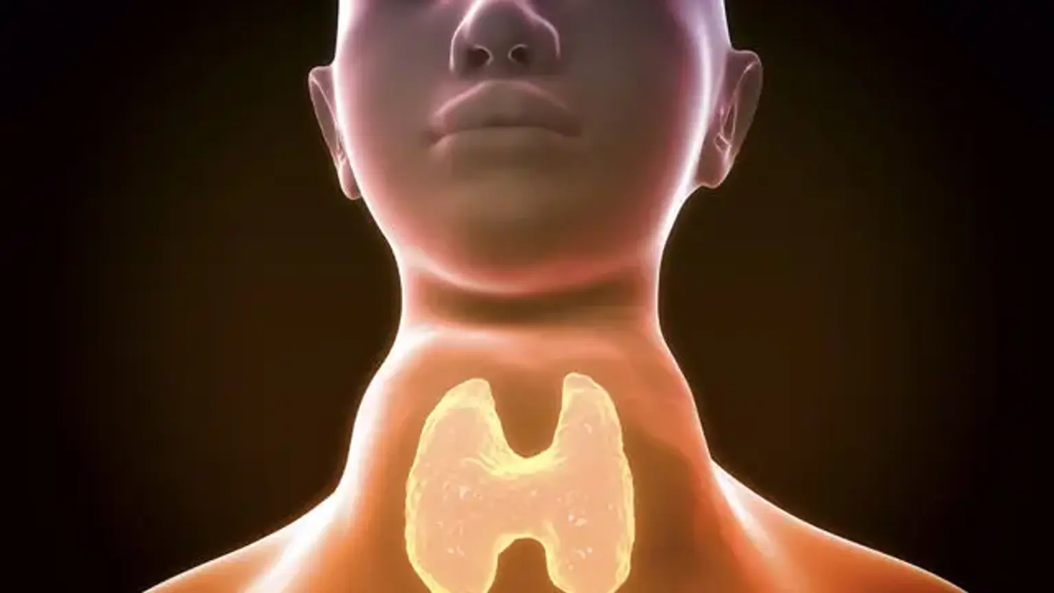
Goiter is a broad word that refers to an enlargement of the thyroid gland that indicates that the volume of the thyroid gland is bigger than usual. Inspection, palpation, or an imaging scan can all be used to detect the existence of a goiter.
Normal thyroid gland dimensions are 4 to 4.8 cm sagittal, 1 to 1.8 cm transverse, and 0.8 to 1.6 cm anteroposterior. On ultrasound estimates, this equates to a volume of 7 to 10 mL and a weight of 10-20 grams. Thyroid size grows in proportion to age and body size. It is greater in males than in females. With increased iodine consumption, the size reduces.
A multitude of physiological or pathological events can cause the thyroid gland to expand. Adolescent goiter and pregnancy are two causes of a physiological goiter. Goiter can occur in conjunction with euthyroidism, hypothyroidism, or hyperthyroidism. It might be diffuse, nodular, or multinodular in appearance.
Because the expanding thyroid is not restrained by the weak anterior cervical muscles, subcutaneous tissue, or skin, the thyroid gland normally expands anteriorly in the neck. The term goiter is most commonly used to refer to cervical goiter. A substernal or retrosternal goiter occurs when the thyroid gland enlarges inferiorly and goes through the thoracic inlet.
Diffuse toxic goiter definition
Goiter is simply the swelling of the thyroid gland. It can be caused by a variety of factors, the most prevalent of which is a lack of dietary iodine. Graves illness and Hashimoto's disease, on the other hand, are more frequent in the United States. Goiter has been divided into several categories, including :
- Morphology (nodular/diffuse),
- Functional state (hyper/hypo/euthyroid),
- Malignancy, and so on.
A "diffuse, toxic" substance A goiter is characterized as a thyroid gland that is diffusely hyperplastic and generates an excess of thyroid hormones.
Epidemiology
Goiter is simply the swelling of the thyroid gland. It can be caused by a variety of factors, the most prevalent of which is a lack of dietary iodine. Graves illness and Hashimoto's disease, on the other hand, are more frequent in the United States.
Women are 7-10 times more likely than males to have a diffuse toxic goiter. It is frequently connected with or immediately following pregnancy. Diffuse toxic goiter can affect people of all ages, however, it is uncommon in children under the age of ten and uncommon in the elderly. The incidence is highest in the third and fourth decades of life. There is an elevated incidence among postpartum women when the condition commonly manifests itself for the first time.
Etiology
Thyroid-stimulating hormone (TSH)-receptor stimulating antibodies produce diffuse toxic goiter and hyperthyroidism. Although the specific reason is unknown, it has been proposed that there is a hereditary absence of suppressor T cells, which results in uncontrolled antibody production, leading to autoimmune illness. The antibody might cross the placenta, causing fetal and neonatal hyperthyroidism.
As with most of these illnesses, a mix of hereditary and environmental variables is frequently present. Immune-regulatory and thyroid autoantigen genes, such as thyroglobulin and TSH-receptor genes, are involved in Graves' illness. The following are examples of non-genetic risk factors:
- Female sex
- Pregnancy
- Mental stress
- Smoking and alcohol
- Infectious diseases
- Iodine administration
- Drugs such as lithium and iodine-containing agents, as well as agents, including amiodarone, interferons and interleukins, and antiretroviral agents
Given that the frequency of Graves disease is ten times higher in women than in males, sex hormones and chromosomal abnormalities, such as skewed X chromosome inactivation, are thought to be triggered. Infection (particularly with Yersinia enterocolitica, due to a molecular mimicry mechanism involving the TSH receptor), vitamin D and selenium deficiencies, thyroid injury, and immunomodulating medications are also implicated.
Associated ophthalmopathy affects roughly 20% of Graves disease patients and is poorly understood. It is assumed to be a similar but distinct autoimmune condition that affects the extraocular muscles. The course of the ailment may be similar to or distinct from that of hyperthyroidism. The presence and severity of clinical ophthalmopathy correlate with the level of anti-TSH receptor antibodies.
Pathophysiology
Diffuse, toxic goiter is characterized by a diffusely swollen, vascular gland with a rubbery consistency. The follicular cells are hypertrophic and hyperplastic under the microscope, with minimal colloid. Lymphocytes and plasma cells enter the gland and can eventually form lymphoid follicles.
All instances of diffuse, toxic goiter are not Graves disease and may be caused by a variety of non-autoimmune causes. However, the vast majority of cases are auto-immune in character. Antibodies are directed against the thyroid-stimulating hormone receptor (TSHr) on follicular cells in Graves disease. Chronic activation of these receptors results in the creation of excess T3, T4, and eventually the growth of the thyroid gland, culminating in a goiter.
It is estimated that 75-80% of Graves disease heritability is caused by probably related genes, variations, and polymorphisms. Although environmental and epigenetic variables may have a role in the pathophysiology of this illness (e.g., onset, progression, and development), the combination of genetic, epigenetic, and immunologic factors is unknown.
Clinical presentation
History
- Patients may come with a history of one or more of the side effects of hyperthyroidism. Weight loss, heat sensitivity (together with other heat-related symptoms such as polydipsia and sweating), tremor, uneasiness, anxiety, weariness, palpitations, shortness of breath, frequent defecation, diarrhea, nausea, vomiting, and so on are examples.
- Patients may also complain of visible neck swelling or a lump in the neck, globus, swallowing trouble, orthopnea, and other symptoms.
Patients with Graves disease may present with additional features like:
- Ophthalmopathy/Graves orbitopathy (seen in 25% of patients): proptosis, diplopia, periorbital edema, excessive lacrimation, etc.
- Thyroid dermatopathy (rare, seen in 4% of patients and usually concurrent with orbitopathy): Slightly thickened pigmented skin, especially over the pretibial area.
- Reproductive system- irregular menstruation
Physical Examination
The common findings in a patient with the diffuse, toxic goiter on physical exam are as follows:
- Constitutional - weight loss
- Head, eyes, ears, neck-neck swelling, proptosis, lid lag, periorbital edema
- Cardiovascular system: tachycardia, irregular heartbeat, systolic hypertension
- Neuromuscular: tremor of extremities, hyperreflexia, hyperactivity, muscle weakness
- Respiratory system - tachypnea
- Skin and extremities - moist, warm skin, pretibial myxedema
Diagnosis
Because serum thyroid-stimulating hormone (TSH) has the best sensitivity and specificity in the diagnosis of thyroid diseases, it should be the first biochemical test performed. If the clinical presentation and serum TSH measurement do not reveal the diagnosis, additional diagnostic tests should be chosen based on available knowledge and resources and may include the following:
- Measurement of TSH-receptor antibodies (TRAb)
- Determination of the radioactive iodine uptake (RAIU)
- Measurement of thyroidal blood flow on ultrasonography
- Thyroid scan (technetium-99m or iodine-123)
The presence of ophthalmopathy implies a Graves disease diagnosis, and no more diagnostic testing is required to determine the source of the hyperthyroidism. If oculopathy is suspected, orbital computed tomography (CT) scanning or magnetic resonance imaging (MRI) may be used.
Diffuse toxic goiter is characterized by a suppressed serum TSH level, an elevated serum free thyroxine level (or T3 if needed), raised TRab titers, or increased radioiodine absorption.
Laboratory Studies
TSH and thyroid hormone levels
TSH (thyroid-stimulating hormone) levels in serum (sensitive or third-generation assay) that are suppressed below normal signal the need for further testing. Graves disease is ruled out by normal serum TSH levels.
Hyperthyroidism is diagnosed when serum free thyroxine (T4) levels are high. In around 5% of instances, levels will be within the normal range. If free thyroxine levels are normal, measure total or free serum triiodothyronine (T3) levels. Hyperthyroidism is diagnosed if the levels are high. Subclinical hyperthyroidism is present if the levels are normal.
Anti-TSH receptor antibodies in serum can be measured. Depending on the test, these antibodies are detected in more than 90% of instances of diffuse toxic goiter. Serum antithyroid antibodies can indicate the existence of Hashimoto thyroiditis
Anabolic steroids, androgens, estrogens, heparin, iodine, phenytoin, rifampin, salicylates, and thyroxine/triiodothyronine are examples of drugs that may affect T4 test findings.
Imaging Studies
Radioactive iodine uptake (RAIU) test
Thyroid radioactive iodine uptake test results are as follows :
- Diffuse toxic goiter (Graves disease): Diffuse high radioiodine uptake.
- Toxic multinodular goiter: Normal or high radioiodine uptake with an asymmetrical pattern
- Toxic adenoma: Normal or high radioiodine uptake with a localized and focal pattern
- Thyroiditis: Low radioiodine uptake
- Thyrotoxicosis from extrathyroidal sources of thyroid hormone: Very low uptake
Ultrasonography
Thyroid ultrasonography and thyroid RAIU have comparable sensitivity for detecting Graves disease. The advantages of ultrasonography include the absence of ionizing radiation exposure, improved accuracy in detecting thyroid nodules, and a lower cost than RAIU. Doppler ultrasonography successfully differentiates diffuse toxic goiter (increased blood flow, diffusely enlarged hypoechogenic) from thyroiditis (decreased blood flow), the most prevalent clinical issue in the differential diagnosis of hyperthyroidism.
Management
The treatment modalities for diffuse toxic goiter include:
Antithyroidthyroid Drugs (ATD)
- The antithyroid drug options are propylthiouracil, thiamazole, and carbimazole.
- Except in individuals who have had a bad reaction to the medicine or who are in the first trimester of pregnancy, thiamazole is a favored treatment for Graves disease. It is favored over propylthiouracil because it is more effective, has a longer half-life and duration of action, and may be used once daily.
- The initial regimen for administering ATD is titration, in which the dosage of ATD is decreased to the lowest achievable dose after the euthyroid condition is attained. The next treatment is block-and-replace, which involves administering a high dosage of ATD in conjunction with thyroxine to maintain euthyroid status.
- One disadvantage of ATD therapy is the chance of recurrence, particularly in the first year after ceasing treatment. Studies show a 50% to 55% chance of recurrence with unfavorable prognostic variables such as severe hyperthyroidism, big goiter, high T3: T4 ratios, chronically suppressed TSH, and high baseline TRAb concentrations.
- Agranulocytosis, hepatotoxicity, vasculitis, and other serious adverse effects of ATD treatment include agranulocytosis, hepatotoxicity, and vasculitis.
Radio-iodine Therapy (I-31, RAI)
- RAI is the most often used therapy approach for Graves disease in the United States, and it is both safe and effective.
- Pregnancy, nursing, and severe uncontrolled thyrotoxicosis are essential contraindications to this treatment.
- It is available in liquid or pill form, and fixed-dosage therapy is just as effective as calculated dose therapy based on gland volume, iodine intake, and other factors.
- To guarantee optimal RAI absorption, patients must cease all iodine-containing medications and follow an iodine-restricted diet.
- ATD treatment should be stopped before using RAI, and it can be restarted after a week of using RAI if necessary.
- The possibility of developing hypothyroidism, as well as transitory radiation hyperthyroidism or aggravation of thyroid-associated ophthalmopathy, are potential adverse effects (TAO).
- While hypothyroidism is routinely tested in subsequent follow-ups, prednisone has been shown to slow the course of moderate TAO.
- Patients must be informed about the necessity for lifetime monitoring for illness recurrence or the development of hypothyroidism, as well as rapid treatment if found.
Surgery
- Thyroidectomy is the most effective treatment for diffuse, toxic goiter, with complete thyroidectomy being more effective than sub-total with comparable adverse effects.
- Because of the risks connected with general anesthesia, such as recurrent laryngeal nerve palsy and hypothyroidism, it is normally reserved as a last resort.
- It is favored in individuals who cannot take antithyroid drugs or RAI, as well as in those with compressive symptoms.
To reduce anesthetic risks, cardiovascular/hemodynamic problems, and the danger of thyroid storm, patients should be made euthyroid prior to surgery. If normalization with antithyroid medications is not achievable, beta-blockers and potassium iodide 4 drops daily for 10 days will reduce thyroid gland vascularity.
Pregnancy and breastfeeding
Radioactive iodine is not recommended during pregnancy or breastfeeding. Pregnancy frequently improves the immunologic disease state during pregnancy but then relapses after delivery. PTU is the preferred treatment because it has less placental transfer than MMI. Rare congenital defects related to MMI (for example, aplasia cutis) are much less likely to be associated with PTU. Overall, the risk of congenital abnormalities with these medicines is comparable to the background normal rate. If there is a difficulty with PTU, MMI may be utilized.
The objective is to keep the free thyroxine level in the higher range of normal in order to limit fetal medication exposure. Monthly monitoring of serum-free thyroxine normally permits the PTU dose to be reduced and, in many cases, stopped in the third trimester. PTU and MMI can both be utilized in nursing moms. Although a tiny quantity of the medication enters the milk, newborn thyroid levels remain normal. PTU and MMI are not harmful during pregnancy or breastfeeding.
Differential Diagnosis
Although diagnosed with the various forms of evaluation, as mentioned above, the differential diagnosis of diffuse, toxic goiter includes:
- Thyrotoxicosis factitia- over prescription/consumption of thyroid hormones
- Subacute thyroiditis
- Multinodular goiter
- TSH secreting pituitary adenoma
- Iatrogenic iodine supplementation
Prognosis
The normal course of diffuse toxic goiter is typically benign and may even spontaneously resolve. The severity of the symptoms and their impact on quality of life vary from person to person and are influenced by age and gender.
Mortality is uncommon, but when it does occur, it is usually related to cardiovascular issues including heart failure, arrhythmias, or myocardial infarction. formalized paraphrase Thyroid storm is uncommon, yet it can be deadly due to dehydration, heat, and organ failure. Myocardial ischemia, congestive failure, or atrial arrhythmias may need anticoagulant therapy. It is possible that you will become illiterate or infected.
Morbidity can be caused by accelerated bone turnover and osteoporosis, particularly in postmenopausal women, or by atrial fibrillation and its sequelae, such as thromboembolism, particularly in older males. Changes in personality and psychopathology, as well as physical weakness and systemic ailments, all contribute to changes in quality of life. Associated oculopathy can be painful, especially if you have double vision. It is possible that it will eventually impair the integrity of the cornea and jeopardize vision.
Associated dermopathy is rare and typically mildly symptomatic, although it can be severe enough to be disabling. Associated hypokalemic periodic paralysis, which is more frequent in Asian males, can be rapid, dramatic, and worrisome, although it normally recovers in a benign manner after a few hours of skeletal muscle paralysis.
There is an increased chance of related immunologic disorders, such as adrenal insufficiency; each condition has its own morbidity and mortality, especially if misdiagnosed.
Complications
- Hyperthyroidism, or thyroid storm due to prolonged untreated excess of thyroid hormone
- Dermatopathy, associated with Graves disease
- Graves ophthalmopathy
Conclusion
Diffuse, toxic goiter may present with hypermetabolic (ex. heat sensitivity, sweating, weight loss, etc.) and adrenergic (ex. palpitations, tremors, emotional lability, etc.) signs of hyperthyroidism, in addition to goiter enlargement. However, older people may not arrive with adrenergic symptoms, but rather with apathy, atrial fibrillation, and other indications of depression, malignancy, or heart problems.
The diagnosis of diffuse, toxic goiter is based on a combination of clinical symptoms and testing. It is critical to evaluate radiographic and blood studies in order to determine the underlying etiology of the condition. Once a patient has been identified, it is the physician's obligation to assist the patient in selecting the appropriate treatment choice based on their profile and with complete awareness of potential adverse effects.
Women must be extra cautious and well-informed since being pregnant or nursing necessitates changing their kind of therapy if it is inappropriate. Because this illness might also cause aesthetic issues. Physicians must be alert and observant to them, as well as educate patients on available treatment options and have a realistic attitude to treatment response.